Systems Biology: Foundational Principles and Transformative Applications in Drug Discovery and Healthcare
This article provides a comprehensive exploration of systems biology, from its core principles to its cutting-edge applications in biomedicine.
Systems Biology: Foundational Principles and Transformative Applications in Drug Discovery and Healthcare
Abstract
This article provides a comprehensive exploration of systems biology, from its core principles to its cutting-edge applications in biomedicine. Tailored for researchers and drug development professionals, it details the foundational concepts of analyzing biological systems as interconnected networks. It further examines quantitative methodological approaches like QSP modeling and constraint-based analysis, addresses common troubleshooting challenges in model complexity and data integration, and validates the discipline's impact through comparative case studies in drug development and regenerative medicine. The synthesis offers a forward-looking perspective on how systems biology is poised to advance personalized therapies and reshape biomedical innovation.
From Reductionism to Holism: Core Concepts and the Rise of Systems Thinking in Biology
Systems biology represents a fundamental paradigm shift in biological science, moving away from the traditional reductionist approach that focuses on isolating and studying individual components, such as a single gene or protein. Instead, it adopts a holistic perspective that investigates complex biological systems as integrated wholes, focusing on the dynamic interactions and emergent properties that arise from these interactions [1]. This interdisciplinary field combines biology, computer science, mathematics, and engineering to develop comprehensive models of biological processes, recognizing that the behavior of a complete biological system cannot be fully understood by examining its parts in isolation [1].
The field formally emerged as a distinct discipline around the year 2000, with the establishment of dedicated institutions such as the Institute for Systems Biology in Seattle [1]. This development was catalyzed by projects like the Human Genome Project, which demonstrated the power of systems-thinking approaches to tackle complex biological challenges [1]. Systems biology acknowledges that biological systems operate through intricate networks of interactions—whether metabolic pathways, cell signaling cascades, or genetic regulatory circuits—and that understanding these networks requires both comprehensive data collection and sophisticated computational modeling [2] [1].
Core Principles of Systems Biology
Foundational Concepts
Systems biology is guided by several core principles that distinguish it from traditional biological research. These principles provide the philosophical and methodological foundation for how systems biologists approach scientific inquiry.
Integration: Systems biology emphasizes the integration of data from multiple sources and scales of biological organization. This includes combining information from genomics, transcriptomics, proteomics, and metabolomics—often referred to collectively as "omics" technologies—to build comprehensive models of biological systems [1]. This integration allows researchers to capture the complexity of biological systems more completely than would be possible by studying any single data type in isolation.
Dynamic Systems Modeling: At the heart of systems biology is the use of mathematical and computational models to simulate the dynamic behavior of biological networks over time [1]. These models enable researchers to make testable predictions about how a system will respond to perturbations, such as genetic modifications or environmental changes, and to identify key control points within complex networks.
Emergent Properties: A central tenet of systems biology is that complex properties of a biological system—such as cellular decision-making, tissue organization, or organismal behavior—arise from the interactions of its simpler components and cannot be predicted by studying those components alone [1]. These emergent properties represent a fundamental aspect of biological complexity that requires systems-level approaches to understand.
Holistic View: In direct opposition to reductionism, systems biology maintains that analyzing the entire system is necessary to understand its structure, function, and response to disturbances [1]. This holistic perspective recognizes that biological function often depends on the coordinated activity of numerous elements working together in network structures.
Contrast with Traditional Approaches
Systems biology differs fundamentally from traditional molecular biology in its approach to scientific investigation [1]. Where traditional molecular biology typically follows a reductionist approach—focusing on a single gene or protein to understand its specific function in isolation—systems biology takes a holistic or integrative approach. It studies how all components (genes, proteins, metabolites, etc.) interact simultaneously as a network to produce the collective behavior of a cell or organism [1]. While molecular biology asks "What does this part do?", systems biology asks "How do all the parts work together?" [1].
This philosophical difference has profound methodological implications. Systems biology has been described as having a mission that "puts it at odds with traditional paradigms of physics and molecular biology, such as the simplicity requested by Occam's razor and minimum energy/maximal efficiency" [2]. Through biochemical experiments on control, regulation, and flux balancing in organisms like yeast, researchers have demonstrated that these traditional paradigms are often "inapt" for understanding biological systems [2].
Quantitative Methodologies and Data Analysis
Mathematical Modeling in Systems Biology
Mathematical modeling is essential in systems biology because biological systems are incredibly complex, with thousands of interacting components that the human mind cannot track simultaneously [1]. Mathematical models provide a framework to organize vast amounts of high-throughput data into a coherent structure, simulate system behavior under different conditions, make testable predictions about system responses, and identify key components or pathways that have the most influence on the system's overall behavior [1].
The process of model development typically follows an iterative cycle of hypothesis generation, experimental design, data collection, model building, and prediction testing. This cycle allows for continuous refinement of our understanding of biological systems. For example, in studying cell migration—a driving force behind many diverse biological processes—researchers have found that "valuable information contained in image data is often disregarded because statistical analyses are performed at the level of cell populations rather than at the single-cell level" [3]. By developing models that can characterize and classify tracked objects from image data at the single-cell level, systems biologists can more accurately interpret migration behavior [3].
Key Quantitative Analytical Approaches
Table 1: Quantitative Analysis Methods in Systems Biology
| Method | Application | Key Features |
|---|---|---|
| Transcriptomics | Measurement of complete set of RNA transcripts | Provides snapshot of gene expression patterns [1] |
| Proteomics | Study of complete set of proteins | Identifies protein expression and post-translational modifications [1] |
| Metabolomics | Analysis of complete set of small-molecule metabolites | Reveals metabolic state and fluxes [1] |
| Glycomics | Organismal, tissue, or cell-level measurements of carbohydrates | Characterizes carbohydrate composition and modifications [1] |
| Lipidomics | Organismal, tissue, or cell-level measurements of lipids | Profiles lipid composition and dynamics [1] |
| Interactomics | Study of molecular interactions within the cell | Maps protein-protein and other molecular interactions [1] |
These analytical approaches generate massive datasets that require sophisticated computational tools for interpretation. The quantification of biological processes from experimental data, particularly image data, involves "automated image analysis followed by rigorous quantification of the biological process under investigation" [3]. Depending on the experimental readout, this quantitative description may include "the size, density and shape characteristics of cells and molecules that play a central role in the experimental assay" [3]. When video data are available, "tracking of moving objects yields their distributions of instantaneous speeds and turning angles, as well as the frequency and duration of contacts between different types of interaction partners" [3].
Experimental Protocols and Methodologies
Framework for Experimental Protocols
Experimental protocols in systems biology require careful structuring to ensure reproducibility and meaningful data integration. The SIRO model (Sample, Instrument, Reagent, Objective) provides a minimal information framework for representing experimental protocols, similar to how the PICO model supports search and retrieval in evidence-based medicine [4]. This model represents the minimal common information shared across experimental protocols and facilitates classification and retrieval without necessarily exposing the full content of the protocol [4].
A comprehensive ontology for representing experimental protocols—the SMART Protocols ontology—has been developed to provide the structure and semantics for data elements common across experimental protocols [4]. This ontology represents the protocol as a workflow with domain-specific knowledge embedded within a document, enabling more systematic representation and sharing of experimental methods [4]. Such formal representations are particularly important in systems biology, where protocols can be extremely complex; for example, the protocol for chromatin immunoprecipitation on a microarray (ChIP-chip) has "90 steps and uses over 30 reagents and 10 different devices" [4].
Key Methodological Approaches
Systems biology employs two main philosophical approaches to investigating biological systems [1]:
Top-down approach: The top-down perspective considers as much of the system as possible and relies primarily on experimental results. Techniques like RNA-Sequencing represent examples of this exploratory top-down perspective, generating comprehensive datasets that can be analyzed to identify patterns and relationships within the system [1].
Bottom-up approach: The bottom-up perspective is used to create detailed models while incorporating experimental data. This approach often starts with well-characterized components and their interactions, building toward a more complete understanding of system behavior through iterative model refinement [1].
Both approaches benefit from the ongoing development of more sophisticated measurement technologies. As noted in research on quantitative analysis of biological processes, "From the viewpoint of the Image-based Systems Biology approach, extracted quantitative parameters are only intermediate results that are exploited as a basis for constructing image-derived models" [3]. This highlights the iterative nature of systems biology, where quantitative measurements feed into model building, which in turn guides further experimental design.
Research Applications and Implications
Applications in Biomedicine and Biotechnology
Table 2: Applications of Systems Biology Across Fields
| Field | Application | Impact |
|---|---|---|
| Personalized Medicine | Patient-specific treatment modeling | Enables tailored therapies based on individual genetic and molecular profiles [1] |
| Drug Discovery | Identification of new drug targets | Accelerates development and predicts potential side effects [1] |
| Agricultural Improvement | Engineering crops with enhanced traits | Develops drought-resistant and higher-yield crops [1] |
| Disease Diagnosis | Development of accurate diagnostic tools | Identifies biomarkers representing overall biological system state [1] |
| Cancer Research | Modeling tumour network disruptions | Identifies key network vulnerabilities and predicts treatment responses [1] |
Systems biology has revolutionized how we approach complex diseases like cancer. Since cancer is "a disease of complex network disruptions, not just a single faulty gene," the systems biology approach is particularly well-suited for studying it [1]. Researchers can create 'systems models' of tumors by integrating patient data on genomics, protein levels, and metabolic activity. These models help identify key network vulnerabilities that drive malignant growth, simulate how a tumor might respond to particular chemotherapy drugs, predict which combination of therapies would be most effective for a specific patient, and discover new biomarkers for early diagnosis and prognosis [1].
Relationship with Bioinformatics
Systems biology and bioinformatics are deeply interconnected and mutually dependent fields [1]. Bioinformatics develops the computational tools, algorithms, and databases needed to collect, store, and analyze massive biological datasets (like DNA sequences or protein structures). Systems biology then uses these bioinformatic tools to interpret the data, build its models, and understand the interactions within the biological system [1]. In essence, "bioinformatics provides the 'how' (the tools and analysis), while systems biology provides the 'why' (the biological understanding and interpretation of the system as a whole)" [1].
This symbiotic relationship extends to the use of specific computational tools and languages in systems biology research. These include "new forms of computational models, such as the use of process calculi to model biological processes and novel approaches for integrated stochastic π-calculus, BioAmbients, Beta Binders, BioPEPA, and Brane calculi and constraint-based modelling" [1]. Additionally, systems biology relies on the "integration of information from the literature, using techniques of information extraction and text mining" [1], as well as programming languages like Python and C++ for building models and analyzing data [1].
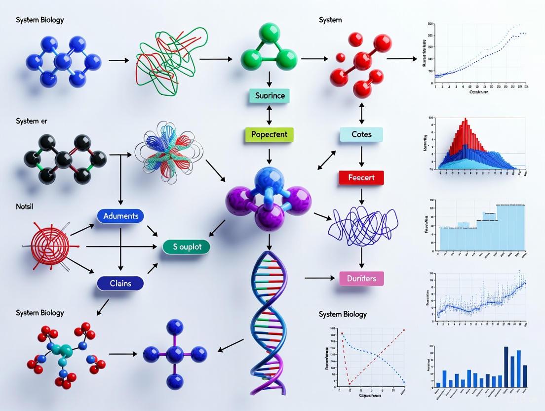
Visualization of Systems Biology Workflows
Integrated Research Workflow
The following diagram illustrates the integrated cyclical process of systems biology research, showing how data generation, integration, modeling, and validation form an iterative feedback loop that drives scientific discovery:
Biological Network Regulation
This diagram visualizes the emergent properties in a biological regulatory network, demonstrating how complex behaviors arise from multiple interacting components:
Essential Research Reagents and Materials
Table 3: Essential Research Reagent Solutions in Systems Biology
| Reagent/Material | Function | Application Examples |
|---|---|---|
| RNA Extraction Kits | Isolation of high-quality RNA from fresh/frozen tissue [4] | Transcriptomics analysis, gene expression studies [1] |
| Antibodies | Detection and quantification of specific proteins | Proteomics, chromatin immunoprecipitation (ChIP) [4] |
| Chemical Entities | Small molecules for metabolic studies | Metabolomics, flux balance analysis [4] |
| Cell Culture Media | Support growth of specific cell types | Cell line maintenance, experimental assays [1] |
| Fluorescent Dyes/Labels | Tagging molecules for detection and tracking | Imaging, flow cytometry, protein localization [3] |
| Enzymes | Catalyze specific biochemical reactions | DNA manipulation, protein modification studies [1] |
| Buffer Systems | Maintain optimal pH and ionic conditions | All experimental protocols requiring specific conditions [4] |
| Computational Tools | Data analysis, modeling, and visualization | Bioinformatics pipelines, network analysis [1] |
The selection of appropriate reagents and materials is critical for generating reliable data in systems biology research. As highlighted in the SIRO model framework, careful documentation of samples, instruments, reagents, and objectives is essential for protocol reproducibility and effective data integration [4]. For example, in a protocol for "Extraction of total RNA from fresh/frozen tissue," specific reagents and their manufacturers must be clearly documented to ensure consistent results across different laboratories [4].
Systems biology continues to evolve as a discipline, developing its own fundamental principles that distinguish it from both traditional biology and physics [2]. As a relatively young field, it has already demonstrated significant value in addressing complex biological questions, though some have noted that as of 2012, it had "not fulfilled everyone's expectations" because many of its applications had not yet been translated into practical use [1]. Nevertheless, proponents maintain confidence that "it may once demonstrate more value in the future" [1].
The future of systems biology will likely involve increasingly sophisticated multi-scale models that integrate data from molecular levels to whole organisms, ultimately leading to more predictive models in medicine and biotechnology. As quantification technologies advance and computational power increases, systems biology approaches will become increasingly central to biological research, potentially transforming how we understand, diagnose, and treat complex diseases. The field continues to discover "quantitative laws" and identify its own "fundamental principles" [2], establishing itself as a distinct scientific discipline with unique methodologies and insights.
The field of biology is undergoing a fundamental paradigm shift, moving away from reductionist, single-target approaches toward a holistic, network-level understanding of biological systems. This transition is driven by the recognition that complex diseases arise from perturbations in intricate molecular networks, not from isolated molecular defects. Supported by advances in high-throughput omics technologies and sophisticated computational models, systems biology provides the framework to analyze these complex interactions. The application of network-level understanding is now increasing the probability of success in clinical trials by enabling a data-driven matching of the right therapeutic mechanism to the right patient population. This whitepaper explores the foundational principles of this paradigm shift, detailing the computational methodologies, experimental protocols, and practical applications that are redefining biomedical research and therapeutic development [5] [6].
Traditional biological research and drug discovery have long relied on a reductionist approach, investigating individual genes, proteins, and pathways in isolation. This "single-target" paradigm operates on the assumption that modulating one key molecular component can effectively reverse disease processes. However, this approach has proven inadequate for addressing complex diseases such as cancer, neurodegenerative disorders, and metabolic conditions, where pathology emerges from dysregulated networks of molecular interactions [6].
The limitations of reductionism have become increasingly apparent in pharmaceutical development, where failure to achieve efficacy remains a primary reason for clinical trial failures. Systems biology has emerged as an interdisciplinary field that addresses this complexity by integrating biological data with computational and mathematical models. It represents a fundamental shift toward understanding biological systems as integrated networks rather than collections of isolated components [6]. This paradigm shift enables researchers to capture the emergent properties of biological systems—characteristics that arise from the interactions of multiple components but cannot be predicted from studying individual elements alone [5].
The Theoretical Foundation of Network Biology
The Complexity of Biological Systems
Biological systems operate through multi-scale interactions that span from molecular complexes to cellular networks, tissue-level organization, and ultimately organism-level physiology. The complexity of these systems is evidenced by phenomena such as incomplete penetrance and disease heterogeneity, even in genetic diseases with defined causal mutations. For example, in conditions like Huntington's disease, Parkinson's disease, and certain cancers, inheritance of causal mutations does not consistently lead to disease manifestation, indicating the influence of broader network dynamics [6].
Network biology facilitates system-level understanding by aiming to: (1) understand the structure of all cellular components at the molecular level; (2) predict the future state of a cell or organism under normal conditions; (3) predict output responses for given input stimuli; and (4) estimate system behavior changes upon component or environmental perturbation [5].
Key Data Types in Network Analysis
Modern systems biology leverages diverse, high-dimensional data types to construct and analyze biological networks:
Table 1: Primary Data Types in Network Biology
| Data Type | Description | Application in Network Biology |
|---|---|---|
| Genomic Sequences | DNA nucleotide sequences preserving genetic information | Identifying genetic variants and their potential network influences [5] |
| Molecular Structures | Three-dimensional configurations of biological macromolecules | Predicting molecular binding interactions and complex formation [5] |
| Gene Expression | mRNA abundance measurements under specific conditions | Inferring co-regulated genes and regulatory relationships [5] |
| Protein-Protein Interactions (PPI) | Binary or complex physical associations between proteins | Constructing protein interaction networks to identify functional modules [5] |
| Metabolomic Profiles | Quantitative measurements of metabolite concentrations | Mapping metabolic pathways and flux distributions [6] |
The integration of these multimodal datasets enables the reconstruction of comprehensive molecular networks that more accurately represent biological reality than single-data-type approaches [6].
Methodological Framework: Constructing Causal Networks
The SiCNet Methodology for Causal Inference
Substantial challenges persist in gene regulatory network (GRN) inference, particularly regarding dynamic rewiring, inferring causality, and context specificity. To address these limitations, the single cell-specific causal network (SiCNet) method has been developed to construct molecular regulatory networks at single-cell resolution using a causal inference strategy [7] [8].
The SiCNet protocol operates in two primary phases:
Phase 1: Reference Network Construction
- Data Preparation: Begin with single-cell RNA sequencing data that has undergone standard quality control, normalization, and batch effect correction.
- Causal Inference: Apply causal inference algorithms (e.g., constraint-based, score-based, or hybrid methods) to construct a reference causal network from a designated reference dataset.
- Network Validation: Validate the reference network using known biological interactions from databases and functional enrichment analyses.
Phase 2: Cell-Specific Network Inference
- Expression Profiling: For each individual cell, obtain the normalized gene expression profile.
- Network Personalization: Utilize the reference network as a scaffold and integrate the cell-specific expression profile to infer cell-specific causal relationships.
- Quantification: Calculate regulatory activity by counting the number of target genes for each regulator in the cell-specific network, generating a network outdegree matrix (ODM) that retains the same dimensions as the original gene expression matrix but represents higher-order regulatory information [7].
The ODM enhances the resolution and clarity of cell type distinctions, offering superior performance in visualizing complex and high-dimensional data compared with traditional gene expression matrices [7].
Bayesian Networks for Causal Discovery
Bayesian Networks (BNs) provide another powerful framework for modeling complex systems under uncertainty. A BN consists of:
- A collection of random variables represented as nodes in a Directed Acyclic Graph (DAG)
- A finite set of mutually exclusive states for each variable
- A Conditional Probability Distribution (CPD) for each variable given its parents
BNs leverage conditional independence to compactly represent the joint probability distribution over a set of random variables, factorized as: P(X₁, X₂, ..., Xₙ) = Πᵢ₌₁ⁿ P(Xᵢ | Pa(Xᵢ)) where Pa(Xᵢ) denotes the parent variables of Xᵢ in the network [9].
Structure learning algorithms for BNs fall into three primary categories:
- Constraint-based algorithms (e.g., PC-Stable) that use conditional independence tests
- Score-based algorithms that optimize scoring functions like BIC or AIC
- Hybrid approaches that combine both methods [9]
Practical Implementation: Research Protocols and Tools
Experimental Workflow for Network Biology
Implementing a systems biology approach requires a structured workflow that integrates experimental and computational components:
Research Reagent Solutions
Table 2: Essential Research Reagents and Tools for Network Biology
| Reagent/Tool | Function | Application Context |
|---|---|---|
| scRNA-seq Platforms | High-throughput measurement of gene expression at single-cell resolution | Generating input data for SiCNet and other single-cell network inference methods [7] [8] |
| Mass Spectrometry Systems | Quantitative profiling of proteins and metabolites | Generating proteomic and metabolomic data for multi-layer network integration [5] [6] |
| gCastle Python Toolbox | End-to-end causal structure learning | Implementing various causal discovery algorithms for network construction [9] |
| bnlearn R Package | Comprehensive Bayesian network learning | Structure learning, parameter estimation, and inference for Bayesian networks [9] |
| Position Weight Matrices (PWMs) | Representation of DNA binding motifs | Identifying transcription factor binding sites for regulatory network inference [5] |
Applications in Drug Discovery and Development
Enhancing Therapeutic Efficacy
Systems biology approaches are revolutionizing drug discovery by enabling the development of multi-target therapies that address the complex network perturbations underlying disease. Unlike single-target approaches that often prove insufficient for complex diseases, network-based strategies can identify optimal intervention points and therapeutic combinations [6].
The systems biology platform for drug development follows a stepwise approach:
- Characterize key pathways contributing to the Mechanism of Disease (MOD)
- Identify and design therapies that reverse disease mechanisms through targeted Mechanisms of Action (MOA)
- Optimize and translate therapies into clinical practice [6]
Clinical Translation and Biomarker Development
Advanced computational methods applied to large preclinical and clinical datasets enable the development of quantitative clinical biomarker strategies. These approaches facilitate:
- Patient stratification through identification of molecular signatures in heterogeneous diseases
- Detection of drug activity and early modulation of disease mechanisms
- Predictive modeling of clinical outcomes for early Go/No-Go decision making [6]
Network-based approaches can identify critical state transitions in disease progression by calculating dynamic network biomarkers (DNBs), providing early warning signals before phenotypic manifestation of disease [7].
Future Perspectives and Challenges
As systems biology continues to evolve, several emerging trends and challenges will shape its future application:
Integration of Spatial Dimensions: Network biology is expanding to incorporate spatial context through technologies like spatial transcriptomics, enabling the construction of spatially-resolved regulatory networks that capture tissue architecture and function [7].
Temporal Network Dynamics: Understanding network rewiring over time remains a fundamental challenge. New methods like SiCNet that can capture dynamic regulatory processes during cellular differentiation and reprogramming represent significant advances in this area [8].
Computational Scalability: As multi-omics datasets continue to grow in size and complexity, developing computationally efficient algorithms for network inference and analysis will be essential. Cloud computing and innovative learning approaches like artificial intelligence are rapidly closing this capability gap [6].
The paradigm shift from linear cause-effect thinking to network-level understanding represents more than just a methodological evolution—it constitutes a fundamental transformation in how we conceptualize, investigate, and intervene in biological systems. By embracing the complexity of biological networks, researchers and drug developers can identify more effective therapeutic strategies that address the true multifaceted nature of human disease.
Systems biology research is fundamentally guided by three core principles: interconnectedness, which describes the complex web of interactions between biological components; dynamics, which focuses on the time-dependent behaviors and state changes of these networks; and robustness, which is the system's capacity to maintain function amidst perturbations [10] [11]. These pillars provide the conceptual framework for understanding how biological systems are organized, how they behave over time, and how they achieve stability despite internal and external challenges. This whitepaper provides an in-depth technical examination of these principles, with particular emphasis on quantitative approaches for analyzing robustness in biological networks, offering methodologies and resources directly applicable to research and drug development.
The Interconnectedness of Biological Networks
Biological interconnectedness refers to the topological structure of relationships between components—genes, proteins, metabolites—within a cell or organism. This network structure is not random but organized in ways that critically influence system behavior and function.
Topological Measures of Network Structure
Quantifying interconnectedness requires specific metrics that describe the network's architecture [10]. The following table summarizes key topological measures used to characterize biological networks:
Table 1: Topological Metrics for Quantifying Network Interconnectedness
| Metric | Description | Biological Interpretation |
|---|---|---|
| Node Degree Distribution | Distribution of the number of connections per node | Reveals overall network architecture (e.g., scale-free, random) |
| Clustering Coefficient | Measures the degree to which nodes cluster together | Indicates local interconnectedness and potential functional modules |
| Betweenness Centrality | Quantifies how often a node lies on the shortest path between other nodes | Identifies critical nodes for information flow and network integrity |
| Network Diameter | The longest shortest path between any two nodes | Characterizes the overall efficiency of information transfer |
| Assortativity Coefficient | Measures the tendency of nodes to connect with similar nodes | Indicates resilience to targeted attacks (disassortative networks are more robust) |
| Edge Density | Ratio of existing connections to all possible connections | Reflects the overall connectivity and potential redundancy |
Functional Structures in Networks
Beyond pure topology, specific functional structures emerge from interconnectedness:
- Network Motifs: Recurring, significant patterns of interconnections that serve as basic circuit elements with defined functions [10]. Examples include feed-forward loops and single-input modules.
- Feedback Loops: Circular chains of dependency where output influences input, contributing profoundly to network stability and responsiveness [10]. Positive feedback amplifies signals, while negative feedback dampens responses.
- Modularity: The organization of networks into semi-autonomous subsystems (modules) that perform specific functions, allowing for compartmentalization of processes and failures [10].
The Dynamics of Biological Systems
Dynamics concerns the time-dependent behavior of biological systems, capturing how network components change their states and interact over time to produce complex behaviors.
Mathematical Frameworks for Modeling Dynamics
Multiple mathematical approaches exist for modeling biological network dynamics, each with specific strengths and data requirements [11]:
Table 2: Comparative Analysis of Dynamic Modeling Approaches
| Model Type | Key Features | Data Requirements | Typical Applications |
|---|---|---|---|
| Boolean Models | Components have binary states (ON/OFF); interactions use logic operators (AND, OR, NOT) [11] | Minimal (network topology, qualitative interactions) | Large networks with unknown parameters; initial qualitative analysis |
| Piecewise Affine (Hybrid) Models | Combine discrete logic with continuous concentration decay; use threshold parameters [11] | Partial knowledge of parameters (thresholds, synthesis/degradation rates) | Systems with some quantitative data available |
| Hill-type Continuous Models | Ordinary differential equations with sigmoid (Hill) functions; continuous concentrations [11] | Detailed kinetic parameters, quantitative time-series data | Well-characterized systems requiring quantitative predictions |
Attractor Landscapes and State Transitions
The dynamic behavior of biological networks can be characterized by attractors—states or patterns toward which the system evolves. These include:
- Fixed Points: Stable steady states where the system remains constant over time, often corresponding to distinct cellular states (e.g., differentiation states) [11].
- Oscillations: Periodic attractors where the system cycles through a repeating sequence of states, fundamental to circadian rhythms and cell cycles [11].
- Chaotic Attractors: Complex, non-repeating patterns sensitive to initial conditions, potentially underlying certain pathological states.
Comparative studies show that while fixed points in asynchronous Boolean models are typically preserved in continuous Hill-type and piecewise affine models, these quantitative models may exhibit additional, more complex attractors under certain conditions [11].
Robustness in Biological Systems
Robustness is defined as the ability of a biological network to maintain its functionality despite external perturbations, internal parameter variations, or structural changes [10] [12]. This property is essential for biological systems to function reliably in unpredictable environments.
Quantitative Metrics for Robustness Assessment
Robustness can be quantified using specific metrics that capture different aspects of system resilience:
Table 3: Metrics and Methods for Quantifying Robustness
| Metric/Index | Description | Application Context |
|---|---|---|
| Robustness Index | A general measure of a system's ability to withstand uncertainties and disturbances [12] | Overall system resilience assessment |
| Sensitivity Coefficient | Measures how sensitive a system's behavior is to parameter changes [12] | Parameter importance analysis; identifying critical nodes |
| Lyapunov Exponent | Quantifies the rate of divergence of nearby trajectories; indicates stability [12] | Stability analysis of dynamic systems |
| Sobol Indices | Measure the contribution of each parameter to output variance in global sensitivity analysis [10] | Comprehensive parameter sensitivity analysis |
Experimental Approaches to Measure Robustness
Experimental validation is crucial for quantifying robustness in biological systems. The following table outlines key perturbation techniques:
Table 4: Experimental Perturbation Techniques for Robustness Analysis
| Technique | Methodology | Measured Outcomes |
|---|---|---|
| Genetic Perturbations | Gene knockouts (CRISPR-Cas9, RNAi), mutations, overexpression [10] | Phenotypic assays, transcriptomics/proteomics profiling, fitness measures |
| Environmental Perturbations | Changes in temperature, pH, nutrient availability, chemical stressors [10] | Growth rates, viability assays, metabolic profiling |
| Chemical Perturbations | Small molecules, inhibitors, pharmaceuticals [10] | Dose-response curves, IC50 values, pathway activity reporters |
| High-Throughput Screening | Systematic testing of multiple perturbations in parallel [10] | Multi-parameter readouts, network response signatures |
Knockout studies represent a particularly powerful approach, ranging from single-gene knockouts that assess individual component importance to double knockouts that reveal genetic interactions and compensatory mechanisms [10]. Experimental validation confirms computational predictions and theoretical models of robustness.
Methodologies for Robustness Analysis
Computational Methods and Simulation Techniques
Computational approaches enable the systematic analysis of robustness without exhaustive experimental testing:
- Sensitivity Analysis: Quantifies how changes in input parameters affect network outputs. Local sensitivity examines small perturbations around nominal values, while global sensitivity explores the entire parameter space [10].
- Topological Attack Simulations: Assess network vulnerability to targeted node or edge removal, identifying critical components whose disruption most impacts function [10].
- Cascading Failure Models: Study how failures propagate through interconnected networks, particularly relevant for understanding disease progression and metabolic deficiencies [10].
- Flux Balance Analysis: Optimizes metabolic networks under steady-state conditions to predict robustness to gene deletions or environmental changes [10].
- Boolean Network Models: Represent gene regulatory networks using binary states, allowing for efficient simulation of network dynamics and attractor identification [10].
Workflow for Comprehensive Robustness Analysis
A systematic approach to robustness analysis integrates both computational and experimental methods, as illustrated in the following workflow:
Research Reagent Solutions Toolkit
Successful experimental analysis of network robustness requires specific research tools and reagents. The following table details essential materials and their applications:
Table 5: Research Reagent Solutions for Robustness Experiments
| Reagent/Tool | Function | Application Context |
|---|---|---|
| CRISPR-Cas9 Systems | Precise genome editing for gene knockouts, knock-ins, and mutations [10] | Genetic perturbation studies; validation of network hubs |
| RNAi Libraries | Gene silencing through RNA interference; high-throughput screening [10] | Functional genomics; identification of essential components |
| Small Molecule Inhibitors | Chemical modulation of specific cellular processes and pathways [10] | Targeted pathway perturbation; drug response studies |
| Fluorescent Reporters | Real-time monitoring of gene expression, protein localization, and signaling events | Dynamic tracking of network responses to perturbations |
| Omics Technologies | Comprehensive profiling (transcriptomics, proteomics, metabolomics) [10] | Systems-level analysis of network responses |
| Conditional Expression Systems | Tissue-specific or time-dependent gene manipulation [10] | Spatial and temporal control of perturbations |
Integrated Analysis Framework
The true power of systems biology emerges from integrating interconnectedness, dynamics, and robustness into a unified analytical framework. This integration enables researchers to move beyond descriptive network maps to predictive models of biological behavior.
Mathematical Representation of Robustness
The robustness of dynamical systems can be formally represented using mathematical frameworks. Consider a biological network described by the equation:
[\dot{x} = f(x,u)]
where (x) is the state vector and (u) is the input vector [12]. Robustness can be analyzed using Lyapunov theory by identifying a Lyapunov function (V(x)) that satisfies:
[\frac{dV}{dt} \leq -\alpha V(x)]
where (\alpha) is a positive constant, ensuring system stability against perturbations [12].
Robust control theory extends this concept to design systems that maintain performance despite uncertainties and disturbances, with applications in both engineering and biological contexts [12].
Robustness Landscapes and Evolutionary Design
Biological networks often exhibit "robustness landscapes" that visualize system performance across different parameter combinations and environmental conditions [10]. These landscapes:
- Map genotype or phenotype space to functional outcomes, revealing regions of high robustness [10]
- Illustrate evolutionary trajectories and fitness peaks, explaining how robustness emerges through natural selection [10]
- Enable prediction of system evolution under selective pressure, with implications for antibiotic resistance and cancer progression
The relationship between network topology and robustness can be visualized to guide both research and therapeutic design:
This integrated perspective reveals how biological systems achieve remarkable resilience through the interplay of their network architecture, dynamic regulation, and evolutionary optimization—providing both fundamental insights and practical strategies for therapeutic intervention in disease networks.
The foundational principles of systems biology research have evolved from a descriptive science to a quantitative, predictive discipline. This shift is underpinned by the integration of computational science and engineering, which provides the frameworks to move from static observations to dynamic, executable models of biological processes [13]. This interdisciplinary approach allows researchers to formalize prior biological knowledge, integrate multi-omics datasets, and perform in silico simulations to study emergent system behaviors under multiple perturbations, thereby offering novel insights into complex disease mechanisms from oncology to autoimmunity [13]. The convergence of these fields is critical for building multicellular digital twins and advancing personalized medicine.
Current Research Landscape and Methodological Trends
The current research landscape is characterized by the application of specific computational intelligence methods to biological problems. Major international conferences in 2025, such as CIBB and CMSB, showcase the breadth of this integration [14] [15].
Table 1: 2025 Research Focus Areas in Computational Biology
| Research Area | Key Computational Methods | Primary Biological Applications |
|---|---|---|
| Bioinformatics & Biostatistics [14] | Machine/Deep Learning, Data Mining, Multi-omics Data Analysis, Statistical Analysis of High-Dimensional Data | Next-Generation Sequencing, Comparative Genomics, Patient Stratification, Prognosis Prediction |
| Systems & Synthetic Biology [14] [15] | Mathematical Modelling, Simulation of Biological Systems, Automated Parameter Inference, Model Verification | Synthetic Component Engineering, Biomolecular Computing, Microbial Community Control, Design of Biological Systems |
| Network Biology & Medical Informatics [14] [13] | Graph Neural Networks (GNNs), Network-Based Approaches, Knowledge-Grounded AI, Biomedical Text Mining | Drug Repurposing, Protein Interaction Networks, Rare Disease Diagnosis, Clinical Decision Support Systems |
A dominant trend is the use of network biology, where graph-based models serve as the backbone for integrating knowledge and data [13]. Furthermore, Generative AI is emerging as a powerful tool for tasks such as molecular simulation, synthetic data generation, and tailoring treatment plans [14]. The emphasis on reproducibility and robust data management is also a critical methodological trend, addressed through platforms that manage experimental data and metadata from protocol design to sharing [16].
Quantitative Frameworks and Data Integration
A core principle of this interdisciplinary field is the rigorous quantification of biological processes, often from image or sequencing data, to serve as the basis for model construction [3].
Table 2: Quantitative Analysis of Biological Processes
| Quantitative Descriptor | Biological Process | Computational/Mathematical Method |
|---|---|---|
| Size, Density, Shape Characteristics [3] | Cell and Molecule Analysis | Automated Image Analysis |
| Instantaneous Speeds, Turning Angles [3] | Cell Migration (Population Level) | Object Tracking from Video Data |
| Frequency & Duration of Contacts [3] | Interaction between Different Cell Types | Object Tracking and Statistical Analysis |
| Parameter-free Classification [3] | Single-Cell Migration Behavior | Automated Characterization of Tracked Objects |
This quantitative description is an intermediate step. From a systems biology viewpoint, these parameters are used to construct image-derived models or to train and validate computational models, moving the research from data collection to mechanistic insight [3]. Effective management of this quantitative data and its associated metadata is paramount, requiring infrastructures that support the entire lifecycle from protocol design to data sharing to ensure reproducibility [16].
Experimental Protocols for Reproducibility
Adhering to standardized experimental protocols is fundamental to ensuring the reproducibility and shareability of research in computational systems biology. The following methodology, inspired by the BioWes platform, outlines a robust framework for managing experimental data and metadata [16].
Protocol: A Framework for Experimental Data and Metadata Management
Objective: To provide a standardized process for describing, storing, and sharing experimental work to support reproducibility and cooperation [16].
Workflow:
- Template Design: Create an electronic template defining the structure and terminology for the experiment. This "empty protocol" standardizes the description of experimental conditions, critical for repeatability [16].
- Protocol Filling: For each specific experiment, instantiate the template by filling in the particular values, creating a protocol. This protocol is directly linked to the resulting experimental data [16].
- Data Acquisition & Processing: Attach raw and processed data to the protocol. The system's modularity allows for the use of custom data processing modules to extract relevant quantitative parameters [16].
- Local Storage & Management: Store the complete protocol (description + data) in a local repository. This maintains all critical information in a single location, enabling efficient organization and retrieval [16].
- Sharing & Collaboration: Share the experimental description (metadata) from the local repository to a central repository. This allows public users to search for specific experimental data without exposing sensitive raw data, fostering collaboration [16].
Essential Materials (Research Reagent Solutions):
- Electronic Protocol System: A platform (e.g., BioWes) to manage templates, protocols, and data [16]. Function: Ensures structural and descriptive standardization.
- Standardized Terminology: A controlled vocabulary for describing experiments. Function: Minimizes ambiguity, enabling effective data mining and re-application of protocols [16].
- Local Database Repository: A secure database (e.g., Microsoft SQL) within an institution's network [16]. Function: Stores all sensitive experimental data and detailed protocols.
- Central Metadata Repository: A public-facing database. Function: Facilitates discovery and collaboration by hosting standardized experiment descriptions [16].
- Data Processing Modules: Custom-built software plug-ins. Function: Automate the extraction and analysis of quantitative parameters from raw data [16].
Diagram 1: Experimental data and metadata management workflow.
Computational Modeling and Toolkits
A critical transition in systems biology is moving from static network representations to dynamic, executable models. This involves applying mathematical formalisms to the interactions within biological networks, enabling the study of system behavior over time and under various perturbations through simulation [13].
Discrete Modeling and Analysis Workflow:
- Network Retrieval: Obtain a disease-specific network or mechanism from a public repository (e.g., Reactome, PANTHER) to use as a model template [13].
- Model Formulation: Use computational modeling software to formalize the network interactions using a discrete modeling approach (e.g., Boolean logic, Petri nets) [13].
- Data Integration: Feed and train the model using available high- or low-throughput data (e.g., transcriptomics, proteomics) to constrain and validate it [13].
- In Silico Simulation & Perturbation: Execute the model to study its emergent behavior. Perform in silico perturbations (e.g., gene knockouts, drug treatments) to generate predictions and novel insights [13].
Diagram 2: From static knowledge to dynamic model simulation.
The integration of biology with computational science and engineering is foundational to the future of systems biology research. This synergy, powered by robust data management, quantitative analysis, and dynamic computational modeling, transforms complex biological systems from opaque entities into understandable and predictable processes. As these interdisciplinary frameworks mature, they pave the way for the development of high-fidelity digital twins of biological processes, ultimately accelerating the discovery of novel therapeutics and enabling truly personalized medicine.
Quantitative Tools and Workflows: Applying Systems Modeling in Biomedical Research and Drug Development
Systems biology seeks to understand complex biological systems by studying the interactions and dynamics of their components. To achieve this, researchers employ computational modeling approaches that can handle the multi-scale and stochastic nature of biological processes. Three core methodologies have emerged as foundational pillars in this field: constraint-based modeling, which predicts cellular functions based on physical and biochemical constraints; kinetic modeling, which describes the dynamic behavior of biochemical networks using mathematical representations of reaction rates; and agent-based simulations, which simulate the behaviors and interactions of autonomous entities to observe emergent system-level patterns. Each methodology offers distinct advantages and is suited to different types of biological questions, spanning from metabolic engineering to drug development and cellular signaling studies. Together, they form an essential toolkit for researchers aiming to move beyond descriptive biology toward predictive, quantitative understanding of living systems [17] [18] [19].
Constraint-Based Modeling
Core Principles and Mathematical Framework
Constraint-based modeling is a computational paradigm that predicts cellular behavior by applying physical, enzymatic, and topological constraints to metabolic networks. Unlike kinetic approaches that require detailed reaction rate information, constraint-based methods focus on defining the possible space of cellular states without precisely predicting a single outcome. The most widely used constraint-based method is Flux Balance Analysis (FBA), which operates under the steady-state assumption that metabolite concentrations remain constant over time, meaning total input flux equals total output flux for each metabolite [17] [20].
The mathematical foundation of FBA represents the metabolic network as a stoichiometric matrix S with dimensions m × n, where m represents metabolites and n represents reactions. The flux vector v contains flux values for each reaction. The steady-state assumption is expressed as Sv = 0, indicating mass balance for all metabolites. Additionally, flux bounds constrain each reaction: αi ≤ vi ≤ β_i, representing physiological limits or thermodynamic constraints. An objective function Z = c^Tv is defined to represent cellular goals, such as ATP production or biomass generation, which is then optimized using linear programming [20].
Methodological Approach and Workflow
The typical workflow for constraint-based modeling involves several key stages. First, network reconstruction involves compiling a comprehensive list of all metabolic reactions present in an organism based on genomic, biochemical, and physiological data. Next, constraint definition establishes the mass balance, capacity, and thermodynamic constraints that bound the solution space. Then, objective function selection identifies appropriate biological objectives for optimization, such as biomass production in microorganisms. Finally, solution space analysis uses computational tools to explore possible flux distributions and identify optimal states [17].
Table: Key Constraints in Flux Balance Analysis
| Constraint Type | Mathematical Representation | Biological Interpretation |
|---|---|---|
| Mass Balance | Sv = 0 | Metabolic concentrations remain constant over time |
| Capacity Constraints | αi ≤ vi ≤ β_i | Physiological limits on reaction rates |
| Thermodynamic Constraints | v_i ≥ 0 for irreversible reactions | Directionality of biochemical reactions |
A significant advantage of constraint-based modeling is its ability to analyze systems without requiring extensive kinetic parameter determination. This makes it particularly valuable for studying large-scale networks, such as genome-scale metabolic models, where comprehensive kinetic data would be impossible to obtain. Advanced FBA techniques include Flux Variability Analysis (FVA), which determines the range of possible flux values for each reaction while maintaining optimal objective function values, and Parsimonious FBA (pFBA), which identifies the most efficient flux distribution among multiple optima by minimizing total flux through the network [20].
Applications and a Representative Case Study
FBA has been successfully applied to predict the metabolic capabilities of various microorganisms, identify essential genes and reactions, guide metabolic engineering efforts, and interpret experimental data. For instance, Resendis-Antonio et al. applied constraint-based modeling to study nitrogen fixation in Rhizobium etli and to investigate the Warburg effect in cancer cells, demonstrating how this approach can provide insights into metabolic adaptations in different biological contexts [17].
A compelling example of constraint-based modeling applied to signaling pathways comes from the analysis of the Smad-dependent TGF-β signaling pathway. Zi and Klipp developed a comprehensive mathematical model that integrated quantitative experimental data with qualitative constraints from experimental analysis. Their model comprised 16 state variables and 20 parameters, describing receptor trafficking, Smad nucleocytoplasmic shuttling, and negative feedback regulation. By applying constraint-based principles to this signaling system, they demonstrated that the signal response to TGF-β is regulated by the balance between clathrin-dependent endocytosis and non-clathrin mediated endocytosis. This approach significantly improved model performance compared to using quantitative data alone and provided testable predictions about pathway regulation [21] [22].
Schematic of Constraint-Based TGF-β Signaling Model: The diagram illustrates how the balance between clathrin-dependent and non-clathrin endocytosis pathways regulates Smad-dependent signal response, as revealed through constraint-based modeling [21] [22].
Kinetic Modeling
Fundamental Concepts and Mathematical Formulations
Kinetic modeling aims to describe and predict the dynamic behavior of biological systems through mathematical representations of reaction rates and molecular interactions. This approach captures the time-dependent changes in species concentrations, making it particularly valuable for understanding signaling pathways, metabolic regulation, and genetic circuits. Two primary mathematical frameworks dominate kinetic modeling: deterministic approaches based on ordinary differential equations (ODEs) and stochastic approaches that account for random fluctuations in molecular interactions [18] [23].
The traditional deterministic approach uses Reaction Rate Equations (RREs) - a set of coupled, first-order ODEs that describe how concentrations of biochemical species change over time. For a simple reaction where substrate S converts to product P with rate constant k, the ODE would be d[S]/dt = -k[S] and d[P]/dt = k[S]. For systems with bimolecular reactions, these equations become nonlinear, capturing the complex dynamics inherent in biological networks [18].
However, when molecular copy numbers are very low, as is common in cellular systems, a deterministic approach may be insufficient. For example, with a typical cellular volume of ~10 femtoliters, the concentration of just one molecule is approximately 160 picomolar - within the binding affinity range of many biomolecules. At these scales, stochastic fluctuations become significant, necessitating discrete stochastic simulation methods. The Stochastic Simulation Algorithm (SSA), developed by Gillespie, provides a framework for modeling these intrinsic fluctuations directly rather than adding noise terms to deterministic equations [18] [23].
Methodological Implementation and Validation
Implementing kinetic models involves several critical steps. First, system definition identifies all relevant molecular species and their interactions. Next, reaction formulation establishes the mathematical representation of each reaction, typically using mass-action kinetics or more complex enzyme kinetic expressions like Michaelis-Menten. Then, parameter estimation determines numerical values for rate constants, often through fitting experimental data. Finally, model simulation and validation compares model predictions with experimental observations to assess accuracy [24].
Table: Comparison of Kinetic Modeling Approaches
| Approach | Mathematical Foundation | Applicable Conditions | Computational Considerations |
|---|---|---|---|
| Deterministic (ODE) | Reaction Rate Equations | Large molecular populations, continuous concentrations | Can become stiff with widely differing timescales |
| Stochastic (SSA) | Chemical Master Equation | Small copy numbers, significant fluctuations | Computationally expensive for large systems |
| Hybrid Methods | Combined ODE and SSA | Systems with both large and small molecular populations | Balance accuracy with computational efficiency |
A significant challenge in kinetic modeling is model validation. Voytik et al. introduced a statistical approach for model invalidation using resampling methods like cross-validation and forecast analysis. Their method compares a kinetic model's predictive power against an unsupervised data analysis method (Smooth Principal Components Analysis), providing a quantitative framework for assessing whether a model structure contains sufficient biological information. If a model without prior biochemical knowledge predicts better than a kinetic model, this suggests inaccuracies or incompleteness in the model's mechanistic description [24].
Experimental Data Integration and Repositories
The construction of reliable kinetic models depends heavily on high-quality experimental data for both parameterization and validation. Platforms like KiMoSys (Kinetic Models of biological Systems) have emerged as web-based repositories that facilitate the exchange of experimental data and models within the systems biology community. KiMoSys provides a structured environment for storing, searching, and sharing kinetic models associated with experimental data, supporting formats such as SBML (Systems Biology Markup Language) and CopasiML. Each dataset and model receives a citable DOI, promoting reproducibility and collaboration in kinetic modeling research [25].
Kinetic Model Development and Validation Workflow: The iterative process of kinetic model development, highlighting the critical role of experimental data and statistical validation methods in creating biologically meaningful models [24] [25].
Agent-Based Simulations
Core Principles and Implementation Framework
Agent-based modeling (ABM) is a computational simulation technique that represents systems as collections of autonomous decision-making entities called agents. Unlike equation-based approaches that describe population-level behaviors, ABM focuses on how system-wide patterns emerge from the aggregate interactions of individual components. In biological contexts, agents may represent molecules, cells, tissues, or even entire organisms, each following relatively simple rules based on their local environment and internal state [19] [26].
The key elements of an ABM include: Agents - autonomous entities with defined states and behaviors; Environments - the spatial context in which agents interact; Rules - the principles governing agent behaviors and interactions; and Stochasticity - random elements that introduce variability into agent decisions. As the simulation progresses through discrete time steps, agents evaluate their state and environment, execute behaviors according to their rules, and interact with other agents and their surroundings. From these individual-level interactions, complex system-level properties emerge that were not explicitly programmed into the model [19].
ABM is particularly well-suited to biological systems due to their inherently decentralized and interactive nature. The technique excels at capturing heterogeneity across individuals, spatial organization effects, and phenomena occurring across multiple temporal and spatial scales. This makes ABM valuable for studying cancer development, immune responses, tissue patterning, and other complex biological processes where population averaging obscures important dynamics [19] [26].
Applications in Biomedical Research
ABM has demonstrated significant utility across multiple domains of biomedical research. In cancer biomedicine, ABMs have been developed to simulate various aspects of tumor development and treatment response, including carcinogenesis, tumor growth, immune cell interactions, and metastatic processes. These models can incorporate cellular heterogeneity, phenotypic switches, and spatial characteristics of the tumor microenvironment that are difficult to capture with differential equation-based approaches [26].
In immunology, ABMs have served as platforms for knowledge integration and hypothesis testing. Meyer-Hermann et al. exploited the emergent properties of ABMs to test different hypotheses regarding B-cell selection in germinal centers, rejecting models that failed to reproduce experimentally observed kinetics. This ABM was further developed to incorporate Toll-like receptor 4 (TLR4) signaling effects, generating novel mechanistic insights into the production of high-affinity antibodies and informing subsequent experimental designs [19].
For patient-specific modeling, ABMs offer the ability to capture individual heterogeneity arising from genetic, molecular, and tissue-level factors. Solovyev et al. combined data on blood flow, skin injury, inflammation, and ulcer formation to study the propensity of spinal cord injury patients to develop ulcers, successfully identifying high-risk patient subsets. Similarly, Li et al. used an ABM approach to optimize treatment strategies for vocal fold injury, where high patient variability complicates treatment prediction [19].
Hybrid Multi-Scale Modeling
A particularly powerful application of ABM is in hybrid multi-scale modeling, where agent-based approaches are integrated with other modeling techniques to capture biological phenomena across distinct organizational levels. For example, ABMs can be coupled with ordinary differential equation (ODE) models to represent intracellular signaling pathways within individual cells, while the ABM component handles cell-cell interactions and spatial organization. Similarly, combining ABM with finite element methods (FEM) enables the simulation of mechanical interactions in tissue environments, as demonstrated in models of glioma development [26].
This hybrid approach allows researchers to address questions that span multiple biological scales - from molecular interactions within individual cells to tissue-level organization and organism-level responses. For drug development, such multi-scale models can predict how molecular interventions translate to cellular behaviors and ultimately to tissue-level treatment outcomes, helping to bridge the gap between animal studies and human clinical trials [19] [26].
Multi-Scale Modeling Framework in Cancer Biomedicine: Agent-based models can be integrated with other modeling approaches to capture biological phenomena from molecular to tissue scales, enabling the simulation of emergent tumor properties [19] [26].
Comparative Analysis and Integration of Methodologies
Strategic Selection of Modeling Approaches
Each of the three core methodologies offers distinct advantages and is suited to different research questions in systems biology. Understanding their complementary strengths enables researchers to select the most appropriate approach for their specific needs or to develop hybrid models that leverage multiple techniques.
Table: Comparative Analysis of Core Modeling Methodologies in Systems Biology
| Methodology | Primary Applications | Data Requirements | Key Strengths | Principal Limitations |
|---|---|---|---|---|
| Constraint-Based Modeling | Metabolic networks, Flux prediction | Network topology, Reaction stoichiometry | No kinetic parameters needed, Genome-scale applications | Cannot capture dynamics, Assumes steady state |
| Kinetic Modeling | Signaling pathways, Metabolic regulation | Rate constants, Concentration measurements | Dynamic predictions, Mechanistic detail | Parameter estimation challenging, Scale limitations |
| Agent-Based Simulations | Cellular interactions, Population heterogeneity | Individual behavior rules, Spatial parameters | Captures emergence, Multi-scale capability | Computationally intensive, Rule specification complex |
Constraint-based modeling excels in analyzing large-scale metabolic networks where comprehensive kinetic data is unavailable. Its ability to predict flux distributions and essential genes without requiring kinetic parameters makes it invaluable for metabolic engineering and systems-level analysis of metabolic functions. However, it cannot capture dynamic behaviors or regulatory effects that occur outside the imposed constraints [17] [20].
Kinetic modeling provides the most detailed description of dynamic behaviors in biological systems, making it ideal for studying signaling pathways, metabolic regulation, and other time-dependent processes. When parameterized with accurate rate constants, kinetic models can make precise quantitative predictions about system behavior under various conditions. However, they require substantial parameter estimation and become computationally challenging for large systems [18] [24] [23].
Agent-based simulations offer unique advantages for systems where spatial organization, heterogeneity, and emergent behaviors are critical. By modeling individual entities rather than population averages, ABM can reveal how system-level properties arise from local interactions. This makes it particularly valuable for studying cancer development, immune responses, and tissue organization. The main limitations include computational demands for large numbers of agents and the challenge of specifying accurate behavioral rules [19] [26].
Methodological Integration and Future Directions
The most powerful applications of systems biology modeling often involve integrating multiple methodologies to overcome their individual limitations. For example, hybrid models might use constraint-based approaches to determine metabolic fluxes within cells, kinetic modeling to describe intracellular signaling networks, and agent-based simulation to capture cell-cell interactions and spatial organization within tissues [19] [26].
Such integrated approaches are particularly valuable in pharmaceutical development, where models must connect molecular-level drug actions to tissue-level and organism-level responses. ABM provides a natural framework for this integration, serving as a platform that can incorporate constraint-based metabolic models or kinetic signaling models within individual agents. This enables simulations that span from molecular mechanisms to physiological outcomes, supporting target evaluation, experimental design, and patient stratification [19].
Future advancements in these methodologies will likely focus on addressing current limitations - improving the scalability of kinetic models, enhancing the computational efficiency of agent-based simulations, and expanding constraint-based approaches to incorporate more types of biological constraints. Additionally, the integration of these modeling approaches with high-throughput experimental data from genomics, transcriptomics, proteomics, and metabolomics will continue to enhance their predictive power and biological relevance [17] [26] [20].
Successful implementation of systems biology modeling approaches requires both computational tools and experimental resources. The following table outlines key research reagents and platforms that support the development and validation of constraint-based, kinetic, and agent-based models.
Table: Essential Research Reagent Solutions for Systems Biology Modeling
| Resource Category | Specific Tools/Reagents | Function and Application |
|---|---|---|
| Model Repositories | KiMoSys, Biomodels Database | Storage, sharing, and citation of models and associated data |
| Modeling Standards | SBML (Systems Biology Markup Language) | Interoperability between modeling tools and simulation platforms |
| Simulation Software | COPASI, Virtual Cell, NetLogo | Simulation of ODE, stochastic, and agent-based models |
| Experimental Data | C13 metabolic flux analysis, Time-course concentration measurements | Parameter estimation and model validation |
| Constraint-Based Tools | COBRA Toolbox, FBA simulations | Flux prediction and analysis of genome-scale metabolic models |
| Validation Approaches | Resampling methods, Cross-validation | Statistical assessment of model predictive power and validity |
Platforms like KiMoSys play a particularly important role in the modeling ecosystem by providing structured repositories for both models and associated experimental data. By assigning digital object identifiers (DOIs) to datasets and models, these platforms support reproducibility and collaboration, enabling researchers to build upon existing work rather than starting anew. The integration of such platforms with scientific journals further enhances the accessibility and transparency of systems biology research [24] [25].
Statistical validation tools, such as the resampling methods described by Voytik et al., provide critical approaches for assessing model quality and avoiding overfitting. These methods enable researchers to distinguish between models that genuinely capture underlying biological mechanisms and those that merely fit noise in the experimental data. As modeling becomes increasingly central to biological research and pharmaceutical development, such rigorous validation approaches will be essential for building trustworthy predictive models that can guide experimental design and therapeutic innovation [24].
Systems biology represents a fundamental shift from traditional reductionist approaches to a holistic perspective that examines complex interactions within biological systems. This paradigm recognizes that biological functions emerge from the dynamic networks of interactions between molecular components across multiple scales, from genes and proteins to metabolites and pathways [27] [28]. The foundational principle of systems biology rests on understanding how these components function collectively as integrated systems, rather than in isolation. As an interdisciplinary field, it combines genomics, proteomics, metabolomics, and other "omics" technologies with computational modeling to construct comprehensive models of biological activity [28].
Multi-omics integration has emerged as a cornerstone of modern systems biology, enabling researchers to move beyond single-layer analyses to gain a more complete understanding of biological systems. The integration of diverse molecular data types—including genomics, transcriptomics, proteomics, and metabolomics—provides unprecedented insights into the complex wiring of cellular processes and their relationship to phenotypic outcomes [29] [30]. This approach is particularly valuable for understanding multifactorial diseases and developing targeted therapeutic strategies, as it can reveal how perturbations at one molecular level propagate through the entire system [29].
Network-based analysis provides a powerful framework for multi-omics integration by representing biological components as nodes and their interactions as edges in a graph structure. This approach aligns with the inherent organization of biological systems, where molecules interact to form functional modules and pathways [29]. Abstracting omics data into network models allows researchers to identify emergent properties, detect key regulatory points, and understand system-level behaviors that cannot be discerned from individual components alone [27]. The network paradigm has proven particularly valuable in drug discovery, where it enables the identification of novel drug targets, prediction of drug responses, and repurposing of existing therapeutics [29].
Foundational Principles and Methodologies
Theoretical Framework: Holism vs. Reductionism
The philosophical foundation of systems biology rests on the tension between holism and reductionism. While reductionism has successfully identified most biological components and their individual functions, it offers limited capacity to understand how system properties emerge from their interactions [27]. Holism, in contrast, emphasizes that "the whole is greater than the sum of its parts" and that unique properties emerge at each level of biological organization that cannot be predicted from studying components in isolation [27]. Systems biology synthesizes these perspectives by acknowledging the necessity of understanding both how organisms are built (reductionism) and why they are so arranged (holism) [27].
The practice of systems biology follows an iterative cycle of theory, computational modeling to generate testable hypotheses, experimental validation, and refinement of models using newly acquired quantitative data [27]. This approach requires the collaborative efforts of biologists, mathematicians, computer scientists, and engineers to develop models that can simulate and predict system behavior under various conditions [28]. Multi-omics technologies have transformed this practice by providing extensive datasets covering different biological layers, enabling the construction of more comprehensive and predictive models [27].
Approaches to Multi-Omics Integration
Multi-stage integration follows a sequential analysis approach where omics layers are analyzed separately before investigating statistical correlations between different biological features. This method emphasizes relationships within each omics layer and how they collectively relate to the phenotype of interest [30].
Multi-modal integration involves simultaneous analysis of multiple omics profiles, treating them as interconnected dimensions of a unified system. This approach can be further categorized into several methodological frameworks [30]:
- Network-based diffusion/propagation methods that spread information across biological networks to identify relevant nodes and subnetworks
- Machine learning-driven approaches that implement network architectures to exploit interactions across different omics layers
- Causality- and network inference methods that model directional relationships and dependencies between molecular entities
Table 1: Classification of Network-Based Multi-Omics Integration Methods
| Method Category | Key Characteristics | Representative Applications |
|---|---|---|
| Network Propagation/Diffusion | Uses algorithms to spread information across network topology | Drug target identification, module detection |
| Similarity-Based Approaches | Leverages topological measures and node similarities | Disease subtyping, biomarker discovery |
| Graph Neural Networks | Applies deep learning to graph-structured data | Prediction of drug response, node classification |
| Network Inference Models | Reconstructs networks from omics data | Gene regulatory network inference, causal discovery |
Biological networks provide the foundational framework for multi-omics integration, with different network types capturing distinct aspects of biological organization:
- Protein-Protein Interaction (PPI) Networks: Map physical and functional interactions between proteins, available from databases such as STRING, BioGRID, and IntAct [29] [31]
- Metabolic Networks: Represent biochemical reaction pathways, accessible through KEGG, Reactome, MetaCyc, and WikiPathways [32] [31]
- Gene Regulatory Networks (GRNs): Model transcriptional regulation relationships, with resources including JASPAR and TRANSFAC [29]
- Drug-Target Interaction (DTI) Networks: Document interactions between pharmaceutical compounds and their biological targets, available in STITCH and Comparative Toxicogenomics Database [29] [31]
Each network type offers unique insights into biological systems, and multi-omics integration often involves combining several network types to create a more comprehensive representation of cellular organization and function [29].
Computational Methods and Workflows
Network-Based Analytical Techniques
Network-based methods for multi-omics integration can be categorized into distinct algorithmic approaches, each with specific strengths and applications in systems biology research:
Network propagation and diffusion methods use algorithms that spread information from seed nodes across the network topology based on connection patterns. These approaches are particularly valuable for identifying disease-relevant modules and subnetworks that might not be detected through differential expression analysis alone [29]. The random walk with restart algorithm is a prominent example that has been successfully applied to prioritize genes and proteins associated with complex diseases [29].
Similarity-based network approaches leverage topological overlap measures and node similarity metrics to identify functionally related modules across omics layers. Methods like Weighted Gene Coexpression Network Analysis (WGCNA) identify clusters of highly correlated genes and relate them to additional data types such as proteomics and clinical outcomes [32]. These approaches are especially powerful for detecting conserved modules across species or conditions [30].
Graph neural networks (GNNs) represent an emerging frontier in network-based multi-omics integration. These deep learning methods operate directly on graph-structured data, enabling them to capture complex nonlinear relationships across omics layers [29] [30]. GNNs can perform node classification, link prediction, and graph-level prediction tasks, making them particularly suited for integrative analysis of heterogeneous multi-omics datasets [30].
Causality and network inference methods aim to reconstruct directional relationships from observational omics data. These approaches can distinguish between correlation and causation, providing insights into regulatory hierarchies and signaling cascades that drive phenotypic changes [30]. Methods like Bayesian networks and causal mediation analysis have been successfully applied to multi-omics data to identify key drivers of disease progression [29].
Diagram 1: Multi-omics network analysis workflow
Experimental Protocols for Multi-Omics Network Analysis
Protocol 1: Multi-layered Network Construction and Analysis
This protocol outlines a comprehensive workflow for constructing integrated networks from genomics, proteomics, and metabolomics data:
Data Preprocessing and Quality Control
- Perform platform-specific normalization for each omics dataset
- Apply quality control metrics: for genomics data, ensure sequencing depth >10 million reads per sample; for proteomics, require protein FDR <1%; for metabolomics, remove features with >20% missing values
- Apply appropriate data transformation (log2 for transcriptomics, arcsinh for CyTOF data, etc.)
- Batch correct using ComBat or similar algorithms to remove technical artifacts
Network Construction and Integration
- Retrieve prior knowledge networks from STRING (protein interactions), KEGG (metabolic pathways), and TRANSFAC (regulatory relationships) [31]
- Construct empirical networks using correlation measures (Pearson for normally distributed data, Spearman for non-parametric data)
- Calculate adjacency matrices for each omics layer using soft thresholding (β = 6 for scale-free topology)
- Integrate multi-omics layers using similarity network fusion or integrative embedding approaches
- Validate network integrity using scale-free topology fit index (R² > 0.8) and mean connectivity measures
Network Analysis and Module Detection
- Identify network modules using hierarchical clustering with dynamic tree cutting (minimum module size = 30 genes)
- Calculate module eigengenes as the first principal component of each module expression matrix
- Correlate module eigengenes with clinical phenotypes to identify relevant modules
- Perform functional enrichment analysis using overrepresentation analysis (hypergeometric test) with FDR correction (q < 0.05)
Visualization and Interpretation
- Visualize integrated networks using Cytoscape with enhancedGraphics and clusterMaker plugins [33]
- Apply functional layout algorithms to position related biological entities proximally
- Annotate nodes with omics-specific symbols and color codes for clear differentiation
- Generate subnetwork views for high-priority modules and pathways
Protocol 2: Network-Based Biomarker Discovery
This protocol specializes in identifying multi-omics biomarker signatures using network approaches:
Differential Analysis Across Omics Layers
- Perform differential expression/abundance analysis for each omics type using appropriate statistical models (limma for genomics, PLS-DA for metabolomics)
- Apply false discovery rate correction (Benjamini-Hochberg, FDR < 0.05) across all tests
- Calculate fold changes and significance values for all measured features
Network Propagation of Differential Signals
- Select significant features (FDR < 0.05, |fold change| > 1.5) as seed nodes
- Implement random walk with restart algorithm (restart probability = 0.7) on integrated network
- Run permutation testing (n = 1000) to establish significance thresholds for propagated scores
- Prioritize nodes with high propagated scores regardless of initial differential significance
Multi-omics Module Identification
- Apply WGCNA to construct consensus modules across omics datasets [32]
- Calculate module preservation statistics (Zsummary > 10 indicates strong preservation)
- Identify driver features within modules using intramodular connectivity measures (kWithin)
- Validate modules in independent datasets when available
Biomarker Signature Validation
- Build multivariate prediction models using module eigengenes or key driver features
- Assess predictive performance through cross-validation (10-fold) and receiver operating characteristic analysis
- Compare multi-omics signatures against single-omics alternatives using DeLong's test
Table 2: Computational Tools for Multi-Omics Network Analysis
| Tool Name | Primary Function | Omics Types Supported | Implementation |
|---|---|---|---|
| WGCNA | Weighted correlation network analysis | Genomics, Proteomics, Metabolomics | R package [32] |
| MixOmics | Multivariate analysis and integration | Multi-omics | R package [32] |
| pwOmics | Time-series multi-omics network analysis | Transcriptomics, Proteomics | R/Bioconductor [32] |
| Grinn | Graph database integration | Genomics, Proteomics, Metabolomics | R package [32] |
| MetaboAnalyst | Integrated pathway analysis | Transcriptomics, Metabolomics | Web application [32] |
| SAMNetWeb | Network enrichment analysis | Transcriptomics, Proteomics | Web application [32] |
| Cytoscape | Network visualization and analysis | Multi-omics | Desktop application [33] |
| MetScape | Metabolic network visualization | Genomics, Metabolomics | Cytoscape plugin [32] |
Research Reagent Solutions
Table 3: Essential Research Resources for Multi-Omics Network Analysis
| Resource Category | Specific Tools/Platforms | Function and Application |
|---|---|---|
| Network Databases | STRING, BioGRID, IntAct | Protein-protein interaction data for network construction [31] |
| Pathway Resources | KEGG, Reactome, WikiPathways | Curated metabolic and signaling pathways for functional annotation [32] [31] |
| Metabolomics Databases | HMDB, MetaboLights | Metabolite identification and reference spectra [32] |
| Genomics Resources | ENSEMBL, NCBI, JASPAR | Genomic annotations and regulatory element predictions [31] |
| Integration Platforms | Cytoscape, MixOmics, Gitools | Multi-omics data integration, visualization, and analysis [32] [33] |
| Statistical Environments | R, Python, Orange | Statistical analysis and custom algorithm development [32] |
Visualization Techniques for Multi-Omics Networks
Effective visualization is critical for interpreting complex multi-omics networks. Key approaches include:
Multi-layered network visualization represents different omics types as distinct layers with intra-layer and inter-layer connections. This approach maintains the identity of each omics type while highlighting cross-omics interactions [30]. Visual attributes (color, shape, size) should be used consistently to encode omics type, statistical significance, and fold change information [33].
Integrated pathway visualization overlays multi-omics data on canonical pathway maps to provide biological context. Tools like MetScape and Reactome enable the simultaneous visualization of genomic, proteomic, and metabolomic data within metabolic pathways and regulatory networks [32].
Interactive visualization systems enable researchers to explore complex multi-omics relationships through filtering, zooming, and detail-on-demand interactions. Platforms such as Cytoscape and the BioVis Explorer provide sophisticated interactive capabilities for exploring biological networks [33] [34].
Advanced visualization techniques include three-dimensional molecular visualization integrated with omics data, virtual reality environments for immersive network exploration, and animated representations of dynamic network changes across conditions or time points [33].
Diagram 2: Multi-omics integration approaches
Applications in Drug Discovery and Biomedical Research
Network-based multi-omics integration has transformative applications across biomedical research, particularly in drug discovery and development:
Drug target identification leverages integrated networks to prioritize therapeutic targets by considering their network position, essentiality, and connectivity across multiple molecular layers. Targets that serve as hubs connecting different functional modules or that bridge complementary omics layers often represent particularly promising candidates [29]. For example, network analysis has revealed that proteins with high betweenness centrality in integrated disease networks make effective drug targets, as their perturbation can influence multiple pathways simultaneously [29].
Drug response prediction uses multi-omics networks to model how genetic, transcriptomic, and metabolic variations influence individual responses to pharmacological interventions. By incorporating patient-specific omics profiles into network models, researchers can identify biomarkers that predict efficacy and adverse events, enabling more personalized therapeutic strategies [29]. Network approaches have been particularly successful in oncology, where they have improved prediction of chemotherapeutic response beyond traditional clinical variables [29].
Drug repurposing applies network-based integration to identify new therapeutic indications for existing drugs by analyzing shared network perturbations between diseases and drug mechanisms. Approaches such as network-based diffusion methods can quantify the proximity between drug targets and disease modules in multi-omics networks, suggesting novel therapeutic applications [29]. This strategy has successfully identified repurposing candidates for conditions ranging from COVID-19 to rare genetic disorders [29] [30].
Disease subtyping utilizes network approaches to identify molecularly distinct subgroups of patients that may benefit from different therapeutic strategies. By clustering patients based on conserved network patterns across omics layers, researchers can define subtypes with distinct pathophysiology, clinical course, and treatment response [30]. This approach has refined classification systems in complex diseases such as cancer, diabetes, and neurological disorders [29].
Future Perspectives and Challenges
As network-based multi-omics integration continues to evolve, several key challenges and future directions emerge:
Computational scalability remains a significant hurdle as the volume and dimensionality of omics data continue to grow. Future method development must focus on efficient algorithms capable of handling millions of molecular features across thousands of samples while maintaining biological interpretability [29]. Approaches leveraging cloud computing, distributed algorithms, and dimensionality reduction will be essential for scaling network analyses to population-level multi-omics datasets [30].
Temporal and spatial dynamics represent an important frontier for multi-omics network analysis. Most current methods treat biological systems as static, yet cellular processes are inherently dynamic and spatially organized [29]. Future approaches must incorporate time-series data to model network rewiring across biological processes and disease progression, while spatial omics technologies will enable the construction of anatomically resolved networks [33].
Interpretability and validation continue to challenge complex multi-omics network models. As methods increase in sophistication, maintaining biological interpretability becomes increasingly difficult [29]. Future work must prioritize developing frameworks for explaining network predictions and validating computational findings through targeted experimental approaches [30]. The integration of prior knowledge and careful hypothesis generation will remain essential for extracting biologically meaningful insights from complex network models [29].
Standardization and reproducibility are critical for advancing the field. Establishing benchmark datasets, standardized evaluation metrics, and best practices for network construction and analysis will enable more rigorous comparison across methods and studies [29]. Community efforts such as the BioVis Explorer provide valuable resources for tracking methodological developments and identifying appropriate tools for specific research questions [34].
The continued evolution of network-based multi-omics integration holds tremendous promise for advancing systems biology research and transforming biomedical discovery. By developing increasingly sophisticated methods for capturing the complexity of biological systems, while maintaining connection to biological mechanism and clinical application, this approach will continue to provide fundamental insights into the organization and dynamics of living systems.
Quantitative Systems Pharmacology (QSP) has emerged as a critical computational discipline that seamlessly bridges the foundational principles of systems biology with the practical applications of model-informed drug development. By constructing mechanistic, mathematical models of biological systems and their interactions with therapeutic compounds, QSP provides a powerful framework for predicting drug behavior and optimizing development strategies [35] [36]. This approach represents a paradigm shift from traditional pharmacological modeling by integrating systems biology concepts—which focus on understanding biological systems as integrated networks rather than isolated components—directly into pharmaceutical development [36].
The genesis of QSP as a formal discipline can be traced to workshops held at the National Institutes of Health (NIH) in 2008 and 2010, which aimed to systematically merge concepts from computational biology, systems biology, and biological engineering into pharmacology [35]. Since then, QSP has matured into an indispensable approach that enables researchers to address a diverse set of problems in therapy discovery and development by characterizing biological systems, disease processes, and drug pharmacology through mathematical computer models [35]. This integration has proven particularly valuable for generating biological hypotheses in silico, guiding experimental design, and facilitating translational medicine [35].
Core Principles and Methodological Framework
Conceptual Foundations and Definitions
At its core, QSP uses computational modeling and experimental data to bridge the critical gap between biology and pharmacology [37]. It employs quantitative mathematical models to examine interactions between drugs, biological systems, and diseases, thereby delivering a robust platform for predicting clinical outcomes [37]. Unlike traditional pharmacokinetic/pharmacodynamic (PKPD) modeling that often relies on phenomenological descriptions, QSP models are typically defined by systems of ordinary differential equations (ODE) that depict the dynamical properties of drug-biological system interactions [35].
A key differentiator of QSP is its mechanistic orientation. Whereas earlier modeling approaches in pharmacology primarily described what was happening, QSP models aim to explain why certain pharmacological effects occur by representing underlying biological processes [36]. This mechanistic understanding enables more confident extrapolation beyond the conditions under which original data were collected—a critical capability for predicting human responses from preclinical data or projecting outcomes in special populations [38].
The QSP Workflow: From Data to Predictive Models
The development and qualification of QSP models follow a progressive maturation workflow that represents a necessary step for efficient, reproducible model development [38]. This workflow encompasses several critical phases:
- Data Programming and Standardization: Raw data from diverse sources are converted into a standardized format that constitutes the basis for all subsequent modeling tasks. This initial step is crucial for accelerating data exploration and ensuring consistency across different experimental settings [38].
- Model Building and Parameter Estimation: Models are constructed using systems of ODEs that integrate features of the drug with target biology and downstream effectors. Parameter estimation presents significant challenges, often requiring multistart strategies to identify robust solutions and profile likelihood methods to investigate parameter identifiability [38].
- Model Qualification and Validation: Through iterative testing against experimental data, models are qualified for their intended purpose. This process includes investigating whether parameters are sufficiently constrained by available data and whether the model can reconcile apparent discrepancies across different datasets [38].
This systematic workflow enables the development of models that can evolve from simplified representations to comprehensive frameworks incorporating population variability and uncertainty [38].
Key Applications in Drug Development
Strategic Decision-Making and De-risking Development
QSP delivers substantial value across the drug development continuum by enabling more informed decision-making and de-risking development programs. A pivotal analysis by Pfizer estimated that Model-Informed Drug Development (MIDD)—enabled by approaches such as QSP, PBPK, and QST modeling—saves companies approximately $5 million and 10 months per development program [37]. These impressive figures represent only direct savings; the additional strategic value comes from QSP's ability to help companies eliminate programs with no realistic chance of success earlier in development, thereby redirecting resources to more promising candidates [37].
Table 1: Quantitative Impact of QSP in Pharmaceutical R&D
| Application Area | Impact Metric | Value | Source |
|---|---|---|---|
| Program Efficiency | Cost Savings | $5 million per program | [37] |
| Program Efficiency | Time Savings | 10 months per program | [37] |
| Regulatory Impact | FDA Submissions | Significant increase over past decade | [37] |
Specific Therapeutic Applications
QSP has demonstrated particular utility in several therapeutic domains:
- Cardio-Renal Metabolic Diseases: QSP-derived mechanistic insights have corroborated novel clinical renal and cardiovascular outcomes for sodium-glucose cotransporter (SGLT) inhibitors and subsequent simulations supported their use in expanded indications such as heart failure [38].
- Immuno-Oncology: QSP approaches have enabled rational selection of immuno-oncology drug combinations by modeling complex interactions between immune cells, tumors, and therapeutic agents [38].
- Rare and Neurodegenerative Diseases: QSP applications in these areas are particularly valuable for simulating clinical trials that would be prohibitively expensive or impractical to conduct experimentally, especially when patient populations are small [37].
The versatility of QSP models extends beyond their initial application. Models developed for a reference indication can continue delivering value to subsequent indications, streamlining clinical dosage optimization and strategic decisions [37]. This long-term utility explains why regulators are increasingly endorsing QSP approaches as standards in drug development [37] [35].
Experimental and Computational Methodologies
QSP Workflow Protocol
The experimental and computational methodology for QSP follows a structured workflow that ensures robust model development and qualification [38]:
Step 1: Data Programming and Structuring
- Convert raw data from various sources into standardized format
- Develop common underlying data structure for QSP and population modeling
- Perform automated data exploration to assess consistency across experimental settings
Step 2: Model Building and Implementation
- Construct systems of ODEs representing drug-disease-biology interactions
- Define model parameters and initial conditions based on prior knowledge
- Implement multiconditional model capable of handling different experimental conditions
Step 3: Parameter Estimation and Identifiability Analysis
- Execute multistart parameter estimation strategy to identify globally optimal solutions
- Assess parameter identifiability using Fisher information matrix and profile likelihood methods
- Evaluate robustness and reliability of estimation convergence
Step 4: Model Simulation and Validation
- Simulate experiments under diverse conditions beyond original data
- Compare model predictions against independent experimental results
- Qualify model for intended purpose through iterative refinement
This workflow serves as a guide throughout the QSP data structuring and modeling process by providing a recipe with minimal ingredients needed for QSP modeling activity to proceed [38].
Table 2: Essential Research Reagents and Computational Tools for QSP
| Category | Specific Tools/Reagents | Function/Purpose |
|---|---|---|
| Computational Environments | MATLAB, R, Python, Julia | Provides flexible environments for model development, simulation, and parameter estimation |
| Modeling Techniques | Ordinary Differential Equations (ODEs), Partial Differential Equations (PDEs), Agent-Based Modeling | Captures dynamical properties of drug-biological system interactions at different scales |
| Parameter Estimation Methods | Multistart optimization algorithms, Profile likelihood methods, Markov Chain Monte Carlo (MCMC) | Enables robust parameter estimation and identifiability analysis |
| Data Types | -Omics data (genomic, transcriptomic, proteomic), PK/PD data, physiological measurements | Provides multi-scale data for model building and validation |
| Specialized Software | PBPK platforms, NLME software, SBML-compatible tools | Supports specific modeling approaches and model exchange |
Educational Foundations and Workforce Development
Building QSP Capability Through Academic-Industry Partnerships
The growing importance of QSP has stimulated significant developments in educational programs and workforce training. Recognizing that QSP requires a unique blend of biological, mathematical, and computational skills, several universities have established specialized programs to cultivate this expertise [39]. These include the University of Manchester's MSc in Model-based Drug Development, Imperial College's MSc in Systems and Synthetic Biology, and the University of Delaware's MSc in Quantitative Systems Pharmacology [39].
A critical success factor in QSP education has been the implementation of industry-academia partnerships that provide students with exposure to real-world applications. These collaborations take various forms, including co-designed academic curricula, specialized training and experiential programs, and structured mentorship initiatives [39]. For instance, AstraZeneca hosts competitive summer internships for MSc and PhD students where participants work alongside multi-disciplinary project teams, potentially leading to joint publications and post-graduation employment [39].
Curriculum Design for Next-Generation QSP Scientists
Effective QSP education requires carefully balanced curricula that integrate foundational knowledge with practical application. Androulakis (2022) presents a detailed framework for constructing courses that bridge computational systems biology and quantitative pharmacology, highlighting specific learning modules including mathematical modeling, numerical simulation, pharmacokinetics and pharmacodynamics [39]. These curricula typically emphasize:
- Interdisciplinary project work that mirrors real-world research challenges
- Authentic, clinically-relevant problem scenarios
- Modular syllabi that build from fundamental principles to advanced applications
- Assessment strategies that evaluate both theoretical understanding and practical implementation skills [39]
Such educational initiatives are essential for developing a workforce capable of advancing the field of systems biology and QSP to meet evolving demands in pharmaceutical research [39].
Future Directions and Emerging Applications
Innovative Frontiers in QSP Research
As QSP continues to evolve, several emerging applications represent particularly promising frontiers:
Virtual Patient Populations and Digital Twins: QSP enables the creation of virtual patient populations and digital twins, which are especially impactful for rare diseases and pediatric populations where clinical trials are often unfeasible [37]. These approaches allow drug developers to explore personalized therapies and refine treatments with unprecedented precision, bypassing dose levels that would traditionally require live trials [37].
Reducing Animal Testing: QSP addresses the limitations of traditional animal models by offering predictive, mechanistic alternatives that optimize preclinical safety evaluations. This aligns with the FDA's push to reduce, refine, and replace animal testing through Certara's Non-Animal Navigator solution and similar approaches [37].
Advanced Clinical Trial Simulations: QSP's hypothesis generation capability enables scientists to simulate clinical trial scenarios that would be prohibitively expensive or impractical to test experimentally. This simulation capability not only builds confidence in efficacy projection but also ensures cost efficiency [37].
Methodological Advancements and Framework Development
Future methodological developments in QSP will likely focus on overcoming current challenges related to model standardization and interoperability. Unlike more mature engineering fields where "modular process simulators" enable automated development of complex structures, QSP models do not yet have comparable quality to their engineering counterparts [36]. The constitutive modules in QSP are often the purpose of the analysis itself, reflecting fundamental differences between complex engineered and complex biological systems [36].
A critical direction for the field involves developing QSP as an integrated framework for assessing drugs and their impact on disease within a broader context that expansively accounts for physiology, environment, and prior history [36]. This framework approach will become increasingly important as the field progresses toward personalized and precision health care delivery [36].
Figure 1: QSP Modeling Workflow: This diagram illustrates the iterative process of Quantitative Systems Pharmacology modeling, from initial data programming through to decision support and hypothesis generation.
Quantitative Systems Pharmacology has firmly established itself as a transformative discipline that effectively bridges the foundational principles of systems biology with the practical demands of model-informed drug development. By providing a mechanistic, quantitative framework for understanding drug-disease interactions, QSP enables more efficient and effective therapeutic development while de-risking critical decisions throughout the process. The continued evolution of QSP methodologies, coupled with growing regulatory acceptance and expanding educational foundations, positions this approach to play an increasingly central role in advancing pharmaceutical research and precision medicine. As the field addresses current challenges related to model standardization and interoperability while embracing emerging applications in virtual patient populations and reduced animal testing, QSP promises to further enhance its impact on bringing safer, more effective therapies to patients.
Quantitative Systems Pharmacology (QSP) has emerged as a transformative, interdisciplinary field that integrates systems biology with pharmacometrics to create dynamic, multi-scale models of drug actions within complex biological systems [40] [41]. Founded on the principles of systems biology, which studies the collective behavior of biological components across multiple scales, QSP provides a mechanistic framework for predicting how therapeutic interventions interact with pathophysiology [42] [41]. By characterizing the dynamic interplay between drugs, biological networks, and disease processes, QSP enables researchers to move beyond reductionist approaches and address the emergent properties that arise from system-level interactions [40] [42].
The foundational principles of systems biology research provide the theoretical underpinning for QSP modeling approaches. Systems biology recognizes that biological entities are complex adaptive systems with behaviors that cannot be deduced solely from individual components, requiring higher-level analysis to understand their evolution through state spaces and attractors [42]. QSP operationalizes these principles by integrating four distinct areas: (a) systems biology, which models molecular and cellular networks; (b) systems pharmacology, which incorporates therapeutic interventions; (c) systems physiology, which describes disease mechanisms in the context of patient physiology; and (d) data science, which enables integration of diverse biomarkers and clinical endpoints [41]. This unified approach positions QSP as an essential methodology for advancing precision medicine across therapeutic areas, particularly in complex diseases like cancer and cardiovascular disorders where nonlinear dynamics and adaptive resistance present significant challenges [40] [43] [42].
QSP in Oncology: Addressing Tumor Heterogeneity and Drug Resistance
Oncology has emerged as the most prominent therapeutic area for QSP applications, with Immuno-Oncology representing the largest segment of recent QSP efforts [40]. The complex, dynamic interactions between tumors, their microenvironment, and therapeutic agents make cancer particularly suited for systems-level approaches. QSP models in oncology typically integrate multiple data modalities—including genomics, transcriptomics, proteomics, and clinical measurements—to simulate tumor behavior and predict response to interventions [42] [44].
Key Applications and Methodologies
Network-Based Target Identification: QSP approaches have enabled the identification of novel therapeutic targets through analysis of signaling networks. For example, network-based modeling of the ErbB receptor signaling network identified ErbB3 as the most sensitive node controlling Akt activation, revealing a potentially superior intervention point compared to traditional targets [44]. These models typically employ ordinary differential equations (ODEs) to represent the dynamics of signaling pathways and their perturbations by targeted therapies.
Combination Therapy Optimization: QSP models have proven valuable in designing effective drug combinations to overcome resistance mechanisms. For HER2-positive breast cancers, QSP approaches have simulated dual targeting strategies (e.g., trastuzumab with pertuzumab or lapatinib) that show improved efficacy in clinical trials by addressing adaptive resistance and feedback mechanisms within signaling networks [42]. Similarly, combinations of BRAF and MEK inhibitors in melanoma have been optimized using QSP models that capture pathway crosstalk and compensatory mechanisms [42].
Immuno-Oncology Applications: Recent QSP efforts have increasingly focused on immuno-oncology, developing models that capture the complex interactions between tumors, immune cells, and immunotherapies. These models incorporate T cell activation dynamics, immune checkpoint interactions (e.g., PD-1/PD-L1), and tumor-immune competition to predict response to immune checkpoint inhibitors and combination immunotherapies [40]. Clinical QSP models that incorporate heterogeneity in patient response have been developed to understand IO combinations beyond "average" tumor responses [40].
Table 1: Representative QSP Applications in Oncology
| Application Area | Specific Example | QSP Contribution | Model Type |
|---|---|---|---|
| Target Identification | ErbB signaling network analysis | Identified ErbB3 as critical node controlling Akt activation | ODE-based signaling network |
| Combination Therapy | HER2-positive breast cancer | Optimized dual targeting strategies to overcome resistance | Multi-scale PK/PD with signaling pathways |
| Immuno-Oncology | IO combination therapies | Incorporated patient heterogeneity to predict combination efficacy | Clinical QSP with immune cell populations |
| Treatment Scheduling | Cyclic cytotoxic chemotherapy | Predicted timing effects on neutropenia and neutrophilia [40] | Cell population dynamics with PK/PD |
| Biomarker Identification | Triple-negative breast cancer | Model predictions for efficacy of atezolizumab and nab-paclitaxel [40] | QSP with tumor-immune interactions |
Experimental Protocol: Developing a QSP Model for Oncology Drug Development
The development of a QSP model for oncology applications follows a systematic workflow that integrates experimental data with computational modeling:
Model Scope Definition: Define therapeutic objectives and identify key biological processes. Construct a physiological pathway map incorporating pharmacological processes. For oncology applications, this typically includes relevant signaling pathways (e.g., MAPK, PI3K/Akt), tumor growth dynamics, immune cell interactions, and drug mechanisms [40] [42].
Data Collection and Integration: Gather prior models, clinical data, and non-clinical data. For a typical oncology QSP model, this includes:
- Genomic data: Mutation status of relevant oncogenes and tumor suppressors
- Transcriptomic data: Gene expression signatures predictive of drug response
- Proteomic and phosphoproteomic data: Protein expression and activation states
- PK/PD data: Drug concentration-time profiles and biomarker responses
- Clinical data: Patient outcomes, tumor response metrics, adverse events [40] [42]
Mathematical Model Development: Convert biological processes into mathematical representations. Oncology QSP models typically employ:
Parameter Estimation and Model Calibration: Estimate unknown parameters using experimental data. Apply optimization algorithms to minimize discrepancy between model simulations and observed data. Utilize sensitivity analysis to identify most influential parameters [40].
Model Qualification and Validation: Calibrate the QSP model against relevant clinical data from target patient populations. Validate model predictions using independent datasets not used in model development. Perform robustness analysis to assess model performance across diverse conditions [40].
The following diagram illustrates the core workflow for QSP model development in oncology:
QSP in Cardiovascular Disease: Modeling Complex Pathophysiology
Cardiovascular disease represents another major therapeutic area where QSP approaches are making significant contributions, particularly in understanding and treating heart failure (HF) [43]. The complex pathophysiology of HF, involving neurohormonal activation, cardiac remodeling, fluid retention, and multi-organ interactions, presents challenges that are ideally suited for systems-level modeling.
Key Applications in Heart Failure
Heart Failure with Reduced vs. Preserved Ejection Fraction: QSP models have been developed to distinguish between heart failure with reduced ejection fraction (HFrEF) and heart failure with preserved ejection fraction (HFpEF) [43]. These models capture the distinct pathophysiological processes in each subtype, including differences in cardiomyocyte hypertrophy, extracellular matrix remodeling, and ventricular stiffness. For HFrEF, models often focus on processes leading to progressive ventricular dilation and systolic dysfunction, while HFpEF models emphasize diastolic dysfunction and vascular stiffening.
Cardiac Remodeling Dynamics: QSP approaches model the complex process of cardiac remodeling following injury, incorporating cellular processes including cardiomyocyte hypertrophy, fibroblast proliferation, extracellular matrix changes, apoptosis, and inflammation [43]. These multi-scale models connect molecular signaling pathways (e.g., neurohormonal activation) to tissue-level changes and ultimately to organ-level dysfunction.
Integrative Fluid Homeostasis: QSP models of cardiovascular disease incorporate the systemic aspects of HF, including fluid retention and congestion mechanisms [43]. These models simulate how reduced cardiac output triggers neurohormonal activation (renin-angiotensin-aldosterone system and sympathetic nervous system), leading to renal sodium and water retention, plasma volume expansion, and ultimately pulmonary and systemic congestion.
Integration of Machine Learning with QSP in Cardiovascular Disease
Recent advances in cardiovascular QSP have incorporated machine learning (ML) methods to enhance model development and calibration [43]. The integration of ML with QSP modeling represents an emergent direction for understanding HF and developing new therapies:
- Supervised Learning Applications: Regression algorithms predict continuous cardiovascular parameters, while classification methods (e.g., support vector machines, random forests) categorize disease subtypes or predict clinical outcomes [43].
- Unsupervised Learning Approaches: Dimensionality reduction techniques (e.g., PCA, t-SNE, UMAP) and clustering methods identify novel patient subgroups with distinct pathophysiological profiles [43].
- Multi-task Deep Learning: These approaches leverage large datasets to improve prediction accuracy for multiple clinical endpoints simultaneously, addressing the multifactorial nature of cardiovascular disease progression [43].
Table 2: QSP Applications in Cardiovascular Disease and Heart Failure
| Application Area | Pathophysiological Focus | QSP Modeling Approach | ML Integration |
|---|---|---|---|
| HF Phenotyping | Distinguishing HFrEF vs HFpEF | Multi-scale models of ventricular function | Unsupervised clustering of patient subtypes |
| Cardiac Remodeling | Cellular and tissue changes post-injury | ODE networks of hypertrophy, fibrosis, apoptosis | Deep learning for biomarker identification |
| Fluid Homeostasis | Neurohormonal activation and renal compensation | Integrative models of RAAS and fluid balance | Reinforcement learning for diuretic dosing |
| Drug Development | Optimizing dosing regimens | PK/PD models linked to disease progression | Multi-task learning for efficacy/safety prediction |
| Disease Progression | Transition from compensation to decompensation | Dynamical systems models of clinical trajectories | Survival analysis with time-varying predictors |
Safety Assessment: QSP as a Predictive Framework for Risk Evaluation
Safety assessment represents a critical application area for QSP, where mechanistic models can predict potential adverse effects and support regulatory decision-making [41]. The integrative nature of QSP allows for the evaluation of drug effects across multiple physiological systems and scales, providing insights into safety concerns that might not be apparent from limited experimental data.
QSP Approaches to Biological Safety
Biosafety and Biosecurity Applications: QSP and systems biology approaches are being deployed within biosafety frameworks to address potential risks associated with advanced biological technologies [45]. These applications include digital sequence screening to control access to synthetic DNA of concern and environmental surveillance for engineered organisms [45]. On the organism level, genetic biocontainment systems create host organisms with intrinsic barriers against unchecked environmental proliferation, representing a biological safety layer informed by systems understanding [45].
Preclinical Safety Evaluation: QSP models are increasingly used to predict potential adverse effects during drug development, reducing reliance on animal testing [37]. By providing predictive, mechanistic alternatives, QSP approaches optimize preclinical safety evaluations and align with regulatory pushes to reduce, refine, and replace animal testing [37]. For example, QSP models of thrombopoiesis and platelet life-cycle have been applied to understand thrombocytopenia based on chronic liver disease, demonstrating how physiological modeling can predict safety concerns [40].
Table 3: QSP Applications in Safety Assessment
| Safety Domain | Specific Application | QSP Methodology | Regulatory Impact |
|---|---|---|---|
| Biosafety | Genetic biocontainment systems | Network models of essential gene functions | Framework for engineered organism safety |
| Cardiotoxicity | Chemotherapy-induced cardiotoxicity | Multi-scale heart model with drug effects | Improved risk prediction for oncology drugs |
| Hematological Toxicity | Chemotherapy-induced neutropenia | Cell population dynamics with PK/PD [40] | Optimized dosing schedules to reduce toxicity |
| Hepatic Safety | Drug-induced liver injury | Metabolic network models with toxicity pathways | Early identification of hepatotoxicity risk |
| Immunotoxicity | Cytokine release syndrome | Immune cell activation models with cytokine networks | Safety forecasting for immunotherapies |
Model Assessment and Regulatory Qualification
The growing use of QSP in safety assessment has highlighted the need for rigorous model evaluation frameworks [41]. Unlike traditional PK/PD models with standardized assessment approaches, QSP models present unique challenges due to their diversity in purpose, scope, and methodology. Key considerations for QSP model assessment include:
- Validation and Verification: Establishing processes to ensure models accurately represent reality (validation) and computational solutions correctly implement mathematical descriptions (verification) [41].
- Credibility Assessment: Developing domain-specific criteria to establish confidence in QSP model predictions, particularly for regulatory submissions [41].
- Model Transparency: Providing comprehensive documentation of model assumptions, structure, parameters, and limitations to enable independent evaluation [41].
- Regulatory Alignment: Increasing engagement with regulatory agencies to establish standards for QSP model acceptance in decision-making contexts [41].
The following diagram illustrates a tumor-immune signaling network, representing the type of complex biological system that QSP models capture for safety and efficacy assessment:
Implementation: Technical Frameworks and Research Tools
Successful implementation of QSP modeling requires appropriate computational tools, software platforms, and research reagents that enable the development and execution of complex multi-scale models.
Computational Tools and Software Platforms
The QSP modeling landscape utilizes diverse software platforms, with MATLAB (including Simbiology) being the most popular environment among QSP modelers [40]. R-based packages including nlmixr, mrgsolve, RxODE, and nlme are also widely used [40]. The choice of software often depends on model complexity, computational requirements, and integration needs with existing research workflows.
Table 4: Essential Research Reagents and Computational Tools for QSP
| Tool Category | Specific Examples | Primary Function | Application Context |
|---|---|---|---|
| Modeling Software | MATLAB/Simbiology, R/nlmixr | ODE model development and simulation | General QSP model implementation |
| PK/PD Platforms | mrgsolve, RxODE, NONMEM | Pharmacometric modeling | Drug-specific PK/PD components |
| Network Analysis | Cytoscape, BioPAX tools | Biological network visualization and analysis | Pathway mapping and network construction |
| Data Integration | R/Bioconductor, Python/Pandas | Multi-omics data integration and preprocessing | Data standardization and exploration |
| Parameter Estimation | MONOLIX, MATLAB optimization | Model calibration and parameter estimation | Parameter optimization against experimental data |
| Sensitivity Analysis | Sobol method, MORRIS | Global and local sensitivity analysis | Identification of influential parameters |
Future Directions and Emerging Applications
The field of QSP continues to evolve with several emerging trends shaping its future development and application:
- Integration of AI and Machine Learning: Combining mechanistic QSP models with data-driven ML approaches to enhance predictive capability and model calibration [43] [37].
- Virtual Patient Populations: Using QSP to generate in silico patient cohorts that capture inter-individual variability, particularly valuable for rare diseases and pediatric populations where clinical trials are challenging [37].
- Model-Informed Drug Development: Expanding the use of QSP across the drug development continuum, from target identification to clinical trial design and regulatory submission [37].
- Digital Twins: Creating patient-specific QSP models that can be used to personalize treatment strategies and optimize therapeutic outcomes [37].
- Regulatory Science Advancement: Increasing engagement with regulatory agencies to establish standards and best practices for QSP model qualification and submission [41].
As QSP continues to mature, its integration with systems biology principles will further enhance its ability to address complex challenges in drug development and personalized medicine across therapeutic areas. The ongoing development of computational tools, experimental technologies, and theoretical frameworks will expand the scope and impact of QSP in biomedical research and clinical practice.
Navigating Complexity: Overcoming Data, Model, and Translational Challenges
Biological systems inherently exhibit multi-scale dynamics, operating across a wide spectrum of spatial and temporal scales, from molecular interactions to cellular networks and organism-level physiology. This complexity presents fundamental challenges for accurate system identification and mathematical modeling, particularly due to the difficulty of capturing dynamics spanning multiple time scales simultaneously [46]. In contrast to traditional reductionist approaches that study biological components in isolation, systems biology employs a holistic framework that analyzes complex interactions within biological systems as integrated networks [1]. This paradigm recognizes that critical biological behaviors emerge from the nonlinear interactions between system components, requiring specialized methodologies that can bridge scales and capture emergent properties.
The Foundational Principles of Systems Biology Research provide context for addressing these challenges, emphasizing integration of multi-omics data, dynamic systems modeling, and understanding of emergent properties [1]. Within this framework, researchers face the specific technical challenge of deriving accurate governing equations directly from observational data when first-principles models are unavailable. This whitepaper examines current computational frameworks that combine time-scale decomposition, sparse regression, and neural networks to address these challenges algorithmically, enabling researchers to partition complex datasets and identify valid reduced models in different dynamical regimes [46].
Computational Framework for Multi-scale System Identification
Foundational Methodologies and Their Integration
A novel hybrid framework has been developed that integrates three complementary methodologies to address the challenges of biological system identification. This approach systematically combines the strengths of Sparse Identification of Nonlinear Dynamics (SINDy), Computational Singular Perturbation (CSP), and Neural Networks (NNs) to overcome limitations of individual methods when applied to multi-scale systems [46].
The SINDy (Sparse Identification of Nonlinear Dynamics) framework operates on the principle that most biological systems can be represented by differential equations containing only a few relevant terms. The method identifies these terms from high-dimensional time-series data by performing sparse regression on a library of candidate nonlinear functions, resulting in interpretable, parsimonious models [46]. For multi-scale systems where SINDy may fail with full datasets, the weak formulation of SINDy improves robustness to noise by using integral equations, while iNeural SINDy further enhances performance through neural network integration [46].
Computational Singular Perturbation (CSP) provides the time-scale decomposition capability essential for handling multi-scale dynamics. This algorithm systematically identifies fast and slow modes within system dynamics, enabling automatic partitioning of datasets into subsets characterized by similar time-scale properties [46]. A critical requirement for CSP is access to the Jacobian (gradient of the vector field), which is estimated from data using neural networks in this framework.
Neural Networks serve as flexible function approximators that estimate the Jacobian matrix from observational data, enabling CSP analysis when explicit governing equations are unavailable [46]. The universal approximation capabilities of NNs make them particularly suitable for capturing the nonlinearities present in biological systems, while their differentiability provides a pathway to robust gradient estimation.
Table 1: Core Components of the Multi-scale Identification Framework
| Methodology | Primary Function | Key Advantage | Implementation Requirement |
|---|---|---|---|
| SINDy | System identification via sparse regression | Discovers interpretable, parsimonious models | Library of candidate functions |
| Computational Singular Perturbation (CSP) | Time-scale decomposition | Automatically partitions datasets by dynamical regime | Jacobian matrix of the vector field |
| Neural Networks | Jacobian estimation | Provides gradients directly from data | Sufficient data coverage for training |
Integrated Workflow Architecture
The integrated framework follows a sequential workflow that leverages the complementary strengths of each component. First, neural networks process the observational data to estimate the Jacobian matrix across the state space. Next, CSP employs these Jacobian estimates to perform time-scale decomposition, identifying distinct dynamical regimes and partitioning the dataset accordingly. Finally, SINDy is applied independently to each data subset to identify locally valid reduced models that collectively describe the full system behavior [46].
This approach is particularly valuable in biological systems where the identity of slow variables may change in different regions of phase space. Traditional global model reduction techniques fail in such scenarios, while the CSP-driven partitioning enables correct local model identification [46]. The entire process is algorithmic and equation-free, making it scalable to high-dimensional systems and robust to noise, as demonstrated in applications to stochastic versions of biochemical models [46].
Experimental Validation and Protocol Design
Michaelis-Menten Model as a Benchmark System
The Michaelis-Menten (MM) model of enzyme kinetics serves as an ideal benchmark for evaluating multi-scale system identification frameworks. Despite its conceptual simplicity, this biochemical system exhibits nonlinear interactions and multi-scale dynamics that present challenges for conventional identification methods [46]. The model describes the reaction between enzyme (E) and substrate (S) to form a complex (ES), which then converts to product (P) while releasing the enzyme.
The full Michaelis-Menten system can be described by the following ordinary differential equations:
dS/dt = -k₁E·S + k₋₁ES dE/dt = -k₁E·S + (k₋₁ + k₂)ES dES/dt = k₁E·S - (k₋₁ + k₂)ES dP/dt = k₂ES
This system exhibits two distinct time scales: fast dynamics during the initial complex formation and slower dynamics as the system approaches equilibrium. Importantly, in different regions of the parametric space, the system displays a shift in slow dynamics that causes conventional methods to fail in identifying correct reduced models [46].
Table 2: Michaelis-Menten Model Parameters and Experimental Setup
| Component | Description | Experimental Role | Measurement Approach |
|---|---|---|---|
| Enzyme (E) | Biological catalyst | Reaction rate determinant | Fluorescence tagging / Activity assays |
| Substrate (S) | Target molecule | System input concentration | Spectrophotometric measurement |
| Complex (ES) | Enzyme-substrate intermediate | Fast dynamics indicator | Rapid kinetics techniques |
| Product (P) | Reaction output | Slow dynamics indicator | Continuous monitoring |
| Rate Constants (k₁, k₋₁, k₂) | Kinetic parameters | Multi-scale behavior control | Parameter estimation from data |
Stochastic Experimental Protocol
To validate the framework's robustness to experimental conditions, researchers implemented a stochastic version of the Michaelis-Menten model, introducing noise levels reflective of biological measurements [46]. The experimental protocol follows these steps:
Data Generation: Simulate the full Michaelis-Menten equations using stochastic differential equations or agent-based modeling to generate synthetic observational data with known ground truth.
Data Preprocessing: Normalize concentration measurements and prepare time-series datasets for each molecular species (E, S, ES, P) across multiple experimental replicates.
Jacobian Estimation: Train neural networks to approximate the system dynamics and compute Jacobian matrices across the state space. The network architecture typically includes 2-3 hidden layers with nonlinear activation functions.
CSP Analysis: Apply Computational Singular Perturbation to the neural network-estimated Jacobians to identify fast and slow modes, automatically partitioning the dataset into regions with similar dynamical properties.
Local SINDy Application: Construct a library of candidate basis functions (polynomial, rational, trigonometric terms) and apply sparse regression to each data subset identified by CSP.
Model Validation: Compare identified models against ground truth using goodness-of-fit metrics and assess predictive capability on held-out test data.
The framework successfully identified the proper reduced models in cases where direct application of SINDy to the full dataset failed, demonstrating its particular value for systems exhibiting multiple dynamical regimes [46].
Essential Research Tools and Implementation
Computational Toolkit for Multi-scale Analysis
Implementing the multi-scale identification framework requires specialized computational tools spanning numerical computation, machine learning, and dynamical systems analysis. The table below summarizes key software resources and their specific roles in the analytical pipeline.
Table 3: Research Reagent Solutions for Multi-scale Systems Biology
| Tool Category | Specific Implementation | Function in Workflow | Resource Link |
|---|---|---|---|
| Neural Network Framework | TensorFlow, PyTorch | Jacobian matrix estimation | tensorflow.org, pytorch.org |
| SINDy Implementation | PySINDy | Sparse system identification | pysindy.readthedocs.io |
| Differential Equations | SciPy, NumPy | Numerical integration and analysis | scipy.org |
| Data Visualization | Matplotlib, ggplot2 | Results visualization and exploration | matplotlib.org, ggplot2.tidyverse.org |
| Symbolic Mathematics | SymPy | Model simplification and analysis | sympy.org |
| Custom CSP Algorithms | GitHub Repository | Time-scale decomposition | github.com/drmitss/dd-multiscale |
Visualization and Color Standards for Scientific Communication
Effective visualization is crucial for interpreting multi-scale biological data. The following standards ensure clarity and accessibility in scientific communications:
Color Palette: Utilize the specified color codes (#4285F4, #EA4335, #FBBC05, #34A853, #FFFFFF, #F1F3F4, #202124, #5F6368) for all visual elements with sufficient contrast between foreground and background [47] [48].
Quantitative Color Scales: Replace rainbow color maps with perceptually uniform alternatives like Viridis or Cividis for representing quantitative data [49].
Chart Selection: Prefer bar charts over pie charts for categorical data comparison, and use scatter plots with regression lines for correlation analysis [49].
Implementation Guidelines and Best Practices
Successful application of the multi-scale identification framework requires careful attention to several implementation considerations. First, data quality and sampling density significantly impact neural network Jacobian estimates; insufficient data coverage, particularly in multi-scale systems, can lead to inaccurate derivative estimates and unreliable partitioning [46]. Researchers should ensure temporal resolution captures the fastest dynamics of interest while maintaining sufficient observation duration to characterize slow modes.
Second, library selection for SINDy requires domain knowledge about potential governing equations. For biological systems, including polynomial, rational, and saturation terms often captures essential nonlinearities. The framework's performance has been validated on systems exhibiting single and multiple transitions between dynamical regimes, demonstrating scalability to increasingly complex biological networks [46].
Finally, integration with experimental design creates a virtuous cycle where initial models inform targeted data collection, refining understanding of biological complexity. This approach moves beyond traditional reductionism to embrace the multi-scale nature of living systems, enabling researchers to derive mechanistic insight directly from observational data while respecting the foundational principles of systems biology [50] [1].
The foundational principles of systems biology research are predicated on a holistic understanding of biological systems, an goal that is fundamentally dependent on the integration of multi-omics datasets. This whitepaper delineates the core technical challenges—data heterogeneity, standardization gaps, and analytical complexity—that impede robust data integration across genomic, proteomic, metabolomic, and transcriptomic domains. Within the context of drug development and clinical research, we outline actionable experimental protocols, quality control metrics, and emerging computational strategies to overcome these hurdles. By providing a structured framework for ensuring data quality, enforcing interoperability standards, and implementing advanced integration architectures, this guide aims to empower researchers and scientists to construct biologically coherent, systems-level models from disparate omics layers.
Systems biology investigates biological systems whose behavior cannot be reduced to the linear sum of their parts' functions, often requiring quantitative modeling to understand complex interactions [51]. The completion of the Human Genome Project and the subsequent proliferation of high-throughput 'omics technologies (transcriptomics, proteomics, metabolomics) have provided the foundational data for these studies [52]. Multi-omics integration is the cornerstone of this approach, enabling researchers to trace relationships across molecular layers and achieve a mechanistic understanding of biology [53]. For instance, in cancer research, integrating genomic and proteomic data has uncovered how specific driver mutations reshape signaling networks and metabolism, leading to new therapeutic targets and more precise patient stratification [53].
However, the path to effective integration is fraught with technical challenges. Each omics platform generates data in different formats, scales, and dimensionalities, creating a deluge of heterogeneous information that must be stored, preprocessed, normalized, and tracked with meticulous metadata curation [53]. The fragmentation of data standards across domains further complicates interoperability, as researchers often must navigate different submission formats and diverse representations of metadata when working with multiple data repositories [54]. This whitepaper addresses these hurdles directly, providing a technical roadmap for ensuring quality, standardization, and interoperability in multi-omics research.
Core Data Integration Challenges
Data Heterogeneity and Volume
The omics landscape encompasses diverse technologies, each producing data with unique characteristics and scales. Next-generation sequencing (NGS) for genomics and transcriptomics generates gigabases of sequence data, while mass spectrometry-based proteomics identifies and quantifies thousands of proteins, and metabolomics profiles small-molecule metabolites using NMR or LC-MS [53]. This inherent technological diversity leads to fundamental data heterogeneity in formats, structures, and dimensionalities, making direct integration computationally intensive and analytically complex [53].
Standardization and Interoperability Gaps
A persistent issue in multi-omics integration is the lack of universal standards for data collection, description, and formatting. The bioinformatics community has responded with several synergistic initiatives to address this fragmentation:
- Content Standards: The Minimum Information for Biomedical or Biological Investigations (MIBBI) project provides a portal for various 'minimum information' checklists, promoting collaborative development of reporting standards [54].
- Semantic Standards: The Open Biological Ontology (OBO) Foundry encourages the development of orthogonal, interoperable ontologies to ensure consistent terminology across domains [54].
- Syntax Standards: Formats like ISA-TAB provide a tabular framework for presenting experimental metadata, using a reference system to complement existing biomedical formats [54].
Despite these efforts, the development of largely arbitrary, domain-specific standards continues to hinder seamless data integration, particularly for multi-assay studies where the same sample is run through multiple omics and conventional technologies [54].
Analytical and Computational Complexity
Biological regulation is not linear; changes at one molecular level do not always predict changes at another. Identifying correlations and causal relationships among omics layers requires sophisticated statistical and machine learning models [53]. Furthermore, multi-omics integration typically follows one of two architecturally distinct paradigms, each with its own computational challenges:
- Horizontal Integration: Combines comparable datasets (e.g., transcriptomes from multiple cohorts) to enable meta-analysis and population-level comparisons. This approach requires careful harmonization of batch effects and study designs [53].
- Vertical Integration: Links distinct omics layers (e.g., genomics, proteomics, metabolomics) from the same biological samples to trace molecular cascades across regulatory layers [53].
The emerging frontier involves hybrid frameworks that bridge both dimensions, uniting population-scale breadth with mechanistic depth through network-based and machine learning algorithms [53].
Quantitative Quality Metrics and Standards
Establishing rigorous quality control (QC) metrics is paramount for ensuring the reliability of integrated omics datasets. The table below summarizes essential QC measures and reference standards across primary omics domains.
Table 1: Essential Quality Metrics and Standards Across Omics Domains
| Omics Domain | Core Quality Metrics | Reference Standards & Controls | Reporting Standards |
|---|---|---|---|
| Genomics | Read depth (coverage), mapping rate, base quality scores (Q-score), insert size distribution | Reference materials (e.g., Genome in a Bottle), positive control samples, PhiX library for sequencing calibration | MIAME (Minimum Information About a Microarray Experiment), standards for NGS data submission to public repositories [54] |
| Transcriptomics | RNA Integrity Number (RIN), library complexity, 3'/5' bias, read distribution across genomic features | External RNA Controls Consortium (ERCC) spike-ins, Universal Human Reference RNA | MIAME, MINSEQE (Minimum Information about a high-throughput Nucleotide SeQuencing Experiment) |
| Proteomics | Protein sequence coverage, peptide spectrum match (PSM) FDR, mass accuracy, retention time stability | Stable isotope-labeled standard peptides, quality control pooled samples | MIAPE (Minimum Information About a Proteomics Experiment), standardized formats for mass spectrometry data [54] |
| Metabolomics | Peak resolution, signal-to-noise ratio, retention time stability, mass accuracy | Internal standards, pooled quality control samples, reference materials from NIST | Chemical Analysis Working Group (CAWG) reporting standards |
Adherence to these metrics and standards ensures that data from individual omics layers is of sufficient quality to support robust integration and downstream biological interpretation.
Experimental Protocols for Integrated Omics
A robust multi-omics study requires a meticulously designed experimental workflow to ensure sample integrity, technical reproducibility, and data alignment. The following protocol outlines a generalized pipeline for a vertically integrated study linking genomics, transcriptomics, and proteomics from the same biological source.
Sample Preparation and Multi-Omics Fractionation
Objective: To process a single biological sample (e.g., tissue biopsy, cell culture) to yield high-quality nucleic acids and proteins for parallel omics analyses.
Materials:
- AllPrep DNA/RNA/Protein Mini Kit: For simultaneous isolation of genomic DNA, total RNA, and protein from a single sample.
- Bioanalyzer or TapeStation: For quality assessment of nucleic acids.
- BCA Assay Kit: For protein quantification.
Methodology:
- Sample Lysis: Homogenize the sample in a denaturing lysis buffer that inactivates RNases and DNases while preserving the integrity of all molecular species.
- Fractionation: Pass the lysate over an AllPrep spin column to selectively bind DNA. RNA and protein are subsequently isolated from the flow-through using silica-based membrane technology and organic precipitation, respectively.
- Quality Control (QC):
- Genomic DNA: Assess integrity via agarose gel electrophoresis and quantify by fluorometry. A260/A280 ratio should be ~1.8.
- Total RNA: Determine RNA Integrity Number (RIN) using a Bioanalyzer. Proceed only with samples having RIN > 8.0.
- Protein: Quantify total protein concentration using the BCA assay and check for degradation via SDS-PAGE.
Data Generation and Pre-processing
Objective: To generate and pre-process raw omics data, converting instrument outputs into analysis-ready datasets.
Table 2: Data Generation and Pre-processing Workflows
| Omics Layer | Core Technology | Primary Data Output | Critical Pre-processing Steps |
|---|---|---|---|
| Genomics | Whole Genome Sequencing (WGS) | FASTQ files | Adapter trimming, read alignment, variant calling, annotation |
| Transcriptomics | RNA Sequencing (RNA-seq) | FASTQ files | Adapter trimming, read alignment, transcript assembly, gene-level quantification |
| Proteomics | Liquid Chromatography-Mass Spectrometry (LC-MS/MS) | Raw spectral data | Peak picking, chromatogram alignment, feature detection, protein inference |
Data Integration and Joint Analysis Workflow
The logical flow of data from individual omics layers through to integrated analysis is depicted below. This workflow ensures that data provenance is maintained and that integration occurs at the appropriate analytical stage.
Data Integration Workflow: From Sample to Analysis
The Scientist's Toolkit: Essential Research Reagents and Materials
Successful multi-omics research relies on a suite of reliable reagents, tools, and platforms. The following table details key solutions for ensuring data quality and integration capability.
Table 3: Essential Research Reagent Solutions for Multi-Omics Studies
| Item | Function | Application Notes |
|---|---|---|
| AllPrep DNA/RNA/Protein Kit | Simultaneous isolation of genomic DNA, total RNA, and protein from a single sample. | Maximizes molecular yield from precious samples and ensures perfect sample pairing across omics layers. |
| ERCC RNA Spike-In Mix | A set of synthetic RNA transcripts used as external controls for RNA-seq experiments. | Monitors technical performance, identifies cross-batch variations, and enables normalization in transcriptomics. |
| Stable Isotope Labeled Amino Acids in Cell Culture (SILAC) | Metabolically labels proteins with heavy isotopes for accurate quantitative proteomics. | Allows for precise quantification of protein abundance and post-translational modifications across samples. |
| Laboratory Information Management System (LIMS) | A centralized software platform for managing sample and data lifecycle [53]. | Tracks sample provenance, standardizes metadata using ontologies, and integrates with analytical pipelines. |
| ISA-TAB File Format | A tabular-based format for communicating experimental metadata [54]. | Structures experimental descriptions to support data integration across public repositories and tools. |
Visualization and Accessibility in Data Presentation
Effective communication of multi-omics findings requires visualizations that are not only informative but also accessible to all readers, including those with color vision deficiencies.
Adherence to Contrast Standards
Visualizations must comply with the WCAG 2.1 guidelines for non-text contrast. Essential non-text imagery, such as SVG icons or data points in a scatter plot, must adhere to a contrast ratio of at least 3:1 against adjacent colors [55]. For text, the enhanced contrast requirement mandates a ratio of at least 4.5:1 for large-scale text and 7:0:1 for other text against its background [56]. The color palette specified for the diagrams in this document (#4285F4, #EA4335, #FBBC05, #34A853, #FFFFFF, #F1F3F4, #202124, #5F6368) has been selected to provide sufficient contrast combinations.
Accessible Visualization Practices
- Color and Pattern: Avoid relying solely on color to convey meaning. Supplement color coding with patterns, shapes, or direct labels to ensure clarity for colorblind users [57] [58].
- SVG Accessibility: For Scalable Vector Graphics (SVGs), ensure that color contrast ratios are maintained and provide alternative text descriptions for complex graphics to support screen reader users [55].
- Dark/Light Mode Support: Implement the
@prefers-color-schemeCSS media query to automatically adapt visualizations to user-selected light or dark themes, benefiting users with photophobia or light sensitivity [55].
Emerging Enabling Technologies
The future of multi-omics integration is being shaped by several technological advancements. Artificial Intelligence and Machine Learning are increasingly embedded in analytical workflows, with deep learning architectures like autoencoders and graph neural networks capable of extracting non-linear relationships across omics layers [59] [53]. Liquid biopsy technologies are advancing toward becoming a standard, non-invasive tool for real-time monitoring, with applications expanding beyond oncology into infectious and autoimmune diseases [60]. Furthermore, cloud computing and data lakes enable the scalable storage and computation required for large-scale multi-omics studies, facilitating collaboration and reproducibility [53].
Multi-omics integration represents a powerful, paradigm-shifting approach within systems biology and precision medicine. However, its potential is gated by significant data integration hurdles related to quality, standardization, and interoperability. Overcoming these challenges requires a concerted effort to implement rigorous QC metrics, adhere to community-driven reporting standards, and leverage robust computational architectures for data fusion. As the field progresses, pairing these foundational data management principles with emerging AI and cloud technologies will be essential for translating multi-omics complexity into actionable biological insight and therapeutic innovation [59] [60] [53]. The foundational principle of systems biology—understanding the whole beyond its parts—can only be realized when its foundational data is integrated upon a solid, traceable, and standardized base.
The advancement of systems biology research is intrinsically linked to our ability to build and analyze complex computational models of biological networks. However, two fundamental bottlenecks persistently challenge this endeavor: parameter uncertainty and model scalability. Parameter uncertainty, stemming from incomplete knowledge of kinetic properties, complicates model validation and reduction, while the increasing complexity of models demands scalable computational infrastructures. This whitepaper examines these interconnected challenges within the broader thesis of establishing robust foundational principles for systems biology research. We detail methodologies for evaluating model reduction under parameter uncertainty and present modern computational frameworks that ensure reproducibility and scalability, providing a structured toolkit for researchers and drug development professionals to build more reliable and efficient biological models.
Systems biology aims to understand the emergent properties of biochemical networks by modelling them as systems of ordinary differential equations (ODEs). These networks, which can encompass dozens of metabolites and hundreds of reactions, represent the dynamic interplay of biological components [61]. The foundational principle here is that the behavior of the system cannot be fully understood by studying compounds in isolation; rather, the network dynamics provide biological insight that is otherwise unattainable. However, the potential high complexity and large dimensions of these ODE models present major challenges for analysis and practical application, primarily manifesting as parameter uncertainty and model scalability issues. This paper addresses these bottlenecks, providing a framework for developing robust, reproducible, and scalable computational biology systems aligned with the core tenets of rigorous scientific research.
The Parameter Uncertainty Bottleneck
The dynamics of biochemical networks are governed by parameters, such as kinetic proportionality constants, which are often poorly characterized. This lack of information on kinetic properties leads to significant parameter uncertainty, which in turn profoundly influences the validity and stability of model reduction techniques [61].
Mathematical Framework of Biochemical Networks
The standard mathematical representation of a biochemical network is given by a system of ODEs [61]: [ \dot{\mathbf{x}} = ZB\mathbf{v} + Z\mathbf{v}{b} ] Here, ( \mathbf{x}(t) ) is the vector of compound concentrations at time ( t ). The network structure is defined by the matrix of complexes, ( Z ), and the linkage matrix, ( B ). The internal reaction fluxes are given by ( \mathbf{v} ), and ( \mathbf{v}{b} ) represents the boundary fluxes. The internal fluxes typically follow a generalized form: [ v{j}(\mathbf{x}) = k{j} d{j}(\mathbf{x}) \exp\left(Z{\mathcal{S}{j}}^{T} \text{Ln}(\mathbf{x})\right) ] where ( k{j} ) is the kinetic parameter for reaction ( j ), and ( d{j}(\mathbf{x}) ) is a function of the concentrations. It is these ( k{j} ) parameters and others in ( \mathbf{v}_{b} ) that are often uncertain.
Model Reduction and Its Dependence on Parameters
Model reduction, including techniques like lumping, sensitivity analysis, and time-scale separation, is essential to simplify large models while retaining their core dynamical behavior [61]. However, the validity of a reduced model is highly sensitive to the chosen parameter set. A reduction that is accurate for one parameter set may be invalid for another, making it difficult to establish a universally applicable simplified model.
One specific reduction procedure involves specifying a set of important compounds, ( \mathcal{M}_{\mathrm{I}} ), and reducing complexes that do not contain any of these compounds by setting their concentrations to constant steady-state values [61]. With ( c ) complexes eligible for reduction, there are ( 2^c ) possible reduced models for a given parameter set.
A Method for Evaluation Under Uncertainty
To evaluate reduced models under parameter uncertainty, a cluster-based comparison method can be employed [61]. The procedure is as follows:
- Sample Parameters: Simulate a large number of parameter sets from the assumed parameter distributions.
- Generate Reduced Models: For each parameter set, generate all possible reduced models (e.g., by reducing different combinations of complexes).
- Compare Dynamics: Compare the dynamics of all reduced models against the original model using a symmetric error measure. For a time interval [0, T] and important compounds ( \mathcal{M}{\mathrm{I}} ), the error between two model outputs ( \mathbf{x}1 ) and ( \mathbf{x}2 ) is: [ E{T}(\mathbf{x}{1},\mathbf{x}{2}) = \frac{1}{2} \left( I{T}(\mathbf{x}{1},\mathbf{x}{2}) + I{T}(\mathbf{x}{2},\mathbf{x}{1}) \right) ] where ( I_{T} ) is the average relative difference integral [61].
- Cluster Analysis: Use cluster analysis on the error measures to group reduced models that exhibit similar dynamics and variability. This identifies the smallest reduced model that best approximates the full model across the parameter uncertainty.
This method reveals that with large parameter uncertainty, models should be reduced further, whereas with small uncertainty, less reduction is preferable [61].
Table 1: Key Concepts in Model Reduction Under Uncertainty
| Concept | Description | Role in Addressing Uncertainty |
|---|---|---|
| Parameter Set | A given set of values for the kinetic and other parameters in the full model. | The baseline for generating and evaluating a specific reduced model instance. |
| Complex Reduction | Setting the concentration of a complex constant equal to its steady-state value. | A primary method for simplifying model structure; its validity depends on parameter values. |
| Symmetric Error Measure ((E_T)) | A measure quantifying the average relative difference between the dynamics of two models. | Allows for unbiased comparison between any two models (full or reduced) across different parameter sets. |
| Cluster Analysis | A statistical method for grouping similar objects based on defined metrics. | Identifies which reduced models behave similarly to the full model across many parameter sets, guiding model selection. |
The Model Scalability Bottleneck
The advent of transformer models and other large-scale AI algorithms in computational biology has exacerbated the need for scalable and reproducible computational systems [62]. The "bitter lesson" of AI suggests that progress often depends on scaling up models, datasets, and compute power, posing significant infrastructure hurdles for researchers.
The Compute Challenge for Foundational Models
Transformer models, such as Geneformer (a relatively small model with 10 million parameters), require substantial computational resources—for instance, training on 12 V100 32GB GPUs for three days [62]. Accessing and effectively using such hardware accelerators requires a complex stack of environment dependencies and SDKs, which can block core research progress.
Frameworks for Scalable and Reproducible Systems
Overcoming scalability and reproducibility issues requires robust computational frameworks. Metaflow, an open-source Python framework originally developed at Netflix, is designed to address these universal challenges in ML/AI [62]. Its value proposition is providing a smooth path from experimentation to production, allowing researchers to focus on biology rather than systems engineering.
Table 2: Computational Framework Solutions for Scalability Bottlenecks
| Challenge | Solution with Metaflow | Benefit for Computational Biology |
|---|---|---|
| Scalable Compute | Use of decorators like @resources(gpu=4) to easily access and scale workloads on cloud or on-premise infrastructure (e.g., AWS Batch, Kubernetes). |
Enables distributed training of large models and batch inference over thousands of tasks without extensive HPC expertise. |
| Consistent Environments | Step-level dependency management using @pypi or @conda to specify Python and package versions. |
Mitigates the "dependency hell" that makes only ~3% of Jupyter notebooks from biomedical publications fully reproducible [62]. |
| Automated Workflows | Packaging notebook code into structured, production-ready workflows that can run without a laptop in the loop. | Enhances reproducibility and allows workflows to run on a shared, robust infrastructure. |
| Collaborative Science | Built-in versioning and artifact tracking for code, data, and models. | Provides tools for sharing and observing work, facilitating collaboration and building on top of existing research. |
Integrated Experimental Protocols
This section provides detailed methodologies for the key experiments and analyses cited in this paper.
Protocol: Model Reduction Evaluation Under Uncertainty
Objective: To identify the most robust reduced model of a biochemical network given uncertain kinetic parameters.
Materials: The full ODE model of the biochemical network (as in Eq. 1), a defined set of important compounds ( \mathcal{M}_{\mathrm{I}} ), and assumed probability distributions for the model's uncertain parameters.
Methodology:
- Parameter Sampling: Draw ( n ) (e.g., 1000) parameter sets from the defined distributions using Latin Hypercube Sampling or Monte Carlo methods.
- Generate Reduced Models: For each parameter set ( i ): a. Simulate the full model to a steady state to obtain reference concentrations. b. Identify all ( c ) complexes not in ( \mathcal{M}_{\mathrm{I}} ) that are eligible for reduction. c. Systematically create ( 2^c ) reduced models by fixing every possible combination of these complexes to their steady-state values.
- Simulate and Calculate Error: For each parameter set ( i ) and each reduced model ( j ): a. Simulate the full and reduced models over a biologically relevant time interval [0, T]. b. Calculate the symmetric error ( ET(\mathbf{x}{ij}, \mathbf{x}{i}) ) between the reduced model ( j ) and the full model, focusing on the compounds in ( \mathcal{M}{\mathrm{I}} ).
- Cluster Analysis: Perform hierarchical clustering on the ( n \times 2^c ) matrix of error values. Select the smallest reduced model from the cluster that consistently groups with the lowest error profiles.
Protocol: Building a Reproducible and Scalable Training Workflow
Objective: To train a transformer model (e.g., Geneformer) in a reproducible and scalable manner.
Materials: A Metaflow-enabled environment, the dataset for training (e.g., genetic sequences), the model architecture definition, and access to a cloud compute resource.
Methodology:
- Define the Workflow: Structure the training code as a Metaflow FlowSpec class, with steps for data preparation, model training, and model validation.
- Declare Dependencies: Use the
@pypidecorator at the step level to pin the versions of all required packages (e.g.,transformers==4.21.0,torch==1.12.0). - Specify Compute Resources: Use the
@resourcesdecorator to request the necessary GPUs for the training step (e.g.,@resources(gpu=4)). - Execute the Flow: Run the workflow from the command line. Metaflow will automatically package the environment and execute the steps on the specified cloud compute, handling distributed training if configured.
The Scientist's Toolkit: Research Reagent Solutions
This table details key computational "reagents" and their functions in tackling the discussed bottlenecks.
Table 3: Essential Computational Tools for Modern Systems Biology Research
| Tool / Reagent | Function | Relevance to Bottlenecks |
|---|---|---|
| Stoichiometric Matrix (Z) | Defines the network structure by representing the stoichiometric coefficients of all complexes. | Foundational for building the mathematical model (Eq. 1); the starting point for all reduction analyses. |
| Cluster Analysis Algorithms | Groups reduced models based on similarity in their dynamical output across parameter space. | Directly addresses parameter uncertainty by identifying robust model reductions. |
| Metaflow Framework | A Python framework for building and managing production-ready data science workflows. | Addresses scalability and reproducibility by managing environments, compute resources, and workflow orchestration. |
| GPU Accelerators (e.g., V100, A100) | Hardware specialized for parallel computations, essential for training large AI models. | Addresses the scalability bottleneck by providing the necessary compute power for foundational biological models. |
| Docker / Conda | Technologies for creating isolated and consistent software environments. | Ensures computational reproducibility, a prerequisite for reliable model reduction and scaling. |
Visualizing Workflows and Relationships
The following diagrams, generated with Graphviz and adhering to the specified color and contrast rules, illustrate core workflows and logical relationships.
Model Reduction Under Uncertainty
Scalable ML System
Parameter uncertainty and model scalability are not isolated challenges but deeply intertwined bottlenecks in the foundational principles of systems biology research. Addressing parameter uncertainty requires sophisticated statistical evaluation of model reduction techniques, ensuring that simplified models are robust to the incomplete knowledge of kinetic parameters. Simultaneously, the scalability bottleneck demands a modern infrastructure approach that prioritizes reproducibility, consistent environments, and seamless access to scalable compute. By integrating rigorous methodological frameworks for model reduction with robust computational platforms like Metaflow, researchers and drug development professionals can build more reliable, efficient, and impactful computational biology systems, ultimately accelerating the translation of systems biology insights into therapeutic discoveries.
The transition of biological discoveries from basic research to clinical applications remains a significant bottleneck in biomedical science. Despite extensive investigations of drug candidates, over 90% fail in clinical trials, largely due to efficacy and safety issues that were not predicted by preclinical models [63]. This translation gap stems from the profound complexity of human biology, which traditional reductionist approaches and animal models often fail to capture adequately. Systems biology, with its holistic framework for understanding biological systems as integrated networks rather than isolated components, provides powerful principles and tools to bridge this gap. This review examines how in silico technologies—computational simulations, modeling, and data integration approaches—are being deployed to enhance the predictability of translational research. We explore the foundational principles of systems biology as applied to drug development, detail specific in silico methodologies and their validation, and present case studies demonstrating successful clinical implementation. Finally, we discuss emerging trends and the future landscape of in silico approaches in biomedical research and development.
The process of translating basic research findings into clinically applicable therapies has been historically challenging. Animal models, long considered essential for evaluating drug safety and efficacy, frequently fail to accurately predict human responses due to fundamental species differences in biology and disease mechanisms [63]. This limitation has driven the development of more human-relevant systems, including advanced cell culture models and computational approaches.
The foundational principles of systems biology offer a transformative framework for addressing these challenges. Systems biology moves beyond reductionism by conceptualizing biological entities as interconnected networks, where nodes represent biological molecules and edges represent their interactions [64]. This perspective enables researchers to understand how perturbations in one part of the system can propagate through the network, potentially leading to disease phenotypes or therapeutic effects. By implementing computational models that simulate these networks, researchers can generate testable hypotheses about disease mechanisms and treatment responses directly relevant to human biology.
The emergence of sophisticated in silico technologies represents a paradigm shift in biomedical research. These approaches include physiologically based pharmacokinetic (PBPK) models that simulate drug disposition in humans, pharmacokinetic/pharmacodynamic (PK/PD) models that quantify exposure-response relationships, and quantitative systems pharmacology (QSP) models that integrate systems biology with pharmacology to simulate drug effects across multiple biological scales [63]. When combined with advanced experimental systems such as organ-on-chip devices and 3D organoids, these computational approaches create powerful platforms for predicting clinical outcomes before human trials begin [65].
Foundational Principles of Systems Biology in Translation
Network Biology as an Organizing Framework
At the core of systems biology lies the principle that biological functions emerge from complex networks of interacting components rather than from isolated molecular pathways. Network representations provide a powerful framework for visualizing and analyzing these interactions, where nodes represent biological entities (genes, proteins, cells) and edges represent their relationships or interactions [64]. This conceptual framework enables researchers to identify critical control points in biological systems and predict how perturbations might affect overall system behavior.
Several network types have proven particularly valuable in translational research:
- Protein-protein interaction networks map physical interactions between proteins, revealing how dysfunctional interactions contribute to disease mechanisms [64].
- Gene regulatory networks illustrate transcriptional regulation relationships, helping identify misregulation patterns in diseases like cancer.
- Metabolic networks model biochemical reaction pathways, enabling the identification of potential drug targets in pathogenic organisms while minimizing host toxicity.
- Drug-target networks map relationships between pharmaceuticals and their protein targets, facilitating drug repurposing and polypharmacology strategies [64].
The Iterative Loop of Systems Biology
Effective application of systems biology in translation relies on an iterative cycle of measurement, model building, prediction, and experimental validation. This approach begins with comprehensive multi-modal datasets that capture biological information at multiple scales, from molecular to organismal levels. Computational models are then constructed to integrate these data and generate hypotheses about system behavior. Finally, these hypotheses are tested through targeted perturbations, with results feeding back to refine the models [66]. This iterative process gradually improves model accuracy and predictive power, ultimately enabling reliable translation from in vitro and in silico observations to clinical outcomes.
Bridging Scales Through Computational Integration
A critical challenge in translational research is connecting molecular-level observations to tissue-level and organism-level phenotypes. Systems biology addresses this through multi-scale modeling approaches that integrate data across biological hierarchies [66]. For example, molecular interactions within a cell can be connected to cellular behaviors, which in turn influence tissue-level functions and ultimately clinical manifestations. This cross-scale integration is essential for predicting how drug effects observed in cellular models will translate to whole-patient responses.
Table 1: Foundational Principles of Systems Biology in Translational Research
| Principle | Description | Translational Application |
|---|---|---|
| Network Analysis | Studying biological systems as interconnected nodes and edges | Identifying key regulatory points for therapeutic intervention |
| Emergent Properties | System behaviors that arise from interactions but are not evident from isolated components | Understanding drug side effects and combination therapies |
| Multi-Scale Integration | Connecting molecular, cellular, tissue, and organism-level data | Predicting organism-level drug responses from cellular assays |
| Iterative Modeling | Continuous refinement of models through experimental validation | Improving predictive accuracy of clinical outcomes over time |
In Silico Technologies and Methodologies
Core Computational Modeling Approaches
Physiologically Based Pharmacokinetic (PBPK) Modeling
PBPK models integrate anatomical, physiological, and compound-specific information to simulate the absorption, distribution, metabolism, and excretion (ADME) of drugs in humans. These models incorporate real physiological parameters such as organ sizes, blood flow rates, and tissue compositions to create a virtual representation of the human body [63]. By accounting for population variability in these parameters, PBPK models can predict inter-individual differences in drug exposure, helping to optimize dosing regimens for specific patient populations before clinical trials begin.
Pharmacokinetic/Pharmacodynamic (PK/PD) Modeling
PK/PD models establish quantitative relationships between drug exposure (pharmacokinetics) and drug effect (pharmacodynamics). These models are extensively utilized in translational pharmacology to bridge the gap between preclinical efficacy measures and clinical outcomes [63]. By characterizing the temporal relationship between drug concentrations and biological responses, PK/PD models help identify optimal dosing strategies that maximize therapeutic effect while minimizing toxicity.
Quantitative Systems Pharmacology (QSP)
QSP modeling represents the most comprehensive integration of systems biology with pharmacology. QSP models incorporate detailed biological pathways, drug-target interactions, and physiological context to simulate how drugs perturb biological systems and produce therapeutic and adverse effects [63]. These models are particularly valuable for evaluating combination therapies, identifying biomarkers of response, and understanding mechanisms of drug resistance.
Integration with Advanced Experimental Systems
In silico technologies achieve their greatest predictive power when integrated with advanced experimental systems that better recapitulate human biology. Microfluidic organ-on-chip systems have emerged as innovative tools that precisely mimic human tissue architecture and function with remarkable accuracy [65]. For instance, researchers have developed biomimetic lung chips that integrate alveolar epithelium, endothelium, and immune cells under fluidic flow, enabling systematic study of infection and immune responses [65].
Three-dimensional (3D) organoid models provide physiologically relevant environments that cannot be replicated by traditional 2D models. These systems recapitulate key aspects of human tissue organization and function, making them valuable for studying disease mechanisms and drug responses [65]. When combined with in silico approaches, organoids generate human-relevant data that significantly improve the accuracy of clinical predictions.
Table 2: In Silico Modeling Approaches and Their Applications
| Model Type | Key Inputs | Outputs/Predictions | Clinical Translation Applications |
|---|---|---|---|
| PBPK | Physiology, anatomy, drug physicochemical properties | Drug concentration-time profiles in different tissues | First-in-human dosing, drug-drug interactions, special population dosing |
| PK/PD | Drug exposure data, in vitro/in vivo efficacy data | Relationship between dose, concentration, and effect | Dose optimization, clinical trial design, therapeutic target validation |
| QSP | Biological pathway data, drug mechanism, systems biology data | Drug effects on cellular networks and emergent phenotypes | Biomarker identification, combination therapy optimization, clinical trial enrichment |
| Virtual Clinical Trials | Virtual patient populations, disease progression models | Clinical outcomes for different treatment strategies | Clinical trial simulation, patient stratification, risk-benefit assessment |
Experimental Protocols and Validation
Protocol: In Silico Prediction of Drug-Induced Cardiac Contractility Changes
Drug-induced changes in cardiac contractility (inotropy) represent a major cause of attrition in drug development. The following protocol, adapted from a recent study [67], outlines an in silico approach for predicting this important safety liability:
1. Input Data Collection
- Collect ion channel inhibition data (IC50 values) from high-throughput screening assays for the compound of interest
- Gather available information on known inotropic mechanisms from literature or structural analogs
- Obtain biomechanical parameters from experimental systems if available
2. Control Population Generation
- Create a population of 323 human ventricular in silico cells representing physiological variability
- Calibrate the model using experimental data from human cardiomyocytes
- Define baseline electrophysiological and contractility parameters
3. Simulation of Drug Effects
- Implement drug effects using a multiscale electromechanical model that combines:
- The ToR-ORd model for human cardiac electrophysiology
- The Land model for cardiomyocyte mechanics description
- Simulate a wide range of drug concentrations (therapeutic to supra-therapeutic)
- For negative inotropes: Simulate effects via ion channel inhibition inputs
- For positive inotropes: Perturb nine key biomechanical parameters
4. Biomarker Extraction and Analysis
- Calculate active tension peak as the primary biomarker for contractility
- Extract additional biomarkers including action potential duration, calcium transient amplitude, and sarcomere shortening
- Compare simulated effects to established thresholds for clinical significance
5. Validation Against Experimental Data
- Compare predictions with in vitro data from optical recordings of sarcomere shortening in human adult primary cardiomyocytes
- Validate against clinical observations of drug-induced inotropy when available
- Refine model parameters based on discrepancies between predicted and observed effects
This protocol successfully predicted drug-induced inotropic changes observed in vitro for 25 neutral/negative inotropes and 10 positive inotropes, with quantitative agreement for 86% of tested drugs [67].
Protocol: Integrated Organ-on-Chip and PK/PD Modeling
The integration of organ-on-chip technology with computational modeling represents a powerful approach for translational prediction:
1. System Development
- Develop a microfluidic organ-on-chip device that recapitulates key aspects of human tissue physiology
- For infection research: Incorporate relevant human cell types (e.g., epithelial, endothelial, immune cells) under fluidic flow
- Validate the system's ability to mimic human tissue responses by comparing to clinical data [65]
2. Experimental Data Generation
- Expose the system to therapeutic interventions across a range of concentrations
- Monitor real-time host-pathogen interactions and treatment responses
- Collect longitudinal data on pathogen load, immune responses, and tissue damage
3. PK/PD Model Development
- Develop a mechanistic PK/PD model that incorporates:
- Drug pharmacokinetics in the chip system
- Drug effects on pathogen replication and host responses
- System-specific parameters (e.g., flow rates, cell numbers)
- Calibrate the model using experimental data from the organ-on-chip system
4. Translation to Human Predictions
- Scale the model to predict human responses by incorporating:
- Human physiological parameters (organ sizes, blood flows)
- Known species differences in drug metabolism and targets
- Simulate clinical dosing regimens and predict efficacy outcomes
- Identify optimal dosing strategies for clinical testing
Researchers have successfully implemented this approach for malaria, integrating malaria-on-a-chip devices with advanced PK/PD modeling to directly predict treatment responses in living organisms [65].
Diagram 1: In Silico Cardiac Contractility Assessment Workflow. This workflow demonstrates the process for predicting drug effects on human cardiac contractility using computational modeling.
Case Studies in Successful Translation
Cardiac Safety Prediction
The Comprehensive in vitro Proarrhythmia Assay (CiPA) initiative represents a landmark case study in regulatory-academia-industry collaboration to advance in silico approaches for cardiac safety assessment. This initiative has demonstrated that human-based electromechanical models can successfully predict drug effects on cardiac electrophysiology and contractility [67].
In a recent validation study, researchers simulated the effects of 41 reference compounds (28 neutral/negative inotropes and 13 positive inotropes) using a population of in silico human ventricular cells. The simulations incorporated ion channel inhibition data for negative inotropes and perturbations of biomechanical parameters for positive inotropes. The results showed that computer simulations correctly predicted drug-induced inotropic changes observed in vitro for 25 neutral/negative inotropes and 10 positive inotropes, with quantitative agreement for 86% of tested drugs [67]. This approach identified active tension peak as the biomarker with highest predictive potential for clinical inotropy assessment.
Network Biology in Drug Repurposing
Network biology approaches have demonstrated significant success in identifying new therapeutic uses for existing drugs. By constructing drug-target networks that map relationships between pharmaceuticals and their protein targets, researchers can systematically identify opportunities for drug repurposing [64].
One notable example comes from analysis of FDA-approved drugs, which revealed that many drugs have overlapping but not identical sets of targets. This network analysis indicated that new drugs tend to be linked to well-characterized proteins already targeted by previously developed drugs, suggesting a shift toward polypharmacology in drug development [64]. This approach has identified novel therapeutic applications for existing drugs, such as:
- Sildenafil: Originally developed for angina, now used for erectile dysfunction based on side effect profile
- Losartan: An antihypertensive drug now investigated for preventing aortic aneurysm in Marfan syndrome
- Fenofibrate: A cholesterol-lowering drug that suppresses growth of hepatocellular carcinoma [64]
Hybrid Computational-Experimental Systems for Infectious Disease
The integration of organ-on-chip technology with computational modeling has created powerful platforms for infectious disease research and therapeutic development. Researchers have successfully combined malaria-on-a-chip devices with advanced PK/PD modeling to directly predict treatment responses in living organisms [65].
This approach represents an early implementation of 'digital twin' technology in infectious disease research, where in vitro systems inform computational models that can simulate human responses. These integrated systems have been particularly valuable for studying complex host-pathogen interactions and for evaluating therapeutic interventions under conditions that closely mimic human physiology [65].
Diagram 2: Systems Biology Framework for Bridging the Translation Gap. This framework illustrates how systems biology principles and in silico technologies address key challenges in translational research.
Table 3: Key Research Reagent Solutions for In Silico-Experimental Integration
| Reagent/Resource | Type | Function in Translational Research | Example Applications |
|---|---|---|---|
| Human-induced Pluripotent Stem Cells (hiPSCs) | Cell Source | Generate human-relevant differentiated cells (cardiomyocytes, hepatocytes, neurons) | Disease modeling, toxicity screening, personalized medicine [63] |
| Organ-on-Chip Systems | Microfluidic Device | Recapitulate human tissue architecture and function under fluidic flow | Host-pathogen interaction studies, drug absorption modeling [65] |
| 3D Organoids | 3D Cell Culture | Self-organizing, three-dimensional tissue models that mimic organ complexity | Disease modeling, drug screening, personalized therapy testing [65] [68] |
| Proximity Ligation Assay (PLA) | Molecular Detection | Sensitive detection of protein interactions and modifications in native context | Validation of protein-protein interactions, post-translational modifications [69] |
| Virtual Physiological Human (VPH) | Computational Framework | Repository of computational models of human physiological processes | Generation of virtual patient populations for in silico trials [70] |
| Human Cell Atlas | Data Resource | Comprehensive reference map of all human cells | Cell-type specific targeting, identification of novel drug targets [68] [66] |
| UK Biobank | Data Resource | Large-scale biomedical database of genetic, health, and lifestyle data | Disease risk prediction, drug target identification [68] |
Challenges and Future Perspectives
Current Limitations
Despite significant advances, several challenges remain in fully realizing the potential of in silico approaches in translational research:
Technical Limitations: Current in silico methods face constraints in accurately capturing the full complexity of human biology. For example, molecular docking methods, while useful for screening compound libraries, can be limited by scoring functions and may not adequately sample protein conformations [70]. Similarly, ligand-based drug design approaches can be computationally demanding, with analysis times often too short to adequately model processes like protein folding that occur over longer timescales [70].
Validation Gaps: Comprehensive clinical validation of in silico models remains a significant challenge. While some studies have shown encouraging results when comparing model predictions to clinical data, broader validation across diverse patient populations is needed [65]. The regulatory acceptance of in silico approaches also requires demonstrated reliability and predictability across multiple contexts.
Data Integration Challenges: Effectively integrating multi-scale, multi-modal data remains technically difficult. Discrepancies between the biological features of human tissues and experimental models—conceptualized as 'translational distance'—can confound insights and limit predictive accuracy [66].
Emerging Trends and Future Directions
AI and Machine Learning Integration: Artificial intelligence is increasingly being deployed to extract meaningful patterns from complex multidimensional data in biomedical research [68]. AI applications in drug discovery can identify novel targets and design more effective compounds with fewer side effects based on predicted interaction profiles. Medical image analysis and EHR interpretation using machine learning algorithms are also expected to reach clinical practice soon.
Digital Twin Technology: The concept of creating virtual replicas of individual patients or biological systems represents an exciting frontier in translational research. Early implementations, such as the integration of organ-on-chip devices with PK/PD modeling [65], demonstrate the potential of this approach to personalize therapies and predict individual treatment responses.
Advanced Multi-omics Integration: New technologies for single-cell and spatial multi-omics are providing unprecedented resolution in measuring biological systems [66]. When combined with computational models, these data offer opportunities to understand human disease complexity at fundamentally new levels, potentially transforming diagnostic processes and therapeutic development.
The integration of in silico technologies with systems biology principles represents a transformative approach to bridging the translation gap in biomedical research. By moving beyond reductionist methodologies to embrace the complexity of biological systems, these integrated approaches offer unprecedented opportunities to improve the predictability of translational research. The foundational principles of systems biology—particularly network analysis and multi-scale integration—provide the conceptual framework necessary to connect molecular-level observations to clinical outcomes.
As the field advances, the continued refinement of in silico models, their validation against clinical data, and their integration with human-relevant experimental systems will be critical to realizing their full potential. The emerging trends of AI integration, digital twin technology, and advanced multi-omics promise to further enhance our ability to translate basic biological insights into effective clinical interventions. Ultimately, these approaches will accelerate therapeutic development, reduce late-stage attrition, and enable more personalized medicine strategies that benefit patients.
Evidence and Impact: Validating Systems Biology Through Comparative Analysis and Clinical Success
In the field of systems biology, model validation represents the critical process of establishing confidence in a model's predictive capability and its biological relevance for a specific context of use. This process provides the foundational assurance that computational and experimental models generate reliable, meaningful insights for scientific research and therapeutic development. Validation frameworks are particularly essential for translating systems biology research into clinically applicable solutions, as they create a structured evidence-building process that bridges computational predictions with biological reality.
The core principle of model validation extends across various applications, from digital biomarkers to epidemiological models and therapeutic target identification. In drug development, the integration of rigorously validated biomarkers has been shown to increase the probability of project advancement to Phase II clinical trials by 25% and improve Phase III success rates by as much as 21% [71]. This underscores the tremendous value of robust validation frameworks in enhancing the efficiency and effectiveness of biomedical research.
This technical guide examines the core principles, methodologies, and applications of model validation frameworks within systems biology research, providing researchers with structured approaches for establishing predictive confidence and biological relevance across diverse model types and contexts of use.
Foundational Validation Frameworks
The V3 Framework for Digital Measures
The V3 Framework (Verification, Analytical Validation, and Clinical Validation) provides a comprehensive structure for validating digital measures, originally developed by the Digital Medicine Society (DiMe) for clinical applications and subsequently adapted for preclinical research [72]. This framework establishes a systematic approach to building evidence throughout the data lifecycle, from raw data capture to biological interpretation.
Table 1: Components of the V3 Validation Framework for Digital Measures
| Component | Definition | Key Activities | Output |
|---|---|---|---|
| Verification | Ensures digital technologies accurately capture and store raw data | Sensor validation, data integrity checks, environmental testing | Reliable raw data source |
| Analytical Validation | Assesses precision and accuracy of algorithms transforming raw data into biological metrics | Algorithm testing, precision/accuracy assessment, reproducibility analysis | Validated quantitative measures |
| Clinical Validation | Confirms measures accurately reflect biological or functional states in relevant models | Correlation with established endpoints, biological relevance assessment | Clinically meaningful biomarkers |
The adaptation of the V3 framework for preclinical research, termed the "In Vivo V3 Framework," addresses unique challenges in animal models, including sensor verification in variable environments and analytical validation that ensures data outputs accurately reflect intended physiological or behavioral constructs [72]. This framework emphasizes replicability across species and experimental setups—an aspect critical due to the inherent variability in animal models.
Regulatory Biomarker Qualification
Regulatory agencies including the FDA and EMA have established rigorous pathways for biomarker qualification that align with validation principles. The Qualification of Novel Methodologies (QoNM) procedure at the EMA represents a formal voluntary pathway toward regulatory qualification, which can result in a Qualification Advice (for early-stage projects) or Qualification Opinion (for established methodologies) [73].
The biomarker qualification process follows a progressive pathway of evidentiary standards:
- Exploratory biomarkers lay the groundwork for validation by identifying potential associations
- Probable valid biomarkers are measured in analytical test systems with well-established performance characteristics with preliminary evidence of physiological, toxicologic, pharmacologic, or clinical significance
- Known valid biomarkers achieve widespread agreement in the scientific community about their significance, often through independent replication [74]
This qualification process requires demonstrating analytical validity (assessing assay performance characteristics) and clinical qualification (the evidentiary process of linking a biomarker with biological processes and clinical endpoints) [74]. The distinction between these processes is critical, with "validation" reserved for analytical methods and "qualification" for clinical evaluation.
Methodological Approaches to Validation
Experimental Protocol for Framework Implementation
Implementing a comprehensive validation framework requires a systematic, phased approach. The following protocol outlines the key methodological steps for establishing predictive confidence and biological relevance:
Phase 1: Context of Use Definition
- Define the specific manner and purpose of use for the model or biomarker
- Specify what is being measured and in what form
- Establish the purpose in testing hypotheses or decision-making
- Document all context of use elements for regulatory alignment [72]
Phase 2: Verification and Technical Validation
- Conduct sensor verification under controlled and variable environmental conditions
- Perform data integrity checks including completeness, accuracy, and consistency assessments
- Establish data acquisition protocols with standard operating procedures
- Implement data storage and security verification [72]
Phase 3: Analytical Validation
- Assess algorithm precision through repeated measurements under identical conditions
- Determine accuracy by comparison to reference standards or established methods
- Evaluate reproducibility across different operators, instruments, and time points
- Establish performance characteristics including sensitivity, specificity, and dynamic range [72] [75]
Phase 4: Biological/Clinical Validation
- Conduct correlation studies with established clinical or biological endpoints
- Perform longitudinal studies to assess predictive capability
- Implement cross-species comparisons for translational relevance (where applicable)
- Assess biological plausibility through mechanistic studies [72]
Phase 5: Independent Replication and Qualification
- Arrange independent validation at different sites
- Conduct cross-validation experiments to establish generalizability
- Submit for regulatory qualification procedures when applicable
- Establish monitoring protocols for ongoing validation [73]
The following workflow diagram illustrates the strategic implementation of a validation framework:
Computational Validation Approaches
For computational models in systems biology, validation incorporates specialized techniques to establish predictive confidence. The Explainable AI Framework for cancer therapeutic target prioritization demonstrates an integrated approach combining network biology with machine learning interpretability [76].
This framework employs:
- Protein-protein interaction (PPI) network centrality metrics (degree, betweenness, closeness, eigenvector centrality)
- Node2Vec embeddings to capture latent network topology
- XGBoost and neural network classifiers trained on CRISPR essentiality scores
- GradientSHAP analysis for feature contribution quantification [76]
This approach achieved state-of-the-art performance with AUROC of 0.930 and AUPRC of 0.656 for identifying essential genes, while providing mechanistic transparency through feature attribution analysis [76]. The framework exemplifies a reduction-to-practice example of next-generation, human-based modeling for cancer therapeutic target discovery.
For epidemiological models, the FDA Validation Framework provides Python-based software for retrospective validation, quantifying accuracy of model predictions including date of peak, magnitude of peak, and time to recovery [75]. This framework uses Bayesian statistics to infer true values from noisy ground truth data and characterizes model accuracy across multiple dimensions.
Validation in Practice: Applications and Case Studies
Biomarker Validation in Drug Development
The implementation of rigorous biomarker validation frameworks has demonstrated significant impact throughout the drug development pipeline. In pain therapeutic development, biomarker categories have been specifically defined to address distinct questions in the development pathway [71]:
Table 2: Biomarker Categories and Applications in Drug Development
| Biomarker Category | Definition | Application in Drug Development |
|---|---|---|
| Susceptibility/Risk | Identifies risk factors and individuals at risk | Target identification, preventive approaches |
| Diagnostic | Confirms presence or absence of disease or subtype | Patient stratification, trial enrichment |
| Prognostic | Predicts disease trajectory or progression | Clinical trial endpoints, patient management |
| Pharmacodynamic/Response | Reflects target engagement directly or indirectly | Proof of mechanism, dose optimization |
| Predictive | Predicts response to a specific therapeutic | Patient selection, personalized medicine |
| Monitoring | Tracks disease progression or therapeutic response | Treatment adjustment, safety assessment |
| Safety | Indicates potential or presence of toxicity | Risk-benefit assessment, safety monitoring |
The validation process for these biomarkers follows a fit-for-purpose approach, where the level of validation is appropriate for the specific context of use and stage of development [74]. This approach acknowledges that validation is an iterative process that evolves as the biomarker progresses through development stages.
Cross-Sector Collaborative Validation
Successful validation often requires collaborative efforts across multiple stakeholders. Industry-academia partnerships have proven particularly valuable for advancing validation frameworks in complex areas such as Quantitative Systems Pharmacology (QSP) [39].
These collaborations take several forms:
- Co-designed academic curricula integrating real-world case studies and industry input
- Specialized training and experiential programs including internships and placements
- Mentorship and career development connecting students with industry experts
- Joint research initiatives addressing specific validation challenges [39]
Such partnerships enhance validation efforts by incorporating diverse perspectives, sharing resources, and aligning academic research with practical development needs. The University of Manchester's MSc in Model-based Drug Development exemplifies this approach, combining theoretical teaching with hands-on modeling and data analysis projects informed by current industry practice [39].
Implementing robust validation frameworks requires specific methodological tools and resources. The following table catalogues essential solutions for researchers establishing predictive confidence and biological relevance:
Table 3: Research Reagent Solutions for Model Validation
| Tool/Category | Specific Examples | Function in Validation |
|---|---|---|
| Network Analysis Tools | STRING database, Node2Vec | Protein-protein interaction network construction and feature extraction [76] |
| Machine Learning Frameworks | XGBoost, Neural Networks, SHAP | Predictive model development and interpretability analysis [76] |
| Validation Software | FDA Epidemiological Validation Framework (Python) | Quantifying predictive accuracy of models [75] |
| Genome Editing Tools | CRISPR/Cas9, Base Editors, Prime Editors | Functional validation of targets and pathways [77] [78] |
| Omics Technologies | Genomics, Transcriptomics, Proteomics, Metabolomics | Comprehensive data for biological validation [77] |
| Regulatory Resources | EMA Qualification of Novel Methodologies, FDA BEST Glossary | Regulatory guidance and standardized definitions [73] [71] |
| Experimental Model Systems | Nicotiana benthamiana, Cell lines, Animal models | Biological validation in relevant systems [77] |
Model validation frameworks provide the essential foundation for establishing predictive confidence and biological relevance in systems biology research. The structured approaches outlined in this guide—from the V3 framework for digital measures to regulatory qualification pathways and computational validation techniques—enable researchers to build robust evidence throughout the model development process.
As systems biology continues to evolve, validation frameworks must adapt to new technologies and applications while maintaining rigorous standards for evidence generation. The integration of explainable AI, cross-sector collaboration, and fit-for-purpose validation approaches will further enhance our ability to translate computational models into meaningful biological insights and therapeutic advancements.
By implementing comprehensive validation frameworks, researchers can ensure that their models not only generate predictions but also provide reliable, biologically relevant insights that advance our understanding of complex biological systems and contribute to improved human health.
The integration of systems biology principles into pharmacological research has catalyzed a significant evolution in model-informed drug discovery and development. Foundational principles of systems biology—emphasizing the emergent behaviors of complex, interconnected biological networks—provide the essential theoretical framework for understanding the distinctions between traditional pharmacokinetic/pharmacodynamic (PKPD) models and quantitative systems pharmacology (QSP) approaches [79]. While PKPD modeling has served as a cornerstone of clinical pharmacology for decades, employing a predominantly "top-down" approach to characterize exposure-response relationships, QSP represents a paradigm shift toward "bottom-up" modeling that explicitly represents the complex interplay between drug actions and biological systems [79] [80]. This whitepaper provides a comprehensive technical comparison of these complementary modeling frameworks, examining their structural foundations, application domains, and implementation workflows to guide researchers in selecting appropriate methodologies for specific challenges in drug development.
The historical progression from basic PKPD models to enhanced mechanistic approaches and ultimately to systems pharmacology reflects the growing recognition that therapeutic interventions must be understood within the full pathophysiological context of disease [79]. As the pharmaceutical industry faces persistent challenges with late-stage attrition due to insufficient efficacy, the need for modeling approaches capable of interrogating biological complexity has never been greater [80]. This analysis situates both PKPD and QSP within the broader thesis of systems biology research, demonstrating how their synergistic application can advance the fundamental goal of understanding drug behavior in the context of the biological system as a whole.
Structural and Conceptual Foundations
Traditional Pharmacokinetic/Pharmacodynamic (PKPD) Models
Traditional mechanism-based PKPD models establish a causal path between drug exposure and response by characterizing specific pharmacological processes while maintaining parsimony as a guiding principle [79] [81]. These models typically integrate three key components: pharmacokinetics describing drug concentration-time courses, target binding kinetics based on receptor theory, and physiological response mechanisms accounting for homeostatic regulation [79]. The classic PKPD framework employs mathematical functions, most commonly the Hill equation (sigmoid Emax model), to quantify nonlinear relationships between drug concentrations and observed effects [79] [81].
Structurally, traditional PKPD models exhibit several distinguishing characteristics. They generally lack explicit representation of physical compartments (tissues, organs) and their associated volumes in the pharmacodynamic components [82]. Consequently, these models do not account for mass transfer between physical compartments, instead describing interactions of variables biologically located in different compartments through functional influences without reference to physiological volumes [82]. This approach results in models with relatively few parameters that are statistically identifiable from typical experimental data, making them well-suited for characterizing input-output relationships at tested dosing regimens and supporting critical decisions on dosing strategies within drug development timelines [83] [84].
Quantitative Systems Pharmacology (QSP) Models
QSP models represent a fundamentally different approach, constructing mathematical representations of the biological system that drugs perturb, with explicit representation of mechanisms across multiple scales of biological organization [82] [80]. These models integrate diverse datasets from molecular, cellular, and physiological contexts into a unified framework that reflects current knowledge of the system [80]. A defining characteristic of QSP is the incorporation of physical compartments and mass transfer between them, with model variables assigned to specific physiological locations and interactions governed by physiological volumes and flow rates [82].
The structural complexity of QSP models enables investigation of emergent system behaviors arising from network interactions rather than focusing exclusively on direct drug-target interactions [85]. Where PKPD models prioritize parsimony, QSP models embrace biological detail to enable prediction of system behaviors in untested scenarios, including the effects of multi-target interventions and combinations of drugs [83] [80]. This mechanistic granularity comes with increased parametric demands, as QSP models typically incorporate numerous parameters with varying degrees of uncertainty, reflecting the current state of biological knowledge [83]. The fundamental objective is not merely to characterize observed data but to build a reusable, extensible knowledge platform that can support diverse applications across multiple drug development programs [80].
Table 1: Fundamental Structural Characteristics of PKPD versus QSP Models
| Characteristic | Traditional PKPD Models | QSP Models |
|---|---|---|
| Structural Approach | Top-down, parsimonious | Bottom-up, mechanism-rich |
| Compartmentalization | Functional compartments (non-physical) | Physical compartments (tissues, organs) with physiological volumes |
| Mass Transfer | Not accounted for between physical compartments | Explicitly represented between physical compartments |
| Model Granularity | Limited biological detail focused on specific processes | Multi-scale representation from molecular to whole-body processes |
| Parameter Identification | High identifiability from available data | Parameters with varying uncertainty; some poorly constrained by data |
| System Representation | Input-output relationships at tested dosing regimens | Network of interacting components exhibiting emergent behaviors |
Methodological Workflows and Experimental Protocols
PKPD Model Development Workflow
The development of traditional PKPD models follows a well-established workflow centered on characterizing specific exposure-response relationships using data from controlled experiments. The process begins with careful experimental design to generate quality raw data, including precise drug administration, frequent sampling for concentration measurement, validated analytical methods, and administration of sufficient drug to elicit measurable effects [84]. Pharmacokinetic data are typically modeled first using compartmental approaches, with polyexponential equations fitted to concentration-time data via nonlinear regression techniques to estimate distribution volumes, clearances, and rate constants [84].
The pharmacodynamic component is subsequently linked to the PK model, with particular attention to temporal dissociations between plasma concentrations and observed effects [81] [84]. The model-building process involves testing various structural models—including direct versus indirect response models and mechanisms accounting for tolerance or rebound effects—to identify the most appropriate representation of the pharmacological system [81]. Throughout development, parsimony remains a guiding principle, with simpler models preferred when they adequately describe the data [83]. The final model is subjected to rigorous validation, including diagnostic checks of goodness-of-fit, visual predictive checks, and bootstrap analysis to quantify parameter uncertainty [84].
Diagram 1: PKPD Model Development Workflow
QSP Model Development Workflow
QSP model development follows a more iterative, knowledge-driven workflow that emphasizes the systematic integration of diverse data sources and biological knowledge. The process begins with a comprehensive definition of the biological system to be modeled, including key pathways, cell types, tissues, and system controls relevant to the disease and drug mechanisms [38] [80]. Model structure is developed based on prior knowledge from literature, databases, and experimental studies, with mathematical representations of key processes implemented using ordinary differential equations or occasionally agent-based or partial differential equation approaches [38].
Parameter estimation presents distinct challenges in QSP due to model complexity and heterogeneous data sources [83] [38]. The workflow employs a multistart estimation strategy to identify multiple potential solutions and assess robustness, complemented by rigorous evaluation of practical identifiability using methods such as profile likelihood [38]. Model qualification involves testing against a diverse set of validation compounds or interventions to ensure the system recapitulates known biology and responds appropriately to perturbations [83]. Unlike PKPD models designed for specific applications, QSP models are developed as platforms that can be reused, adapted, and repurposed for new therapeutic questions, with staged development allowing resource investment to be distributed over time with incremental returns [80].
Diagram 2: QSP Model Development Workflow
Exemplar Experimental Protocol: Transformation of PK/PD to QSP Model
A published case study demonstrating the transformation of a mechanism-based PK/PD model of recombinant human erythropoietin (rHuEPO) in rats to a QSP model illustrates key methodological differences [82]. The original PK/PD model included a two-compartment PK sub-model and a PD component describing effects on red blood cell maturation, with all variables located in a single volume of distribution (Vd) [82].
The transformation to a QSP model involved several critical modifications: (1) replacement of the single physical compartment with two distinct physiological compartments (plasma and bone marrow) with corresponding physiological volumes; (2) introduction of a new variable representing reticulocyte count in bone marrow; (3) implementation of a mass transfer process representing reticulocyte transport from bone marrow to plasma with rate law v4=Q*Rp; and (4) establishment of new steady-state constraints reflecting the multi-compartment physiology [82].
This structural transformation reduced the number of parameters requiring estimation (from Smax, SC50, TP1, TP2, TR, Vd to Smax, SC50, TP1, TP2, Q) while enhancing physiological relevance [82]. The resulting QSP model demonstrated improved translational utility, enabling allometric scaling from rats to monkeys and humans with satisfactory prediction of PD data following single and multiple dose administration across species [82].
Comparative Analysis of Applications and Outputs
Application Domains and Use Cases
The distinctive structural characteristics of PKPD and QSP models make them uniquely suited to different application domains within drug discovery and development. Traditional PKPD models excel in contexts requiring efficient characterization of exposure-response relationships and optimization of dosing regimens for specific populations [79] [84]. Their statistical efficiency and parameter identifiability make them particularly valuable for late-stage development decisions, including dose selection for Phase 3 trials, dosing adjustments for special populations, and supporting regulatory submissions [79] [86].
QSP models find their strongest application in early research and development stages where mechanistic insight is paramount for program decisions [38] [80]. They are particularly valuable for exploring emergent system behaviors, understanding multi-target interventions, identifying knowledge gaps, generating mechanistic hypotheses, and supporting target selection and validation [85] [80]. Disease-scale QSP platforms enable comparative assessment of different therapeutic modalities and combination therapies across diverse virtual patient populations, providing a quantitative framework for strategic decision-making before substantial experimental investment [38] [80].
Table 2: Application Domains and Representative Use Cases
| Application Domain | Traditional PKPD Models | QSP Models |
|---|---|---|
| Dose Regimen Selection | Primary application; optimal dosing for specific populations | Secondary application; mechanistic context for dosing |
| Target Validation | Limited application | Primary application; quantitative assessment of target modulation |
| Translational Prediction | Limited to pharmacokinetic scaling and exposure matching | Allometric scaling of pathophysiology and drug response |
| Combination Therapy | Empirical assessment of combined exposure-response | Mechanistic evaluation of network interactions and synergies |
| Biomarker Strategy | Exposure-biomarker relationships | Biomarker identification and validation in disease context |
| Clinical Trial Design | Sample size optimization, endpoint selection | Virtual patient populations, endpoint modeling, regimen comparison |
Comparative Strengths and Limitations
Each modeling approach exhibits distinct strengths and limitations that determine their appropriate application contexts. Traditional PKPD models offer high statistical reliability with parameters that are typically well-identified from available data, computational efficiency enabling rapid simulation and evaluation of multiple scenarios, established methodologies with standardized software tools and regulatory familiarity, and proven utility for specific development decisions including dose selection and trial design [83] [79] [84]. Their primary limitations include limited biological mechanistic detail, constrained predictive capability beyond tested scenarios, minimal representation of biological variability and system controls, and reduced utility for complex, multi-scale biological questions [79] [80].
QSP models provide complementary strengths, including enhanced biological realism through multi-scale mechanistic representation, capability to simulate emergent system behaviors not explicitly encoded, integration of diverse data types and knowledge sources, support for hypothesis generation and testing of biological mechanisms, and reusability across projects and development stages [38] [80]. These advantages come with significant challenges, including high resource requirements for development and maintenance, parameter identifiability issues with uncertain parameter estimates, limited standardization of methodologies and platforms, and greater regulatory unfamiliarity compared to established PKPD approaches [83] [38].
Implementation Considerations and Research Reagents
The Scientist's Toolkit: Essential Research Reagents and Solutions
Successful implementation of PKPD and QSP modeling approaches requires specific computational tools, data resources, and methodological competencies. The following table details key components of the modern pharmacometrician's toolkit.
Table 3: Essential Research Reagents and Computational Solutions
| Tool/Resource Category | Specific Examples | Function and Application |
|---|---|---|
| PKPD Modeling Software | NONMEM, Monolix, Phoenix NLME | Population parameter estimation using nonlinear mixed-effects framework |
| QSP Modeling Platforms | MATLAB, SimBiology, R, Julia | Flexible model construction and simulation of complex biological systems |
| PBPK Platforms | GastroPlus, Simcyp Simulator | Physiologically-based pharmacokinetic prediction for absorption and distribution |
| Data Programming Tools | R, Python, SAS | Data curation, standardization, and exploration for modeling inputs |
| Model Qualification Tools | PsN, Xpose, Piraha | Diagnostic testing, model evaluation, and visualization |
| Experimental Data | In vitro binding assays, in vivo efficacy studies, clinical biomarker data | Parameter estimation, model calibration, and validation |
| Literature Knowledge | Curated databases, pathway resources, quantitative biology archives | Prior knowledge for model structure and parameter initialization |
Implementation Roadmaps and Decision Frameworks
Selecting between PKPD and QSP modeling approaches requires careful consideration of multiple factors, including the specific research question, available data, program timeline, and resources. PKPD approaches are generally preferred when the primary objective is efficient characterization of exposure-response relationships for dose selection, when development timelines are constrained, when high-quality PK and PD data are available from relevant studies, and when the biological context is sufficiently understood that mechanistic simplification does not compromise predictive utility [83] [84].
QSP approaches become advantageous when addressing questions involving complex, multi-scale biological systems, when mechanistic insight is required to understand drug actions or explain unexpected outcomes, when exploring therapeutic interventions beyond the scope of available clinical data, when evaluating multi-target therapies or combination regimens, and when developing reusable knowledge platforms to support multiple projects across a therapeutic area [38] [80]. The most impactful pharmacological modeling strategies often involve the complementary application of both approaches, using QSP to generate mechanistic hypotheses and PKPD to refine specific exposure-response predictions [38].
Within the foundational principles of systems biology research, both traditional PKPD and QSP modeling approaches provide distinct but complementary value for understanding drug behavior in biological systems. PKPD models offer statistical rigor and efficiency for characterizing specific input-output relationships, making them indispensable for late-stage development decisions and regulatory applications. QSP models provide the mechanistic depth and biological context needed to understand emergent behaviors, optimize multi-target interventions, and support strategic decisions in early research. The continuing evolution of both methodologies—including the emergence of hybrid approaches that incorporate systems pharmacology concepts into population frameworks—promises to enhance their synergistic application across the drug development continuum [38].
The progressive maturation of QSP workflows, standardization of model qualification practices, and increasing regulatory acceptance suggest a future where model-informed drug development will increasingly leverage both approaches in parallel [38] [80]. For researchers and drug development professionals, developing competency in both modeling paradigms—and understanding their appropriate application contexts—will be essential for maximizing their impact on addressing the fundamental challenges of modern therapeutics development. As the field advances, the integration of these modeling approaches within a comprehensive systems biology framework will undoubtedly play an increasingly central role in bridging the gap between empirical observation and mechanistic understanding in pharmacological research.
Systems biology provides a crucial framework for modern drug discovery by emphasizing the interconnectedness of biological components within living organisms. This holistic perspective enables researchers to move beyond a reductionist approach to understand complex disease networks, biological pathways, and system-level perturbations. By applying systems biology principles, the pharmaceutical industry has gained profound insights into complex biological processes, helping to address persistent challenges in disease understanding, treatment optimization, and therapeutic design [87]. The integration of artificial intelligence (AI) and machine learning (ML) with systems biology has created a transformative paradigm shift, offering data-driven, predictive models that enhance target identification, molecular design, and clinical development [88].
The foundational principles of systems biology are particularly valuable in addressing the inherent challenges of traditional drug discovery, which remains a complex, resource-intensive, and time-consuming process often requiring more than a decade to progress from initial target identification to regulatory approval [88]. Despite technological advancements, high attrition rates and escalating research costs remain significant barriers, with success rates of drug candidates progressing from preclinical studies to market approval remaining below 10% [88]. The emergence of AI-powered approaches utilizing deep learning (DL), generative adversarial networks (GANs), and reinforcement learning algorithms to analyze large-scale biological and chemical datasets has significantly accelerated the discovery of novel therapeutics [88].
Table 1: Core Challenges in Drug Discovery and Systems Biology Solutions
| Challenge Area | Traditional Approach Limitations | Systems Biology & AI-Enabled Solutions |
|---|---|---|
| Target Identification | Limited understanding of complex disease networks; single-target focus | Network analysis of genomic, proteomic, and transcriptomic data to identify novel druggable targets within biological systems [88] |
| Lead Optimization | Trial-and-error approaches; limited data interpretation | AI-driven analysis of molecular structures and biological datasets; predictive models for bioavailability, efficacy, and safety [88] [89] |
| Clinical Trial Design | Patient heterogeneity; poor recruitment; high failure rates | AI-powered patient stratification; digital biomarker collection; predictive analytics for site selection and monitoring [89] |
| Time and Cost | >10 years and >$2.6 billion per approved drug [88] | AI-driven approaches can reduce discovery timelines from years to months [89] |
Success Stories in Target Identification
Target identification represents the crucial first step in drug discovery, where systems biology and AI have demonstrated remarkable success by analyzing complex biological networks and vast datasets to uncover novel therapeutic targets. Companies like BenevolentAI have leveraged AI to mine extensive biomedical literature, omics data, and clinical trial results to identify promising new therapeutic targets for complex diseases, significantly accelerating this initial drug development stage [89]. This approach aligns with systems biology principles that highlight the intricate interconnectedness of biological components, enabling researchers to identify key leverage points within disease networks rather than focusing on isolated targets [87].
Case Study: AI-Driven Target Discovery for Autoimmune Diseases
The pioneering work of Dr. Mary Brunkow at the Institute for Systems Biology exemplifies the power of systems biology approaches in target identification. Research began with investigating a mysterious mutant mouse known as "scurfy," which led to the identification of the FOXP3 gene and unlocked the understanding of how regulatory T cells prevent autoimmune disease [90]. These discoveries, recognized with the 2025 Nobel Prize in Physiology or Medicine, have pointed to new treatments in cancer and autoimmunity by revealing fundamental control mechanisms within the immune system [90]. This work demonstrates how systems biology approaches can decipher complex regulatory networks to identify high-value therapeutic targets.
The experimental protocol for this groundbreaking research involved several key methodologies:
- Genetic Mapping: Positional cloning techniques were used to identify the FOXP3 gene locus from the scurfy mouse model [90].
- Immune Cell Characterization: Flow cytometry and single-cell analysis were employed to characterize regulatory T cell populations and their functional properties [90].
- Functional Validation: In vitro and in vivo models were used to validate the role of FOXP3 in immune tolerance and autoimmune prevention [90].
- Network Analysis: Systems biology approaches were applied to understand FOXP3's position within broader immune regulatory networks [90].
Case Study: BenevolentAI's JAK Inhibitor Identification for COVID-19
During the COVID-19 pandemic, BenevolentAI successfully applied its AI-platform to identify Janus kinase (JAK) inhibitors as potential treatments [88]. The approach leveraged knowledge graphs integrating multiple data sources to repurpose existing drugs, demonstrating how AI-driven target identification can rapidly address emerging health threats. The platform analyzed scientific literature, clinical trial data, and omics datasets to identify baricitinib as a potential therapeutic candidate, which subsequently received emergency use authorization for COVID-19 treatment [88].
Table 2: Quantitative Outcomes of AI-Driven Target Identification
| Success Metric | Traditional Approach | AI/Systems Biology Approach | Documented Example |
|---|---|---|---|
| Timeline | 2-5 years | Months to 1-2 years | Exscientia's DSP-1181: from concept to human trials in under 12 months [89] |
| Data Processing Scale | Limited dataset analysis | Millions of molecular structures and vast biological datasets [89] | BenevolentAI's knowledge graph mining scientific literature, omics data, and clinical results [89] |
| Novel Target Yield | Low, biased toward established biology | High, identification of previously unexplored targets | FOXP3 identification through systems analysis of immune regulation [90] |
| Cost Efficiency | High resource requirements | Estimated 20% reduction compared to traditional methods [89] | Exscientia's reduced discovery costs through machine learning algorithms [89] |
Success Stories in Lead Optimization
Lead optimization has been revolutionized by AI and systems biology approaches that enhance the efficiency of molecular design and improve compound properties. AI-driven models enable faster target identification, molecular docking, lead optimization, and drug repurposing, offering unprecedented efficiency in discovering novel therapeutics [88]. These approaches utilize deep learning (DL), generative adversarial networks (GANs), and reinforcement learning algorithms to analyze large-scale biological and chemical datasets, significantly accelerating the optimization process [88].
Case Study: Exscientia's DSP-1181 - AI-Designed Drug Candidate
Exscientia's collaboration with Sumitomo Dainippon Pharma produced DSP-1181, the first AI-generated drug to enter human trials [89]. Using machine learning algorithms, Exscientia decreased the discovery timeline from years to months, reducing costs by an estimated 20% compared to traditional methods [89]. This achievement demonstrates how AI-driven lead optimization can dramatically compress development timelines while maintaining quality and efficacy standards.
The experimental protocol for AI-driven lead optimization typically involves:
- Data Curation: Assembling high-quality datasets of molecular structures, properties, and biological activities [88].
- Model Training: Implementing machine learning algorithms (CNNs, RNNs, GNNs) to learn structure-activity relationships [88].
- Generative Design: Using generative adversarial networks (GANs) or reinforcement learning to propose novel compound structures with optimized properties [88].
- Synthesis Prediction: AI algorithms help chemists design more efficient and sustainable routes for synthesizing new compounds, moving from discovery to manufacturability faster [89].
- In Silico Validation: Predicting ADMET (Absorption, Distribution, Metabolism, Excretion, Toxicity) properties and off-target effects before synthesis [88].
Case Study: Insilico Medicine's AI-Driven Fibrosis Drug
Insilico Medicine successfully designed and validated AI-generated drug candidates for fibrosis, demonstrating the potential of generative AI in pharmaceutical innovation [88]. The company's approach leveraged generative adversarial networks to design novel molecular structures with optimal properties for fibrosis treatment, with the resulting drug candidate entering clinical trials in under 18 months—significantly faster than traditional approaches [88]. This case exemplifies how AI-driven lead optimization can accelerate the entire drug development pipeline while maintaining rigorous scientific standards.
Table 3: Quantitative Improvements in Lead Optimization Through AI
| Optimization Parameter | Traditional Methods | AI-Enhanced Approach | Documented Impact |
|---|---|---|---|
| Timeline | 3-6 years | 12-18 months | Insilico Medicine's fibrosis drug: concept to clinical trials in <18 months [88] |
| Compound Synthesis | High numbers synthesized; low success rates | Targeted synthesis of AI-designed candidates | Exscientia's precise molecular design reducing synthetic efforts [89] |
| Property Prediction | Limited computational accuracy | High-accuracy prediction of binding affinity, toxicity, and pharmacokinetics | AI models predicting protein-ligand interactions and optimizing molecular structures [88] |
| Success Rate | High attrition during development | Improved candidate quality through multi-parameter optimization | AI fine-tuning drug candidates by improving bioavailability, efficacy, and safety profiles [88] |
Success Stories in Clinical Trial Design
Clinical trial design has been transformed by AI and systems biology approaches that enhance patient recruitment, engagement, and overall trial efficiency. AI is redefining risk-based monitoring and overall operational efficiency in clinical trials, with companies like IQVIA implementing machine learning systems that flag issues such as low adverse event reporting rates at trial sites, uncovering staff training gaps that can be quickly addressed to preserve trial integrity [89]. By predicting site performance issues, AI has reduced on-site visits while improving data accuracy, demonstrating how predictive analytics can optimize trial management [89].
Case Study: YPrime's NLP-Enhanced Patient Engagement Platform
YPrime, an eCOA provider, used natural language processing (NLP) in a Parkinson's disease study to detect inconsistencies in patient responses, enabling coordinators to refine questionnaire wording in real-time [89]. Their hybrid AI translation approach also cut translation times for patient diaries from months to weeks, supporting global trial deployment and boosting patient engagement by 15% [89]. This application demonstrates how AI can enhance both the quality and efficiency of patient-reported outcomes in clinical research.
The experimental protocol for AI-enhanced clinical trials includes:
- Patient Recruitment Optimization: Using AI platforms like Antidote.me to match eligible patients with suitable trials by analyzing vast databases of medical records, social determinants of health, and real-world data [89].
- Digital Biomarker Collection: Implementing AI analysis of continuous data from wearables (e.g., Apple Watch, FitBit, smart patches) to detect subtle changes in patient health [89].
- Risk-Based Monitoring: Deploying machine learning systems to flag site performance issues and protocol deviations in real-time [89].
- Operational Optimization: Utilizing AI for intelligent site selection, supply chain management, and automated document review [89].
Case Study: AI-Driven Patient Recruitment and Digital Biomarkers
Platforms specializing in precision patient recruitment use AI to match eligible patients with suitable trials by analyzing vast databases of medical records, social determinants of health, and real-world data, significantly reducing recruitment bottlenecks and screen failure rates [89]. This approach addresses one of the most persistent challenges in clinical research—timely patient enrollment—while ensuring that trial populations better represent target patient groups.
Beyond recruitment, AI enables the collection of digital biomarkers through continuous data from wearables, providing richer, more objective real-world data than traditional intermittent clinic visits [89]. These digital biomarkers can detect subtle changes in patient health status, such as alterations in sleep patterns, activity levels, and heart rate variability, offering a more comprehensive understanding of treatment effects in real-world settings [89].
Table 4: Quantitative Impact of AI on Clinical Trial Efficiency
| Trial Parameter | Traditional Performance | AI-Optimized Performance | Documented Evidence |
|---|---|---|---|
| Patient Recruitment | Slow enrollment; high screen failure rates | Precision matching reducing bottlenecks | AI platforms significantly reducing screen failure rates through better patient-trial matching [89] |
| Data Quality | Manual entry errors; limited oversight | Real-time inconsistency detection; automated monitoring | YPrime's NLP detecting response inconsistencies in Parkinson's study [89] |
| Operational Efficiency | High monitoring costs; protocol deviations | Predictive analytics reducing site visits; early issue detection | IQVIA's machine learning flagging site issues, reducing monitoring visits [89] |
| Patient Engagement | Low compliance; high dropout rates | Personalized interfaces; real-time feedback | 15% boost in patient engagement through AI-optimized eCOA platforms [89] |
| Global Deployment | Lengthy translation processes | AI-translation cutting time from months to weeks | YPrime's hybrid AI translation supporting faster global trial deployment [89] |
Experimental Protocols and Methodologies
AI-Driven Target Identification Protocol
The successful application of AI and systems biology to target identification requires a structured methodological approach. Based on documented success stories, the following protocol provides a framework for implementing these technologies:
Phase 1: Data Assembly and Curation
- Collect multi-omics datasets including genomic, proteomic, and transcriptomic data from public repositories and proprietary sources [88]
- Mine biomedical literature using natural language processing (NLP) to extract relationships between biological entities [89]
- Integrate clinical data from electronic health records, clinical trials, and real-world evidence [88]
- Ensure data quality through normalization, batch effect correction, and outlier detection [88]
Phase 2: Network Biology Analysis
- Construct knowledge graphs integrating diverse data types to represent biological systems [89]
- Apply graph theory algorithms to identify key network nodes and vulnerabilities [87]
- Implement machine learning approaches including graph neural networks (GNNs) to predict novel target-disease associations [88]
- Validate network predictions using siRNA screens or CRISPR-based functional genomics [90]
Phase 3: Druggability Assessment
- Predict binding pockets and assess tractability using structure-based approaches [88]
- Evaluate safety profiles by analyzing tissue-specific expression and genetic constraint [88]
- Assess chemical tractability through analogy to known drug targets [89]
- Prioritize targets based on multi-criteria optimization including novelty, druggability, and business alignment [88]
AI-Enhanced Lead Optimization Protocol
Phase 1: Molecular Representation
- Encode molecular structures using extended-connectivity fingerprints (ECFPs), graph representations, or 3D molecular descriptors [88]
- Augment data with experimental measurements of binding affinity, solubility, and cytotoxicity [88]
- Apply transfer learning from large unlabeled molecular datasets to improve predictive performance [88]
Phase 2: Generative Molecular Design
- Implement generative adversarial networks (GANs) or variational autoencoders (VAEs) to explore chemical space [88]
- Use reinforcement learning with multi-property optimization objectives [88]
- Apply transformer-based architectures trained on reaction databases to predict synthetic accessibility [89]
- Employ genetic algorithms with structure-based scoring functions [88]
Phase 3: Multi-parameter Optimization
- Predict binding affinity using deep learning models trained on structural and sequence data [88]
- Forecast ADMET properties using quantitative structure-activity relationship (QSAR) models [88]
- Optimize for synthetic accessibility using retrosynthesis prediction tools [89]
- Balance multiple properties through Pareto optimization or weighted scoring functions [88]
The Scientist's Toolkit: Research Reagent Solutions
Table 5: Essential Research Reagents and Computational Tools for AI-Driven Drug Discovery
| Tool Category | Specific Examples | Function and Application |
|---|---|---|
| AI/ML Platforms | BenevolentAI Platform, Exscientia CentaurAI, Insilico Medicine PandaOmics | Target identification and validation through analysis of genomic, proteomic, and transcriptomic data [88] [89] |
| Data Resources | Public omics databases (TCGA, GTEx, DepMap), literature corpora, clinical trial databases | Provide structured biological and chemical data for AI model training and validation [88] |
| Computational Modeling Tools | AlphaFold for protein structure prediction, molecular docking software, QSAR modeling platforms | Predict protein-ligand interactions and optimize molecular structures [88] |
| Laboratory Validation Technologies | CRISPR screening libraries, high-content imaging, mass cytometry, single-cell RNA sequencing | Experimental validation of AI-derived hypotheses and targets [90] |
| Clinical Trial Optimization Platforms | AI-powered eCOA platforms (YPrime), patient matching systems (Antidote.me), risk-based monitoring tools (IQVIA) | Enhance patient engagement, recruitment, and trial operational efficiency [89] |
The integration of artificial intelligence with the foundational principles of systems biology is producing remarkable success stories across the drug discovery continuum, from target identification to clinical trial design. These approaches have demonstrated quantifiable improvements in efficiency, success rates, and cost-effectiveness, with documented cases reducing discovery timelines from years to months [89], improving patient engagement by 15% [89], and cutting costs by an estimated 20% compared to traditional methods [89]. The convergence of AI and systems biology represents a paradigm shift in pharmaceutical research, enabling a more comprehensive understanding of biological complexity and its translation into innovative therapeutics.
Despite these promising developments, challenges remain in the widespread adoption of AI-driven approaches, including data quality and bias, regulatory hurdles, and the interpretability of AI models [88]. Future advancements will likely focus on standardizing biological datasets, integrating multi-omics data, developing explainable AI (XAI) models, and establishing regulatory frameworks for AI-generated discoveries [88]. As these technologies continue to evolve, they promise to further accelerate the development of personalized and highly effective therapeutics, ultimately transforming the landscape of pharmaceutical innovation and patient care.
A new paradigm, known as Integrative and Regenerative Pharmacology (IRP), is emerging at the nexus of pharmacology, regenerative medicine, and systems biology. This field represents an essential advancement of pharmacology by applying the principles of regenerative medicine and the toolkit of cell and molecular biology into drug discovery and therapeutic action [91]. IRP aims not merely to manage pathophysiologic symptoms but to restore the physiological structure and function of tissues through targeted therapies, marking a fundamental shift from traditional pharmacology's focus on symptom reduction and disease course alteration [91]. The convergence of these disciplines creates a transformative approach to therapeutic development that emphasizes multi-level, holistic interventions designed to repair, renew, and regenerate rather than merely block or inhibit biological processes.
This paradigm shift challenges the traditional drug discovery model and points toward systems-based, healing-oriented therapeutic approaches [91]. The foundational principle of IRP lies in its unifying nature—it envisions achieving therapeutic outcomes that are not possible with pharmacology or regenerative medicine alone by emphasizing both the improvement of tissues' functional outcomes and the restoration of their structural integrity [91]. This approach is inherently interdisciplinary, requiring collaboration between academia, industry, clinics, and regulatory authorities to realize its full potential [91].
Conceptual Framework and Foundational Principles
Defining the Integrated Approach
Integrative pharmacology constitutes the systematic investigation of drug-human interactions across molecular, cellular, organ, and system levels [91]. This field combines traditional pharmacology with signaling pathways and networks, bioinformatic tools, and multi-omics approaches (transcriptomics, genomics, proteomics, epigenomics, metabolomics, and microbiomics) [91]. The primary objectives include improving our understanding, diagnosis, and treatment of human diseases by elucidating mechanisms of action at the most fundamental pharmacological level, while facilitating the prediction of potential targets, pathways, and effects that could inform the development of more effective therapeutics [91].
Regenerative pharmacology has been defined as "the application of pharmacological sciences to accelerate, optimize, and characterize (either in vitro or in vivo) the development, maturation, and function of bioengineered and regenerating tissues" [91]. This represents the fusion of pharmacological techniques with regenerative medicine principles to develop therapies that promote the body's innate healing capabilities [91]. The complementary and synergistic nature of these research areas enables two-way development: pharmaceutical innovations can improve the safety and efficacy of regenerative therapies, while regenerative medicine approaches offer new platforms (e.g., 3D models, organ-on-a-chip) for both drug development and testing [91].
Core Principles of Systems Biology in IRP
The integration of systems biology provides the foundational framework that enables IRP's transformative potential. Several core principles guide this integration:
Network-Based Understanding: Systems biology approaches enable the mapping of intricate molecular interactions within biological systems, providing insights into the complex interplay between genes that confer complex disease phenotypes [92]. This network perspective moves beyond single-target approaches to understand emergent properties of biological systems.
Multi-Scale Integration: The integration of various large-scale biomedical omics data helps unravel molecular mechanisms and pathophysiological roots that underpin complex disease systems at personalized network levels [92]. This approach connects molecular-level events with tissue and organ-level outcomes.
Dynamic Modeling: Computational models can simulate the behavior of biological systems over time, predicting how interventions might affect the trajectory of tissue regeneration and repair [91]. This is particularly valuable for understanding the temporal aspects of regenerative processes.
The conceptual relationship between these disciplines and their evolution into IRP can be visualized as an integrated workflow:
Computational and Visualization Methodologies
Data Integration and Analysis Frameworks
Systems biology approaches in IRP rely on sophisticated computational platforms that can integrate and analyze complex, multi-dimensional datasets. These platforms are guided by novel systems biology concepts that help unlock the underlying intricate interplay between genes that confer complex disease phenotypes [92]. Several advanced computational tools have been developed specifically for this purpose, including:
NetDecoder: A network analysis tool that helps identify context-specific signaling networks and their key regulators.
Personalized Mutation Evaluator (PERMUTOR): Enables evaluation of mutation significance at the individual patient level.
Regulostat Inferelator (RSI): Deduces regulatory networks from multi-omics data.
Machine Learning-Assisted Network Inference (MALANI): Leverages machine learning approaches to infer biological networks.
Hypothesis-driven artificial intelligence (HD-AI): Combines AI with hypothesis-driven research approaches [92].
Genomic coordinates function as a common key by which disparate biological data types can be related to one another [93]. In computational biology, heterogeneous data are joined by their location on the genome to create information-rich visualizations yielding insight into genome organization, transcription and its regulation [93]. The Gaggle Genome Browser exemplifies this approach—it is a cross-platform desktop program for interactively visualizing high-throughput data in the context of the genome, enabling dynamic panning and zooming, keyword search, and open interoperability through the Gaggle framework [93].
Advanced Visualization Techniques
Biological data visualization represents a critical branch of bioinformatics concerned with the application of computer graphics, scientific visualization, and information visualization to different areas of the life sciences [33]. This includes visualization of sequences, genomes, alignments, phylogenies, macromolecular structures, systems biology, microscopy, and magnetic resonance imaging data [33]. An emerging trend is the blurring of boundaries between the visualization of 3D structures at atomic resolution, the visualization of larger complexes by cryo-electron microscopy, and the visualization of the location of proteins and complexes within whole cells and tissues [33].
Several specialized visualization techniques are particularly relevant to IRP research:
Sequence Visualization: Tools like sequence logos provide graphical representations of sequence alignments that display residue conservation at each position as well as the relative frequency of each amino acid or nucleotide [33].
Multiple Sequence Alignment: Viewers such as Jalview and MEGA provide interactive platforms for visualizing and analyzing multiple sequence alignments, offering features for highlighting conserved sequence regions, identifying motifs, and exploring evolutionary relationships [33].
Structural Visualization: Tools like PyMOL and UCSF Chimera enable visualization of sequence alignments in the context of protein structures, allowing researchers to analyze spatial arrangements of conserved residues and functional domains [33].
Interactive 3D Visualization: Offers hands-on engagement with macromolecules, allowing manipulation such as rotation and zooming to enhance comprehension [33].
The field of bioinformatics data visualization faces several grand challenges, with the overarching goal being Intelligence Amplification—helping researchers manage and understand increasingly complex data [94]. This is particularly important as life scientists become increasingly reliant on data science tools and methods to handle rapidly expanding volumes and complexity of biological data [94].
The following diagram illustrates a representative data integration and visualization workflow for systems biology data:
Experimental Design and Methodological Approaches
Integrative Pharmacology Strategies
The grand challenge for IRP implementation involves convergent strategies spanning multiple research approaches [91]. These strategies include studies ranging from in vitro and ex vivo systems to animal models that recapitulate human clinical conditions, all aimed at developing novel pharmacotherapeutics and identifying mechanisms of action (MoA) [91]. A critical component is the development of cutting-edge targeted drug delivery systems (DDSs) capable of exerting local treatment while minimizing side or off-target effects [91]. These approaches should be leveraged to develop transformative curative therapeutics that improve symptomatic relief of target organ disease or pathology while modulating tissue formation and function [91].
Advanced experimental models are essential for IRP research, including:
3D Tissue Models: Providing more physiologically relevant environments for studying tissue regeneration and drug responses.
Organ-on-a-Chip Systems: Microfluidic devices that simulate the activities, mechanics, and physiological responses of entire organs and organ systems.
Stem Cell-Derived Models: Patient-specific cellular models that enable personalized therapeutic screening and development.
Stem cells can be considered as tunable combinatorial drug manufacture and delivery systems, whose products (e.g., secretome) can be adjusted for different clinical applications [91]. This perspective highlights the integrative nature of IRP, where biological systems themselves become therapeutic platforms.
Protocol for Network Pharmacology Analysis
Network pharmacology provides a powerful methodological approach for IRP research. The following detailed protocol outlines a standard workflow for network-based analysis of therapeutic interventions:
Data Collection and Preprocessing
- Gather compound and target information from public databases (e.g., ChEMBL, BindingDB)
- Collect disease-associated genes from OMIM, DisGeNET, and similar resources
- Obtain protein-protein interaction data from STRING, BioGRID, or IntAct
- Normalize and standardize identifiers across all datasets
Network Construction
- Generate compound-target networks using bipartite graph representation
- Construct protein-protein interaction networks with confidence scores
- Integrate multiple network types to create comprehensive interaction maps
- Apply quality control metrics to ensure network reliability
Topological Analysis
- Calculate centrality measures (degree, betweenness, closeness) to identify key nodes
- Perform module detection to identify functional clusters
- Analyze network robustness and vulnerability to node removal
- Compare network properties against appropriate null models
Functional Enrichment Analysis
- Conduct Gene Ontology (GO) enrichment analysis for biological processes
- Perform KEGG pathway enrichment to identify affected pathways
- Analyze disease ontology associations for clinical relevance
- Apply multiple testing correction (e.g., Benjamini-Hochberg) to control false discovery rate
Validation and Experimental Design
- Prioritize key targets and pathways for experimental validation
- Design appropriate in vitro and in vivo experiments to test predictions
- Develop specific assays to measure network-level effects of interventions
- Establish criteria for success based on network perturbation patterns
This methodological approach enables researchers to move beyond single-target thinking to understand system-level effects of therapeutic interventions, which is essential for both regenerative medicine and pharmacology integration.
Key Research Reagent Solutions
The implementation of IRP research requires specialized reagents and materials that enable the study of complex biological systems. The following table details essential research reagent solutions used in this field:
Table 1: Key Research Reagent Solutions for IRP Studies
| Reagent Category | Specific Examples | Research Application | Function in IRP Studies |
|---|---|---|---|
| Multi-Omics Platforms | RNA-seq kits, mass spectrometry reagents, epigenetic profiling kits | Comprehensive molecular profiling | Enable systems-level understanding of therapeutic mechanisms and regenerative processes [91] [92] |
| Stem Cell Culture Systems | Pluripotent stem cells, differentiation kits, organoid culture media | Development of regenerative models | Provide tunable combinatorial drug manufacture and delivery systems [91] |
| Advanced Biomaterials | Smart biomaterials, stimuli-responsive polymers, scaffold systems | Targeted drug delivery and tissue engineering | Enable localized, temporally controlled release of bioactive compounds [91] |
| Network Analysis Tools | NetDecoder, PERMUTOR, RSI, MALANI | Computational systems biology | Facilitate novel network tools for data integration and personalized network analysis [92] |
| Visualization Software | PyMOL, Chimera, Jalview, Gaggle Genome Browser | Structural and data visualization | Enable exploration of complex biological data from molecular to systems level [33] [93] |
Translation and Clinical Applications
Therapeutic Development Workflow
The translation of IRP concepts into clinical applications follows a structured workflow that integrates computational, experimental, and clinical components. This workflow ensures that systems biology insights are effectively incorporated into therapeutic development:
Quantitative Market and Clinical Impact
The translation of IRP research into clinical practice is demonstrated by the growing market for regenerative biologic injectables, which represents a critical segment within the regenerative medicine and biologic therapeutics industry [95]. The following table summarizes key quantitative data reflecting this translation:
Table 2: Regenerative Biologic Injectables Market Analysis (2025-2035)
| Metric | 2025 Value | 2035 Projection | Growth Analysis | Key Segment Details |
|---|---|---|---|---|
| Overall Market Value | USD 8.8 billion [95] | USD 19 billion [95] | 115.9% absolute increase; 8% CAGR [95] | Market expansion driven by minimally invasive regenerative treatments [95] |
| Product Type Segmentation | Platelet-Rich Plasma (PRP) dominates with 34% market share [95] | Continued PRP leadership expected [95] | PRP growth due to healing characteristics and cost-effectiveness [95] | Includes autologous cell/BMAC/stem-cell derived, amniotic/placental allografts, exosome/EV & other biologic [95] |
| Therapeutic Application Segmentation | Orthopedics & MSK represents 38% market share [95] | Strong continued presence in musculoskeletal applications [95] | Growth supported by established performance characteristics and therapeutic precision [95] | Includes aesthetics/anti-aging, wound & ulcer care, and other specialized applications [95] |
| Growth Period Analysis | 2025-2030: Projected to grow from USD 8.8B to USD 12.9B [95] | 2030-2035: Projected to grow from USD 12.9B to USD 19B [95] | 2025-2030: 40.2% of total decade growth; 2030-2035: 59.8% of total decade growth [95] | Later period characterized by specialty applications and enhanced biologic materials [95] |
Challenges and Future Perspectives
Implementation Barriers
Despite its significant promise, IRP faces substantial implementation challenges that must be addressed for successful clinical translation. These barriers can be systematized as follows [91]:
Investigational Obstacles: Unrepresentative preclinical animal models impact the definition of therapeutic mechanisms of action and raise questions about long-term safety and efficacy.
Manufacturing Issues: Challenges with scalability, automated production methods and technologies, and the need for Good Manufacturing Practice (GMP) compliance.
Regulatory Complexity: Diverse regulatory pathways with different regional requirements (e.g., EMEA and FDA) without unified guidelines for these advanced therapies.
Ethical Considerations: Concerns regarding patient privacy and data security, particularly with the use of sensitive biological materials like embryonic stem cells.
Economic Factors: High manufacturing costs and reimbursement challenges, especially in low- and middle-income countries where accessibility is ultimately limited by the high cost of Advanced Therapy Medicinal Products (ATMPs).
These translational barriers rank among the most pressing issues facing IRP advancement, as evidenced by the numerous preclinical studies but limited number of clinical trials [91]. Additionally, the field faces the challenge of fully capturing holistic principles of biological systems while applying reductionist experimental approaches [96].
Emerging Technologies and Future Directions
Several emerging technologies and approaches show significant promise for addressing current limitations in IRP:
Artificial Intelligence Integration: AI holds the promise of addressing IRP challenges and improving therapeutic outcomes by enabling more efficient targeted therapeutics, predicting drug delivery system effectiveness, and anticipating cellular response [91]. The development of hypothesis-driven artificial intelligence (HD-AI) represents a particularly promising approach [92].
Advanced Biomaterials: The development of 'smart' biomaterials that can deliver locally bioactive compounds in a temporally controlled manner is expected to be key for future therapeutics [91]. Stimuli-responsive biomaterials, which can alter their mechanical characteristics, shape, or drug release profile in response to external or internal triggers, represent transformative therapeutic approaches [91].
Improved Drug Delivery Systems: Advanced DDSs, such as nanosystems (nanoparticles, nanofibers) and scaffold-based approaches, when combined with imaging capabilities, enable real-time monitoring of physiological response to released compounds or even of the regeneration process itself [91].
Personalized Medicine Approaches: Utilizing patient-specific cellular or genetic information, advanced therapies can be tailored to maximize effectiveness and minimize side or off-target effects [91]. The growing emphasis on personalized medicine and alternative therapeutic approaches is contributing to increased adoption of regenerative biologic injectable solutions that can provide authentic functional benefits and reliable regenerative characteristics [95].
Long-term follow-up clinical investigation is required to assess regenerative drugs and biologics beyond initial clinical trials [91]. There is an urgent need to increase the robustness and rigor of clinical trials in regenerative medicine, which will require interdisciplinary clinical trial designs that incorporate pharmacology, bioengineering, and medicine [91]. As the field advances, "regeneration today must be computationally informed, biologically precise, and translationally agile" [91].
Conclusion
Systems biology represents a fundamental shift in biomedical research, providing a powerful, holistic framework to decipher the complexity of living organisms. The foundational principles of interconnected networks, combined with robust quantitative methodologies like QSP modeling, are already demonstrating significant impact by improving decision-making in drug discovery and development. While challenges in data integration, model complexity, and translation remain, the continued evolution of this field—fueled by AI, advanced biomaterials, and multi-omics technologies—is paving the way for a new era of predictive, personalized, and regenerative medicine. The future of systems biology lies in its deeper integration with clinical practice and industrial bioprocesses, ultimately enabling the development of transformative therapeutics that restore health by understanding and intervening in the full complexity of disease.
References
- 1. : Concepts, Applications & Bioinformatics Systems Biology [https://www.vedantu.com/biology/systems-biology]
- 2. : the elements and Systems of life biology principles [https://pubmed.ncbi.nlm.nih.gov/19913018/]
- 3. of Quantitative processes - Leibniz-HKI analysis biological [https://www.leibniz-hki.de/en/quantitative-analysis-of-biological-processes.html]
- 4. Using semantics for representing | Journal of... experimental protocols [https://jbiomedsem.biomedcentral.com/articles/10.1186/s13326-017-0160-y]
- 5. in the Context of Big Data and Systems - PMC Biology Networks [https://pmc.ncbi.nlm.nih.gov/articles/PMC4058291/]
- 6. Frontiers | Systems biology platform for efficient development and translation of multitargeted therapeutics [https://www.frontiersin.org/journals/systems-biology/articles/10.3389/fsysb.2023.1229532/full]
- 7. SiCNet enables causal construction at individual cell resolution network [https://www.news-medical.net/news/20250919/SiCNet-enables-causal-network-construction-at-individual-cell-resolution.aspx?trk=article-ssr-frontend-pulse_little-text-block]
- 8. Constructing Cell-Specific Causal of Individual Cells for... Networks [https://pubmed.ncbi.nlm.nih.gov/40585960/]
- 9. Frontiers | A GUIDE TO BAYESIAN NETWORKS SOFTWARE FOR... [https://www.frontiersin.org/journals/systems-biology/articles/10.3389/fsysb.2025.1631901/abstract]
- 10. Quantifying and measuring robustness in biological networks | Fiveable [https://fiveable.me/systems-biology/unit-9/quantifying-measuring-robustness-biological-networks/study-guide/FlwXSPZfRTj2umZf]
- 11. A comparative study of qualitative and quantitative dynamic models... [https://epjnonlinearbiomedphys.springeropen.com/articles/10.1140/epjnbp/s40366-016-0031-y]
- 12. Mastering Robustness in Dynamical Systems [https://www.numberanalytics.com/blog/ultimate-guide-robustness-dynamical-systems]
- 13. GitHub - WCSCourses/Comp_Sys_ Bio : From data to models to... 2025 [https://github.com/WCSCourses/Comp_Sys_Bio2025]
- 14. CIBB | Politecnico di Milano [https://www.bioinformatics.polimi.it/CIBB2025/]
- 15. International Conference on Computational Methods in Systems ... [https://cmsb2025.sciencesconf.org/]
- 16. BioWes-from design of experiment, through protocol to repository, ... [https://ncbi.nlm.nih.gov/pmc/articles/PMC4959364]
- 17. - Constraint | SpringerLink based Modeling [https://link.springer.com/rwe/10.1007/978-1-4419-9863-7_1143]
- 18. Kinetic Modeling of Biological Systems - PMC [https://pmc.ncbi.nlm.nih.gov/articles/PMC2877599/]
- 19. Agent‐Based Modeling in Systems Pharmacology - PMC [https://pmc.ncbi.nlm.nih.gov/articles/PMC4716580/]
- 20. - Constraint and flux balance analysis | based ... modeling Systems [https://fiveable.me/systems-biology/unit-11/constraint-based-modeling-flux-balance-analysis/study-guide/Oo6YYJECiE8REn7w]
- 21. - Constraint and based analysis of the Smad... modeling kinetic [https://pubmed.ncbi.nlm.nih.gov/17895977/]
- 22. - Constraint and Based Analysis of the... | PLOS One Modeling Kinetic [https://journals.plos.org/plosone/article?id=10.1371/journal.pone.0000936]
- 23. Kinetic modeling of biological systems - PubMed [https://pubmed.ncbi.nlm.nih.gov/19381542/]
- 24. How informative is your kinetic model?: using resampling methods for model invalidation | BMC Systems Biology | Full Text [https://bmcsystbiol.biomedcentral.com/articles/10.1186/1752-0509-8-61]
- 25. KiMoSys: a web-based repository of experimental data for Kinetic Models of biological Systems [https://www.kimosys.org/]
- 26. Agent-based modeling in cancer biomedicine: applications and tools for calibration and validation - PMC [https://pmc.ncbi.nlm.nih.gov/articles/PMC11057625/]
- 27. Systems biology - Wikipedia [https://en.wikipedia.org/wiki/Systems_biology]
- 28. What is Systems Biology? - Institute for Systems Biology (ISB) [https://isbscience.org/what-is-systems-biology/]
- 29. -based Network - multi in drug... omics integrative analysis methods [https://biodatamining.biomedcentral.com/articles/10.1186/s13040-025-00442-z]
- 30. Frontiers | Computational approaches for network -based integrative ... [https://www.frontiersin.org/journals/molecular-biosciences/articles/10.3389/fmolb.2022.967205/full]
- 31. -Based Approaches for Network - Multi | Springer... omics Integration [https://experiments.springernature.com/articles/10.1007/978-1-0716-0239-3_23?error=cookies_not_supported&code=5e906713-c7b9-43c3-b1fc-077aedc3ea86]
- 32. , Genomic , and Proteomic Integration Strategies... Metabolomic Data [https://pmc.ncbi.nlm.nih.gov/articles/PMC4562606/]
- 33. Biological data visualization - Wikipedia [https://en.wikipedia.org/wiki/Biological_data_visualization]
- 34. BioVis Explorer: A visual guide for biological data visualization techniques | PLOS One [https://journals.plos.org/plosone/article?id=10.1371/journal.pone.0187341]
- 35. Quantitative systems pharmacology - Wikipedia [https://en.wikipedia.org/wiki/Quantitative_systems_pharmacology]
- 36. Quantitative Systems Pharmacology: A Framework for Context - PMC [https://pmc.ncbi.nlm.nih.gov/articles/PMC4996481/]
- 37. Summit QSP : A New Standard in the Future of 2025 Drug Development [https://www.certara.com/blog/qsp-summit-2025-a-new-standard-in-the-future-of-drug-development/]
- 38. Quantitative Systems Pharmacology: An Exemplar Model‐Building... [https://pmc.ncbi.nlm.nih.gov/articles/PMC6617832/]
- 39. Frontiers | Bridging academia and industry: advancing systems ... [https://www.frontiersin.org/journals/systems-biology/articles/10.3389/fsysb.2025.1627214/full]
- 40. Recent applications of quantitative systems pharmacology and... [https://pmc.ncbi.nlm.nih.gov/articles/PMC8528185/]
- 41. Towards a comprehensive assessment of QSP models: what would it... [https://pmc.ncbi.nlm.nih.gov/articles/PMC9922790/]
- 42. Can Systems Biology Advance Clinical Precision Oncology? - PMC [https://www.ncbi.nlm.nih.gov/pmc/articles/PMC8699328/]
- 43. Review of applications and challenges of quantitative systems... [https://link.springer.com/article/10.1007/s10928-021-09785-6]
- 44. Cancer systems biology - Wikipedia [https://en.wikipedia.org/wiki/Cancer_systems_biology]
- 45. Safety by design: Biosafety and biosecurity in the age of synthetic genomics - PubMed [https://pubmed.ncbi.nlm.nih.gov/36895643/]
- 46. Data-driven identification of biological using systems - multi ... scale [https://journals.plos.org/ploscompbiol/article?id=10.1371/journal.pcbi.1013193]
- 47. Color palette - Partner Marketing Hub [https://partnermarketinghub.withgoogle.com/brands/google-news/visual-identity/color-palette/]
- 48. Google New Color Palette [https://www.color-hex.com/color-palette/51381]
- 49. Research data tools - Peer Recognized visualization [https://peerrecognized.com/dataviz/]
- 50. Analysis of Multiscale | Acta Biotheoretica Biological Systems [https://link.springer.com/article/10.1007/s10441-013-9170-z]
- 51. Systems biology - Latest research and news | Nature [https://www.nature.com/subjects/systems-biology]
- 52. Systems Biology: The Next Frontier for Bioinformatics - PMC [https://pmc.ncbi.nlm.nih.gov/articles/PMC3038413/]
- 53. The Role of a Genomics LIMS in Tackling Multi- Omics ... Data [https://freelims.org/the-role-of-a-genomics-lims-in-tackling-multi-omics-data-integration-challenges/]
- 54. Towards Interoperable Reporting Standards for Omics : Hopes... Data [https://pmc.ncbi.nlm.nih.gov/articles/PMC3041584/]
- 55. Accessible SVGs: Inclusiveness Beyond Patterns — Smashing Magazine [https://www.smashingmagazine.com/2020/03/accessible-svgs-inclusiveness-beyond-patterns/]
- 56. Text has enhanced contrast | ACT Rule | WAI | W3C [https://www.w3.org/WAI/standards-guidelines/act/rules/09o5cg/]
- 57. The Ultimate Data Visualization Handbook for Designers | by UX Magazine | Medium [https://uxmag.medium.com/the-ultimate-data-visualization-handbook-for-designers-efa7d6e0b6fe]
- 58. Designing Effective Data Visualizations - Data Visualization - Guides at Johns Hopkins University [https://guides.library.jhu.edu/datavisualization/design]
- 59. Insights from NextGen Omics - Ardigen | Top AI-Powered CRO... 2025 [https://ardigen.com/insights-from-nextgen-omics-2025/]
- 60. and Future Trends in Biomarker Analysis for Clinical... Challenges [https://blog.crownbio.com/challenges-and-future-trends-in-biomarker-analysis-for-clinical-research-whats-ahead-in-2025]
- 61. Evaluating model reduction under parameter uncertainty [https://bmcsystbiol.biomedcentral.com/articles/10.1186/s12918-018-0602-x]
- 62. Build Reproducible and Scalable Computational Biology Systems [https://outerbounds.com/blog/reproducible-scalable-computational-biology-systems]
- 63. The Combination of Cell Cultured Technology and In Model to... Silico [https://pmc.ncbi.nlm.nih.gov/articles/PMC8151315/]
- 64. Bridging the gap between clinicians and systems biologists: from network biology to translational biomedical research - PMC [https://www.ncbi.nlm.nih.gov/pmc/articles/PMC5120462/]
- 65. Frontiers | Editorial: Advancements in translational models: bridging basic infection research and clinical applications [https://www.frontiersin.org/journals/medicine/articles/10.3389/fmed.2025.1730195/full]
- 66. Adapting systems biology to address the complexity of human disease in the single-cell era | Nature Reviews Genetics [https://www.nature.com/articles/s41576-025-00821-6]
- 67. Frontiers | In of silico -induced changes in human... predictions drug [https://www.frontiersin.org/journals/pharmacology/articles/10.3389/fphar.2025.1500668/full]
- 68. The translational gap: bridging basic research, clinical practices and society [https://www.nature.com/articles/d42473-023-00253-y]
- 69. Proximity Ligation Assay: From a Foundational Principle to a Versatile Platform for Molecular and Translational Research [https://www.mdpi.com/2218-273X/15/10/1468]
- 70. Limitations of In Methods | proventainternational.com Silico [https://proventainternational.com/a-critical-evaluation-of-the-advantages-and-limitations-of-in-silico-methods-in-clinical-research/]
- 71. Discovery and validation of biomarkers to aid the development of safe and effective pain therapeutics: challenges and opportunities | Nature Reviews Neurology [https://www.nature.com/articles/s41582-020-0362-2]
- 72. for in vivo digital measures - PMC Validation framework [https://pmc.ncbi.nlm.nih.gov/articles/PMC11751004/]
- 73. Biomarkers in Medicines Development—From Discovery to Regulatory Qualification and Beyond - PMC [https://pmc.ncbi.nlm.nih.gov/articles/PMC9086587/]
- 74. Validation of Analytical Methods for Biomarkers Employed in Drug Development - PMC [https://pmc.ncbi.nlm.nih.gov/articles/PMC2744124/]
- 75. for Epidemiological Validation | Center for... Framework Models [https://cdrh-rst.fda.gov/validation-framework-epidemiological-models]
- 76. Explainable deep learning framework for cancer therapeutic target... [https://arxiv.org/html/2511.12463v1]
- 77. Frontiers | Plant synthetic biology: from knowledge to biomolecules [https://www.frontiersin.org/journals/plant-science/articles/10.3389/fpls.2025.1562216/full]
- 78. Frontiers | Genetic circuits in synthetic biology: broadening the toolbox of regulatory devices [https://www.frontiersin.org/journals/synthetic-biology/articles/10.3389/fsybi.2025.1548572/full]
- 79. Moving from Basic towards Systems Pharmacodynamic Models - PMC [https://pmc.ncbi.nlm.nih.gov/articles/PMC3743951/]
- 80. Quantitative Systems Pharmacology: A Case for Disease Models - PMC [https://pmc.ncbi.nlm.nih.gov/articles/PMC5217891/]
- 81. Basic concepts of pharmacokinetic/pharmacodynamic (PK/PD) modelling - PubMed [https://pubmed.ncbi.nlm.nih.gov/9352388/]
- 82. Exploration of structural differences between mechanism based PK / PD ... [https://insysbio.com/blog/exploration-of-structural-differences-between-mechanism-based-pkpd-and-qsp-models-implication-in-translational-modeling]
- 83. Methodologies for Quantitative Systems Pharmacology ( QSP ) Models ... [https://pmc.ncbi.nlm.nih.gov/articles/PMC5572127/]
- 84. PK/PD Models - an overview | ScienceDirect Topics [https://www.sciencedirect.com/topics/pharmacology-toxicology-and-pharmaceutical-science/pk-pd-models]
- 85. What is Quantitative Systems Pharmacology ( QSP ) Modeling ? [https://www.news-medical.net/life-sciences/What-is-Quantitative-Systems-Pharmacology-(QSP)-Modeling.aspx]
- 86. Nca pk, pkpd , pbpk, QSP , MIDD, wtf ? [https://www.linkedin.com/pulse/nca-pk-pkpd-pbpk-qsp-midd-wtf-ang%C3%A9lique-perrillat-mercerot-phd]
- 87. Frontiers | iGEM 2025 /2026: Systems in Biomedical... Biology [https://www.frontiersin.org/research-topics/73776/igem-20252026-systems-biology-in-biomedical-innovations-and-healthcare]
- 88. Artificial Intelligence Meets Drug Discovery... | Preprints.org [https://www.preprints.org/manuscript/202503.0912/v1]
- 89. AI in Action: Three Real World Success Transforming Pharma... Stories [https://www.linkedin.com/pulse/ai-action-three-real-world-success-stories-pharma-cros-stokkan-bray-eytwe]
- 90. Home - Institute for Systems (ISB) Biology [https://isbscience.org/]
- 91. Frontiers | Unifying pharmacology, systems , and... biology [https://www.frontiersin.org/journals/pharmacology/articles/10.3389/fphar.2025.1729610/full]
- 92. Overview - Integrated and... - Mayo Clinic Systems Biology Research [https://www.mayo.edu/research/labs/integrated-systems-biology-ai/overview]
- 93. Integration and visualization of systems biology data in context of the genome | BMC Bioinformatics | Full Text [https://bmcbioinformatics.biomedcentral.com/articles/10.1186/1471-2105-11-382]
- 94. Frontiers | Grand Challenges in Bioinformatics Data Visualization [https://www.frontiersin.org/articles/10.3389/fbinf.2021.669186/full]
- 95. Injectables Market Regenerative Biologic [https://www.futuremarketinsights.com/reports/regenerative-biologic-injectables-market]
- 96. Drug repurposing in traditional Chinese medicine: from empirical... [https://pubmed.ncbi.nlm.nih.gov/40822477/]