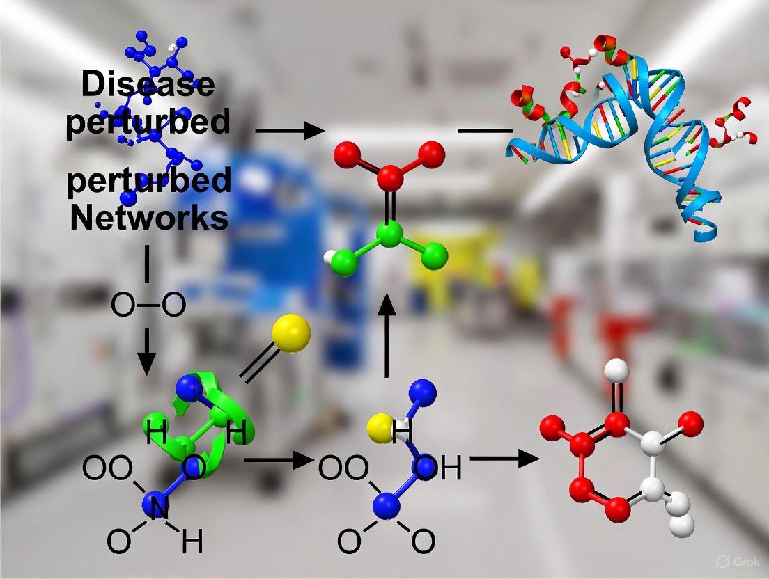
Molecular Fingerprints of Disease-Perturbed Networks: From AI-Driven Analysis to Clinical Translation
This article explores the transformative role of molecular fingerprints in characterizing disease-perturbed biological networks for drug discovery.
Molecular Fingerprints of Disease-Perturbed Networks: From AI-Driven Analysis to Clinical Translation
Abstract
This article explores the transformative role of molecular fingerprints in characterizing disease-perturbed biological networks for drug discovery. It provides a comprehensive overview for researchers and drug development professionals, covering foundational concepts of network biology and perturbation theory, modern AI-driven methodologies for fingerprint generation and analysis, strategies to overcome computational and biological challenges, and rigorous validation frameworks. By synthesizing recent advances in network medicine, multi-omics integration, and artificial intelligence, we demonstrate how molecular fingerprints serve as powerful computational tools for decoding complex disease mechanisms, predicting drug synergy, and accelerating the development of targeted therapies and drug repurposing strategies.
Decoding Biological Networks: The Foundation of Disease Perturbation Analysis
Biological networks describe the complex relationships within biological systems, representing entities such as genes, proteins, or metabolites as nodes (vertices) and their functional or physical interactions as connections (edges) [1]. The visual and computational analysis of these networks enables researchers to integrate multiple sources of heterogeneous data to probe complex biological hypotheses and validate mechanistic models [1]. In the context of disease, these networks are not static; they can be disrupted or "perturbed" by various factors, including genetic mutations, environmental exposures, or pharmacological interventions. Controlled perturbation experiments are fundamental in elucidating the underlying causal mechanisms that govern cellular behavior, as they measure changes in experimental readouts (e.g., gene expression) resulting from introducing a specific perturbation to a biological system [2].
The theory of network targets represents a paradigm shift in understanding drug-disease relationships. Instead of focusing on single molecules, this theory posits that diseases emerge from perturbations in complex biological networks, and therefore, effective therapeutic interventions should target the disease network as a whole [3]. This holistic, systems-based approach combines computational biology, pharmacology, and systems biology to explore how drugs act on multiple targets within biological systems to modulate disease progression [3].
Core Concepts of Perturbation Theory in Biology
Perturbation theory in biology provides a framework for understanding how systems respond to disturbances. The core principle is that introducing a controlled change (perturbation) to a biological network reveals causal relationships between its components.
Types of Perturbations and Experimental Readouts
Biological perturbations can be broadly categorized by their nature and the scale at which their effects are measured. The table below summarizes the primary types.
Table 1: Types of Biological Perturbations and Their Readouts
| Perturbation Type | Examples | Common Readouts | Key Characteristics |
|---|---|---|---|
| Genetic Perturbations | CRISPR-based gene knockout or knockdown [2] [4] | Transcriptomics (single-cell or bulk RNA-seq) [2] | Targets specific genes to infer function and causality. |
| Chemical Perturbations | Small-molecule drugs, inhibitors [2] [5] | Transcriptomics, cell viability assays [2] | Used for drug discovery and mechanism of action studies. |
| Combination Perturbations | Pairwise CRISPRi, drug combinations [2] [3] | Viability, transcriptomic changes [2] [3] | Reveals synergistic or antagonistic interactions. |
Formalizing Perturbations: The Causal Framework
From a computational perspective, a perturbation can be formalized as an intervention that alters the underlying data-generating process of a biological system. Given a system of random variables ( X ) (e.g., gene expression levels) with an observational distribution ( PX ), an intervention on a variable ( Xi ) assigns a new conditional distribution ( \tilde{P}(Xi \mid X{\pii}) ), where ( \pii ) denotes the parents of ( Xi ) in the causal graph ( G ) [4]. The goal of perturbation analysis is often to identify the set of intervention targets ( I ) responsible for the shift from ( PX ) to the interventional distribution ( \tilde{P}_X ) [4].
Computational Methodologies for Analyzing Perturbed Networks
The scale and heterogeneity of modern perturbation data—spanning thousands of perturbations across diverse readout modalities and biological contexts—make computational approaches indispensable for deriving generalizable insights [2]. Several advanced deep-learning models have been developed to address this challenge.
Key Computational Models
Table 2: Computational Models for Perturbation Analysis
| Model Name | Core Architecture | Primary Function | Reported Performance |
|---|---|---|---|
| Large Perturbation Model (LPM) [2] | PRC-disentangled, decoder-only deep learning | Integrates heterogeneous perturbation data; predicts outcomes and infers mechanisms. | State-of-the-art in predicting unseen perturbation transcriptomes; outperforms CPA and GEARS [2]. |
| Causal Differential Networks (Cdn) [4] | Joint causal structure learner + attention-based classifier | Identifies root-cause variables intervened upon from observational/interventional data pairs. | Outperforms baselines on seven single-cell transcriptomics datasets; generalizes to unseen cell lines [4]. |
| Network Target Theory Model [3] | Transfer learning integrated with biological molecular networks | Predicts drug-disease interactions (DDIs) and synergistic drug combinations. | AUC of 0.9298, F1 score of 0.6316 for DDI prediction; F1 of 0.7746 for drug combinations after fine-tuning [3]. |
| RNAsmol [5] | Sequence-based deep learning with data perturbation & augmentation | Predicts interactions between RNA and small molecules. | Outperforms other methods in cross-validation and unseen evaluation benchmarks [5]. |
Workflow for Causal Perturbation Target Identification
The following diagram illustrates the integrated workflow of the Causal Differential Networks (Cdn) approach for identifying perturbation targets.
Applications in Drug Discovery and Therapeutic Development
Computational models of biological networks and perturbations are revolutionizing drug discovery by providing new ways to identify and validate therapeutic targets.
Drug Repurposing and Combination Therapy
The network target theory facilitates drug repurposing by revealing novel drug-disease interactions within the network context. For instance, a model integrating diverse biological networks identified 88,161 drug-disease interactions involving 7,940 drugs and 2,986 diseases [3]. Furthermore, these models can predict synergistic drug combinations. After fine-tuning, one algorithm achieved an F1 score of 0.7746 for predicting effective combinations and identified two previously unexplored synergistic drug combinations for distinct cancer types, which were subsequently validated through in vitro cytotoxicity assays [3].
Elucidating Mechanisms of Action
Large Perturbation Models (LPMs) can map chemical and genetic perturbations into a unified latent space, revealing shared molecular mechanisms. In one study, pharmacological inhibitors were clustered in close proximity to CRISPR interventions targeting the same genes (e.g., MTOR inhibitors near MTOR perturbations) within the LPM's learned embedding space [2]. Intriguingly, this approach can also reveal off-target activities; for example, pravastatin was placed near anti-inflammatory drugs targeting PTGS1, corroborating known anti-inflammatory effects of this statin [2].
Experimental and Computational Protocols
To ensure reproducibility and facilitate adoption of these advanced techniques, this section outlines key methodological details.
Protocol: Building a Large Perturbation Model (LPM)
Objective: To train a deep learning model that integrates multiple, heterogeneous perturbation experiments by representing Perturbation, Readout, and Context (PRC) as disentangled dimensions [2].
Input Data:
- Data Sources: Pooled data from perturbation experiments such as LINCS [2], which includes both genetic (e.g., CRISPR) and pharmacological perturbations across multiple cellular contexts.
- Data Representation: Each experiment is symbolized as a (P, R, C) tuple, where P is the perturbation identity, R is the readout type (e.g., transcriptomics, viability), and C is the biological context (e.g., specific cell line).
Procedure:
- Data Integration: Assemble a diverse set of perturbation experiments without requiring full overlap in P, R, or C dimensions.
- Model Architecture: Implement a decoder-only architecture that conditions on the symbolic P, R, and C inputs. This design avoids the limitations of encoder-based models in low signal-to-noise scenarios.
- Model Training: Train the model to predict the outcome of in-vocabulary (P, R, C) combinations. The training objective is to learn generalizable perturbation-response rules disentangled from specific contextual details.
- Validation: Evaluate the model on held-out experiments, predicting post-perturbation outcomes for unseen perturbations. Performance is typically measured by accuracy in predicting transcriptomic changes or other relevant readouts.
Protocol: Identifying Targets via Causal Differential Networks (Cdn)
Objective: Given an observational dataset and an interventional dataset, identify the root-cause variables that were the targets of the intervention [4].
Input Data:
- Observational Dataset ((D{obs})): Samples from the natural distribution (PX) of the system (e.g., single-cell transcriptomics of untreated cells).
- Interventional Dataset ((D{int})): Samples from the perturbed distribution (\tilde{P}X) (e.g., single-cell transcriptomics after a specific drug treatment).
Procedure:
- Causal Graph Inference: Train a causal structure learning module to infer a causal graph (G{obs}) from (D{obs}) and a graph (G{int}) from (D{int}).
- Feature Extraction: Compute differences between the inferred graphs (G{obs}) and (G{int}), along with other statistical features derived from the two datasets.
- Target Prediction: Feed the graph differences and statistical features into an attention-based classifier. This module is trained to map these inputs to a set of variables (I) that were intervened upon.
- Joint Training: Train both the causal learner and the classifier jointly in a supervised framework on thousands of synthetic or real datasets to amortize inference and improve robustness to data noise and sparsity.
Table 3: Key Research Reagents and Databases for Network Perturbation Studies
| Resource Name | Type | Primary Function in Research | Key Features |
|---|---|---|---|
| LINCS Data [2] | Dataset | Provides a vast collection of perturbation-response signatures. | Genetic and pharmacological perturbations across multiple cell lines; used for training models like LPM. |
| Perturb-seq Datasets [4] | Dataset | Provides single-cell transcriptomic readouts of genetic perturbations. | Enables causal inference of gene regulatory networks and identification of intervention targets. |
| DrugBank [3] | Database | Source of drug-target interaction data and drug structures. | Provides known interactions and SMILES notations for pharmaceutical agents. |
| STRING [3] | Database | Provides a comprehensive protein-protein interaction (PPI) network. | Serves as a prior biological network for network propagation and feature extraction. |
| Comparative Toxicogenomics Database (CTD) [3] | Database | Curates known drug-disease and chemical-gene interactions. | Used as a benchmark for validating predicted drug-disease interactions. |
| ROBIN Dataset [5] | Dataset | Benchmark for RNA-small molecule interaction prediction. | Used for training and evaluating models like RNAsmol. |
The integration of biological network analysis with perturbation theory provides a powerful, systems-level framework for understanding disease mechanisms and accelerating therapeutic discovery. Computational approaches like Large Perturbation Models, Causal Differential Networks, and Network Target Theory models are at the forefront of this effort. They enable the integration of heterogeneous data, the prediction of perturbation outcomes, the identification of causal intervention targets, and the discovery of novel drug-disease interactions and synergistic combinations. As these methodologies continue to evolve, they hold the promise of systematically deriving therapeutic insights from the growing universe of perturbation data, ultimately paving the way for more effective and personalized treatments for complex diseases.
Defining Molecular Fingerprints for Network States and Perturbations
In the evolving landscape of systems biology and drug discovery, the concept of molecular fingerprints has expanded beyond characterizing simple chemical structures to capturing the complex states of biological networks and their responses to perturbation. Molecular fingerprints, traditionally defined as vectors representing the presence or absence of specific molecular substructures, provide a machine-readable format for computational analysis of chemical compounds [6] [7]. Within the context of disease-perturbed networks research, this concept extends to encoding network-level states and perturbation signatures that reflect pathological changes and therapeutic interventions.
The integration of molecular fingerprinting techniques with network biology represents a paradigm shift in understanding disease mechanisms. Where traditional approaches examined molecular entities in isolation, network fingerprinting captures the systemic properties that emerge from interactions between cellular components. This technical guide explores the theoretical foundations, computational methodologies, and practical applications of molecular fingerprints for characterizing network states and perturbations, with particular emphasis on advancing therapeutic discovery for complex diseases.
Theoretical Foundations
Evolution from Chemical to Network Fingerprints
Traditional molecular fingerprints encode structural information using several predominant methodologies. Path-based fingerprints (e.g., Atom Pair fingerprints) analyze paths through molecular graphs by storing unique paths starting from each atom [6]. Circular fingerprints (e.g., Extended Connectivity Fingerprints - ECFP) iteratively capture local atomic environments by aggregating information from neighboring atoms at increasing radii [6]. Substructure-based fingerprints (e.g., MACCS keys) use predefined structural patterns, while pharmacophore fingerprints encode interaction capabilities like hydrogen bonding [6]. String-based fingerprints operate directly on SMILES representations, fragmenting them into substrings for analysis [6].
The transition to network fingerprints requires abstracting these principles to higher-order biological systems. Where chemical fingerprints capture structural motifs, network fingerprints encode functional motifs - recurrent patterns of interaction that define network behavior. These include feedback loops, regulatory modules, and signaling pathways whose states vary between physiological and pathological conditions.
Network Perturbation Theory
Biological networks exist in defined states stabilized by regulatory interactions. The concept of Inhibitory-Stabilized Networks (ISNs) illustrates how cortical networks maintain stability through strong recurrent inhibition that balances excitatory connections [8]. In such networks, perturbations produce characteristic signatures - for instance, exciting inhibitory neurons in ISNs paradoxically decreases their activity due to network-level feedback [8]. Similar principles apply to molecular networks, where perturbation fingerprints capture these system-level responses.
Disease states represent persistent perturbations that alter network topology and dynamics. Molecular fingerprints of disease-perturbed networks encode these alterations, providing a quantitative basis for identifying therapeutic interventions that revert networks to healthy states.
Computational Methodologies
Fingerprint Generation for Network States
Table 1: Molecular Fingerprint Types and Network Applications
| Fingerprint Type | Key Characteristics | Network Application |
|---|---|---|
| Extended Connectivity (ECFP) | Circular topology, radius-dependent, hashed bits | Capturing local network motifs and domains |
| MACCS Keys | 166 predefined structural fragments | Standardized network feature detection |
| Morgan Fingerprints | Neighborhood atoms, radius and size parameters | Mapping connectivity patterns in networks |
| Pharmacophore Fingerprints | Interaction capabilities (H-bond, charge) | Protein-ligand interaction networks |
| Atom Pair | Atom types and shortest path distance | Long-range connections in networks |
| MinHashed (MHFP) | SMILES substrings via MinHash | Network similarity assessment |
Generating fingerprints for network states begins with representing the network as a multiscale graph where nodes represent biomolecules and edges represent interactions. For each node, a feature vector captures its dynamic state (expression, modification, localization) and network context (connectivity, centrality). The network fingerprint emerges from integrating these node-level descriptors through approaches such as:
- Graph neural networks that learn embeddings capturing both node attributes and topological position [9]
- Subgraph aggregation methods that extract local neighborhoods around each node
- Spectral methods that capture global network properties through eigenvector analysis
For small molecules operating within these networks, traditional fingerprinting methods remain relevant. The RDKit library in Python provides robust implementations, with Morgan fingerprints generated through code such as [7]:
Perturbation Fingerprinting
Perturbation fingerprints encode network responses to interventions, capturing both intended and off-target effects. The methodology involves:
- Baseline fingerprinting: Establishing pre-perturbation network state using the approaches described above
- Controlled perturbation: Applying defined perturbations (genetic, chemical, or environmental)
- Response quantification: Measuring post-perturbation changes in network components
- Differential fingerprinting: Computing the difference between pre- and post-perturbation states
In gene regulatory networks, tools like TopNet enable inference of network structure from perturbation data, modeling interdependence between genes when nodes are both perturbed and measured [10]. For chemical perturbations, fingerprint transfer strategies integrate structural motifs with bioactivity data, enabling design of molecules with desired network effects [11].
Experimental Protocols
Protocol 1: Gene Regulatory Network Inference from Perturbation Data
This protocol details network inference using TopNet, adapted from established methodologies [10]:
Step 1: Initial Gene Perturbations
- Select target genes based on disease relevance
- Design perturbation agents (siRNA, CRISPR, or small molecules)
- Transfer cells to collagen-coated tissue culture dishes (e.g., 1 μg/cm² rat tail collagen type I)
- Implement perturbations at appropriate multiplicities of infection for viral delivery methods
Step 2: Expression Measurement
- Harvest cells at multiple time points post-perturbation (e.g., 6, 12, 24, 48 hours)
- Extract RNA using standardized kits (e.g., Qiagen RNeasy)
- Prepare sequencing libraries (e.g., Illumina TruSeq)
- Sequence with sufficient depth (minimum 30 million reads per sample)
Step 3: Data Preparation
- Perform quality control (FastQC)
- Align reads to reference genome (STAR aligner)
- Generate expression matrices (featureCounts)
- Normalize data (TPM or DESeq2 normalization)
Step 4: Network Modeling with TopNet
- Input formatted expression data
- Set algorithm parameters (regularization strength, convergence threshold)
- Execute network inference
- Validate model stability through bootstrap resampling
Step 5: Network Summarization and Visualization
- Extract significant regulatory interactions (FDR < 0.05)
- Annotate edges with directionality and strength
- Generate network diagrams (Cytoscape)
- Identify key network hubs and bottlenecks
Protocol 2: AI-Driven Theranostic Probe Design
This protocol enables design of single-molecule theranostics targeting specific network nodes, adapted from recent advances [11]:
Step 1: Passive Targeting Identification
- Curate dataset of known subcellular localization molecules
- Compute molecular fingerprints (ECFP, Morgan)
- Train machine learning classifiers to identify localization patterns
- Extract key substructural fingerprints associated with target localization
Step 2: Active Targeting Design
- Obtain 3D structure of target protein (e.g., Grp78 for ER stress)
- Implement deep learning-based molecular generation model (e.g., PM-1)
- Generate candidate structures with high predicted binding affinity
- Filter for synthetic accessibility and drug-likeness
Step 3: Fingerprint Transfer and Integration
- Transfer identified passive targeting fingerprints to generated structures
- Incorporate fluorescent motifs for imaging capabilities
- Optimize structures for multifunctionality
Step 4: Validation
- Synthesize top candidates (e.g., ABT-CN2)
- Validate targeting capability (e.g., Pearson's correlation coefficient = 0.93)
- Assess therapeutic potential (e.g., IC50 = 53.21 μM)
- Confirm mechanism through dynamic simulations
Data Presentation and Analysis
Fingerprint Performance Benchmarking
Table 2: Fingerprint Performance on Natural Product Bioactivity Prediction
| Fingerprint Category | Representative Examples | Accuracy Range | Best Use Cases |
|---|---|---|---|
| Path-based | Atom Pairs, DFS | 0.72-0.89 | Synthetic compounds |
| Circular | ECFP, FCFP | 0.75-0.92 | Diverse chemotypes |
| Substructure | MACCS, PUBCHEM | 0.68-0.85 | Rapid screening |
| Pharmacophore | PH2, PH3 | 0.79-0.94 | Target-focused design |
| String-based | LINGO, MHFP | 0.77-0.91 | Natural products |
Systematic evaluation of fingerprint performance is essential for method selection. Recent benchmarking on over 100,000 unique natural products from COCONUT and CMNPD databases revealed substantial differences in fingerprint performance [6]. While Extended Connectivity Fingerprints represent the de-facto standard for drug-like compounds, other fingerprints matched or outperformed them for natural product bioactivity prediction [6].
For perturbation encoding, differential fingerprints that capture network state changes before and after intervention provide the most discriminative power. These can be optimized through multi-fingerprint ensembles that leverage complementary strengths of different encoding methods.
Visualization of Workflows
Network Perturbation Fingerprinting Workflow
Diagram 1: Network perturbation fingerprinting workflow
AI-Driven Molecule Design for Network Perturbation
Diagram 2: AI-driven molecule design workflow
The Scientist's Toolkit
Table 3: Essential Research Reagents and Resources
| Resource | Function/Application | Example Sources |
|---|---|---|
| RDKit | Open-source cheminformatics toolkit for fingerprint generation | RDKit.org |
| COCONUT Database | Natural product compounds for fingerprint benchmarking | COCONUT collection |
| CMNPD | Marine natural products with bioactivity annotations | Comprehensive Marine NP Database |
| ChEMBL | Bioactive molecule properties for model training | EMBL-EBI |
| Young Adult Mouse Colon (YAMC) cells | Model system for perturbation studies | Material Transfer Agreement |
| ΦΝΧ-E packaging cells | Retroviral vector production for genetic perturbations | ATCC |
| Collagen-coated dishes | Extracellular matrix support for cell culture | Corning, Becton Dickinson |
| TopNet algorithm | Gene regulatory network inference from perturbation data | McMurray et al. protocol |
Applications in Disease-Perturbed Networks
Endoplasmic Reticulum Stress Targeting
In a demonstration integrating molecular fingerprints with network perturbation, researchers developed ABT-CN2, a multidimensional fluorescent agent targeting Grp78, a key regulator of ER stress [11]. This approach combined:
- Machine learning-based fingerprint transfer for passive ER targeting
- Deep learning-based 3D molecular generation (PM-1 model) for active Grp78 binding
- Multifunctional design unifying targeting, imaging, and inhibition in a single molecule
The resulting molecule exhibited a compact structure (MW < 400), robust targeting (Pearson's correlation = 0.93), and antitumor activity (IC50 = 53.21 μM), demonstrating the potential of fingerprint-based approaches for designing network-directed therapeutics [11].
Natural Product Network Pharmacology
Natural products present particular challenges for fingerprint encoding due to structural complexity, including wider molecular weight distributions, multiple stereocenters, and higher sp³-hybridized carbon fractions [6]. Systematic evaluation of 20 fingerprinting algorithms revealed that different encodings provide fundamentally different views of the natural product chemical space [6]. This has profound implications for understanding how natural products perturb biological networks, as accurate structural representation is prerequisite for predicting network effects.
Future Directions
The field of molecular fingerprints for network states and perturbations is rapidly evolving along several trajectories:
- Multimodal learning frameworks that integrate structural, interaction, and dynamic data into unified fingerprint representations [9]
- Geometric deep learning extending fingerprinting to 3D molecular and network conformations
- Temporal fingerprinting capturing network dynamics across multiple timescales
- Causal inference methods distinguishing correlative from causative network perturbations
- Federated learning approaches enabling network fingerprinting across distributed datasets while preserving data privacy
As these methodologies mature, molecular fingerprints for network states and perturbations will increasingly guide therapeutic discovery, enabling precise interventions that restore diseased networks to healthy states.
In the field of molecular systems biology, representing and analyzing complex cellular interactions is fundamental to understanding disease mechanisms. Two distinct computational paradigms have emerged: knowledge-based networks and data-driven networks. Knowledge-based networks are constructed from curated, prior biological knowledge found in databases, emphasizing interpretability and grounding in established science [12]. In contrast, data-driven networks are inferred directly from high-throughput experimental data (e.g., imaging, genomics) using algorithms, prioritizing the discovery of novel patterns and relationships without heavy reliance on pre-existing models [13]. This guide provides an in-depth technical comparison of these approaches, framed within cutting-edge research on molecular fingerprints of disease-perturbed networks.
Core Conceptual Differences
The table below summarizes the fundamental distinctions between knowledge-based and data-driven network approaches.
Table 1: Fundamental Characteristics of Knowledge-Based and Data-Driven Networks
| Characteristic | Knowledge-Based Networks | Data-Driven Networks |
|---|---|---|
| Primary Data Source | Curated knowledge from scientific literature and databases (e.g., KEGG, protein-protein interactions) [12] [14] | Raw, high-dimensional experimental data (e.g., high-content imaging, gene expression) [13] |
| Construction Basis | Integration of established facts and pathway models | Algorithmic inference, machine learning, and statistical analysis of datasets [13] [15] |
| Typical Representation | Knowledge Graphs; manually drawn pathway maps [12] [14] | Network models derived from data correlations or model perturbations [13] |
| Key Strength | Interpretability, clear biological context, familiarity to biologists [12] [16] | Potential for novel discovery, adaptability to new data, ability to model complex, unforeseen interactions [13] [15] |
| Inherent Limitation | Limited to current knowledge, may miss novel biology [12] | Can be a "black box"; difficult to interpret and integrate with existing knowledge [17] [16] |
Construction Methodologies
Knowledge-Based Network Construction
Knowledge-based networks are built through the systematic assembly of established biological interactions. A prime example is the creation of a network fingerprint for disease characterization [12] [18].
Protocol: Constructing a Network Fingerprint [12]
- Define Basic Networks: Select a set of well-annotated, basic biological networks (e.g., 93 KEGG signaling pathways) that serve as a reference library [12].
- Represent the Target Network: Obtain the molecular network of the disease or biological state of interest (e.g., the Type 1 Diabetes Mellitus network from KEGG).
- Calculate Similarity Metrics: For the target network and each basic network, compute a similarity score. This score should integrate both:
- Topological Similarity: Based on network structure, often using algorithms like Affinity Propagation (AP) clustering.
- Functional Similarity: Based on the biological functions of components, using annotations like Gene Ontology (GO).
- Normalize Scores: Normalize the similarity scores using a random simulation procedure to account for network size and connectivity.
- Form the Fingerprint: The vector of normalized similarity scores to all basic networks constitutes the network fingerprint. This multidimensional vector provides an intuitive, knowledge-based characterization of the target network [12].
The following diagram illustrates this workflow:
Figure 1: Workflow for Constructing a Knowledge-Based Network Fingerprint.
Data-Driven Network Construction
Data-driven approaches infer networks directly from large-scale experimental data. A representative method involves mapping the perturbome—the network of interactions between cellular perturbations—from high-content imaging data [13].
Protocol: Mapping the Perturbome from Morphological Profiles [13]
- Perturbation and Feature Extraction: Treat cells with a library of individual drugs and their pairwise combinations. Use high-content microscopy to image the cells and extract quantitative morphological features (e.g., cell shape, organelle distribution). Each perturbation is represented as a vector in this high-dimensional morphological space.
- Define Expected Non-Interaction: The expected effect of a non-interacting drug combination is defined as the vector sum of the two individual drug perturbation vectors.
- Quantify Deviation: Measure the deviation between the observed morphological vector for the drug combination and the expected vector. This deviation is quantified to classify the interaction.
- Classify Interaction Type: Use a mathematical framework to classify the interaction into one of 12 specific types based on the direction and magnitude of the deviation. This captures whether one drug enhances, suppresses, or alters the effect of the other.
- Construct the Perturbome Network: Build a network where nodes represent individual drugs and edges represent the classified interaction between them, resulting in a data-driven perturbome network [13].
The diagram below outlines this data-driven process:
Figure 2: Data-Driven Workflow for Perturbome Network Construction.
Hybrid and Advanced Approaches
Modern research often blends these paradigms. Knowledge graphs integrate diverse biological data (genes, drugs, diseases, side effects) into a unified, structured network, enabling the application of machine learning for tasks like drug repurposing [14]. Furthermore, frameworks like MoCL enhance data-driven graph neural networks for molecules by incorporating domain knowledge at both local and global levels, guiding model learning to be more semantically meaningful [17].
Experimental Protocols and Applications
Key Experiment: Disease Classification via Network Fingerprinting
This experiment demonstrates the application of knowledge-based networks to reveal disease relationships [12].
- Objective: To classify 44 human disease networks from KEGG based on their biological relatedness.
- Method:
- Fingerprint Extraction: Network fingerprints for all 44 disease networks were extracted against 93 KEGG signaling pathways, as per the protocol in Section 3.1.
- Clustering: Hierarchical clustering (complete linkage, Euclidean distance) was applied to the fingerprint vectors.
- Result: Diseases were significantly classified into four coherent groups: a cancer-enriched group, an infectious disease-enriched group, a group with neurodegenerative and cardiovascular diseases, and an immune disease-enriched group. This classification showed substantial agreement (Kappa = 0.70) with manual KEGG classifications while revealing suggestive new relationships, such as clustering prion disease with other infectious diseases [12].
Key Experiment: Predicting Drug Interactions from the Perturbome
This experiment exemplifies a data-driven approach to understanding how drug perturbations interact [13].
- Objective: To systematically understand how different cellular perturbations (drugs) influence each other's effects.
- Method:
- Interactome Compilation: A comprehensive human protein-protein interactome (309,355 interactions) was compiled.
- Perturbation Library: A library of 267 clinically approved compounds with diverse mechanisms of action was used.
- Perturbome Mapping: The perturbome was mapped by profiling all 35,611 pairwise drug combinations using the high-content imaging protocol from Section 3.2.
- Analysis: The interactome localization of a drug's targets ("perturbation module") was calculated using Glass' ∆. The relationship between interactome distance and drug interaction type was analyzed.
- Result: A direct link was found between drug similarities on the cell morphology level and the proximity of their protein targets within the interactome. The distance between drug targets was also predictive of the type of interaction (synergistic, antagonistic, etc.) observed in the perturbome network [13].
The Scientist's Toolkit
The table below lists essential resources for constructing and analyzing knowledge-based and data-driven networks.
Table 2: Essential Research Reagents and Resources
| Resource Name | Type | Primary Function in Research |
|---|---|---|
| KEGG Pathway Database [12] | Knowledgebase | Source of manually curated basic networks and disease pathways for knowledge-based fingerprinting and validation. |
| Protein-Protein Interactome [13] | Knowledgebase/Network | A unified network of protein interactions used as a scaffold to map drug targets and understand perturbation modules. |
| Gene Ontology (GO) [12] | Knowledgebase | Provides standardized functional annotations for genes/proteins, used to calculate functional similarity between networks. |
| Chemical Compound Library [13] | Experimental Reagent | A diverse set of chemical perturbagens (e.g., 267 approved drugs) used to experimentally probe the perturbome. |
| High-Content Imaging System [13] | Experimental Platform | Automated microscopy used to generate high-dimensional morphological profiles for single and combined drug perturbations. |
| Graph Neural Networks (GNNs) [17] | Computational Tool | A class of deep learning models for data-driven learning on graph-structured data, such as molecular graphs. |
Visualizing Signaling Pathways and Workflows
The following diagram synthesizes the logical relationship between the two network approaches and their contribution to the broader research context of molecular fingerprinting in disease.
Figure 3: Two Paradigms Converging on the Study of Disease Networks.
The perturbome represents a systematic framework for understanding how cellular systems respond to perturbations, such as drug treatments or genetic changes. It maps the complex interactions between these disturbances and their high-dimensional effects on the cell, linking molecular-level changes to phenotypic outcomes [19]. This guide details the core principles, analytical frameworks, and experimental methodologies for mapping perturbomes, with a focus on applications in drug development and network biology. The ability to classify perturbation interactions into distinct types provides a powerful tool for predicting drug combination effects, understanding side mechanisms, and identifying molecular fingerprints within disease-perturbed networks [19] [20] [13].
In systems biology, a perturbation is any intervention that disrupts a cell's normal state, such as a small molecule drug, a genetic knockout, or an environmental stressor. The perturbome conceptualizes the complete set of functional influences that result from systematically perturbing a biological system and measuring the outcomes [21]. It is the network of networks that captures how individual disturbances propagate through the molecular interactome to produce complex phenotypic effects.
The central thesis of perturbome research is that disease states and therapeutic interventions can be understood as perturbations to the intricate network of cellular components. Mapping these relationships provides a principled way to understand how independent perturbations influence each other—a fundamental challenge in developing combination therapies and explaining adverse drug reactions [19] [13]. The perturbome framework connects three essential maps: the interactome (physical network of molecular interactions), the perturbation modules (localized neighborhoods within the interactome that are affected by a specific perturbation), and the phenotypic landscape (the resulting high-dimensional cellular phenotypes) [19].
Theoretical and Mathematical Framework
Classifying High-Dimensional Perturbation Interactions
Traditional models of perturbation interactions (e.g., drug combinations) typically focus on single readouts like cell survival, limiting observations to simple synergy, antagonism, or non-interaction. The perturbome framework utilizes high-dimensional readouts—such as cell morphological profiles or gene expression patterns—to enable a much more detailed classification of interaction types [19] [13].
In this framework, a cellular state is represented as a point in a high-dimensional feature space. A perturbation is represented as a vector that moves the system from its unperturbed state to a new state. For two perturbations (\vec{A}) and (\vec{B}), the expected independent combination is the vector sum (\vec{A} + \vec{B}). Any deviation from this expectation indicates an interaction, which can be decomposed into distinct components that capture the direction and nature of the interference [19]. This mathematical approach allows for the classification of any interaction between perturbations into 12 distinct interaction types, moving beyond the traditional ternary classification [19] [13].
Network Propagation and the Neuronal Perturbome
The perturbome concept extends to neuronal networks, where the neuronal perturbome describes the functional influence of perturbing individual neurons on the activity of others in the network. Computational models of neuronal networks reveal that the relationship between the physical connectome (structural connectivity) and the functional perturbome is complex in strongly recurrent networks [21].
In simplified models, the influence (\psi(E1 \rightarrow E2)) of perturbing neuron E1 on neuron E2 can be analytically derived from the network's weight matrix. The analysis shows that strong excitatory-inhibitory connectivity is necessary for feature-specific suppression effects observed experimentally. This theoretical framework helps interpret how different connectivity motifs shape the perturbome and influence sensory information processing [21].
Experimental Methodologies for Perturbome Mapping
High-Content Imaging and Morphological Profiling
Overview: This approach uses high-content microscopy to capture changes in cell morphology induced by perturbations, followed by computational image analysis to extract quantitative morphological features [19] [13].
Detailed Protocol:
- Cell Culture and Perturbation: Treat human cell lines with individual compounds or their pairwise combinations. Include controls (e.g., DMSO-treated cells) [19].
- High-Content Imaging: Fix cells at determined time points and acquire images using automated microscopy systems.
- Feature Extraction: Process images to segment individual cells and extract morphological features (e.g., cell size, shape, texture, organelle distribution). A typical profile may encompass hundreds to thousands of quantitative descriptors [19].
- Vector Representation: For each perturbation, represent its effect as a vector in the multidimensional morphological space, pointing from the unperturbed control state to the perturbed state [19].
- Interaction Calculation: For combination perturbations, calculate the expected vector sum of individual effects and compare it to the observed effect using the mathematical framework to classify the interaction type [19].
Key Applications: Systematic mapping of drug-drug interactions, identification of unexpected side effects, and linking drug-induced morphological changes to their targets in the molecular interactome [19] [13].
Proteomic Perturbation Profiling
Overview: This method identifies changes in protein abundance or stability following perturbations to infer mechanisms of action, particularly for drugs with unknown targets [22].
Detailed Protocol:
- Treatment Optimization: Determine the Delayed Cytocidal Concentration (DCC25), defined as the drug concentration that, after a 6-hour treatment followed by wash-off, results in 25% reduction in proliferation after 48 hours. This ensures comparable sublethal stress levels across different compounds [22].
- Sample Preparation: Treat cells (e.g., Trypanosoma brucei for anti-parasitic drug studies) with compounds at DCC25 for 6 hours. Include appropriate controls.
- Proteomic Analysis: Lyse cells and perform quantitative mass spectrometry-based proteomics to measure global protein abundance changes.
- Data Analysis: Identify proteins with significantly reduced stability or abundance. Compare profiles across different compounds to hypothesize about mechanisms of action and polypharmacology [22].
Key Applications: Target deconvolution for phenotypically-identified drug leads, understanding polypharmacology, and comparing mechanisms of action between candidate compounds [22].
Machine Learning Identification of Core Perturbome Genes
Overview: This computational approach integrates multiple transcriptomic datasets from various perturbations to identify a core set of genes consistently involved in stress response across multiple conditions [20].
Detailed Protocol:
- Data Collection: Compile microarray or RNA-seq datasets from public repositories (e.g., GEO) representing diverse perturbation conditions for the organism of interest (e.g., Pseudomonas aeruginosa).
- Data Normalization: Apply robust multi-array average (RMA) or similar normalization to make datasets comparable.
- Feature Selection: Implement multiple machine learning algorithms (Support Vector Machine, Random Forest, K-Nearest Neighbors) using both single partition and multiple partition methods to rank genes by their importance in classifying perturbed vs. control samples [20].
- Core Gene Identification: Select genes that are consistently highly ranked across multiple algorithms and perturbation types as the core perturbome.
- Network Analysis: Construct interaction networks from the core genes and analyze topological properties to identify key regulatory hubs [20].
Key Applications: Identification of universal stress response pathways, discovery of novel drug targets in pathogenic bacteria, and understanding central regulatory mechanisms in stress response [20].
Data Presentation and Analysis
Quantitative Analysis of Perturbation Interactions
Table 1: Classification and Frequency of Drug Perturbation Interaction Types from a Large-Scale Imaging Screen [19]
| Interaction Type | Description | Frequency | Molecular Predictability |
|---|---|---|---|
| Additive | Combined effect equals vector sum of individual effects | 36.2% | High (based on target proximity) |
| Synergy | Enhanced effect in same direction | 15.7% | Moderate |
| Antagonism | Reduced effect compared to expected | 22.1% | Moderate |
| Directional | One perturbation changes direction of another | 8.3% | Low |
| Emergent | New phenotype not seen with individual perturbations | 4.9% | Very Low |
| Other Types | Remaining interaction classes | 12.8% | Variable |
Table 2: Core Perturbome Genes Identified in Pseudomonas aeruginosa Using Machine Learning Approaches [20]
| Gene Category | Count | Primary Functions | Network Properties |
|---|---|---|---|
| DNA Damage Repair | 14 | Nucleotide excision repair, recombination | High betweenness centrality |
| Aerobic Respiration | 9 | Electron transport, ATP synthesis | Modular hubs |
| Biosynthesis | 12 | Amino acid, cofactor production | Peripheral connectivity |
| Unknown Function | 11 | Not yet characterized | Various topological roles |
The Scientist's Toolkit: Essential Research Reagents
Table 3: Key Research Reagents and Computational Tools for Perturbome Mapping
| Reagent/Tool | Function | Application Example |
|---|---|---|
| High-Content Imaging Systems | Automated microscopy and image acquisition | Quantifying morphological changes in drug-treated cells [19] |
| Compound Libraries | Collections of chemically diverse perturbations | Screening individual drugs and combinations [19] [13] |
| Protein-Protein Interaction Networks | Comprehensive maps of molecular interactions | Mapping perturbation modules and their overlaps [19] |
| Mass Spectrometry Platforms | Global protein quantification | Identifying protein abundance changes after perturbations [22] |
| Machine Learning Algorithms (SVM, RF, KNN) | Feature selection and classification | Identifying core perturbome genes from transcriptomic data [20] |
| Network Analysis Software | Graph theory and topological analysis | Characterizing perturbome network properties [19] [20] |
Visualizing Perturbome Concepts and Workflows
Core Perturbome Mapping Workflow
Mathematical Framework for Perturbation Interactions
Interactome-Based Prediction Model
Applications in Disease Network Research
Drug Discovery and Combination Therapy
Perturbome mapping directly addresses a central challenge in pharmacology: the systematic understanding of how complex cellular perturbations induced by different drugs influence each other [19] [13]. By classifying drug-drug interactions into specific types based on their high-dimensional effects, researchers can rationally design combination therapies that maximize therapeutic synergy while minimizing adverse effects [19].
The framework has demonstrated practical utility in predicting clinically relevant interactions. For instance, the proximity between different drug perturbation modules in the interactome successfully predicts both therapeutic synergies and adverse reaction potentials. Anti-protozoal drugs associated with psychoactive side effects were found to overlap perturbation space with analeptics that stimulate the central nervous system, while anti-gout medications showed proximity to diuretics—reflecting the clinically observed side effect of hyperuricemia with diuretic use [19] [13].
Target Deconvolution and Polypharmacology
For drugs discovered through phenotypic screening, the perturbome framework enables mechanistic insights without requiring prior knowledge of molecular targets. The proteomic perturbation approach has successfully differentiated mechanisms of action between trypanocidal compounds NEU-4438 and SCYX-7158 (acoziborole), showing that while NEU-4438 prevents DNA biosynthesis and basal body maturation, acoziborole destabilizes CPSF3 and inhibits polypeptide translation [22]. This target-agnostic method is particularly valuable for understanding polypharmacology—when drugs interact with multiple cellular targets—which is increasingly recognized as common rather than exceptional in drug action [22].
Universal Stress Response Signatures
The identification of core perturbome genes across multiple stress conditions reveals conserved molecular circuits that respond to diverse perturbations. In Pseudomonas aeruginosa, machine learning approaches identified 46 core response genes associated with multiple perturbations, with functional enrichment in DNA damage repair and aerobic respiration processes [20]. These core perturbome elements represent central control points in the cellular stress response and potential targets for novel antimicrobial strategies that would be less prone to resistance development.
Perturbome mapping represents a paradigm shift in how we understand cellular responses to interventions, moving beyond single-target models to embrace the complexity of biological networks. The integration of high-dimensional readouts with network biology and machine learning creates a powerful framework for predicting how perturbations interact and propagate through cellular systems.
Future developments will likely focus on multi-scale perturbome mapping that integrates molecular, cellular, and tissue-level responses, as well as dynamic perturbome tracking that captures temporal evolution of perturbation responses. The application of perturbome concepts to clinical medicine holds promise for personalized combination therapies tailored to individual disease network states.
The consistent finding that perturbation targets aggregate in specific interactome neighborhoods, and that the overlap between these neighborhoods predicts functional interactions, provides a principled foundation for network-based pharmacology [19] [13]. As molecular network maps become more comprehensive and perturbation profiling technologies more scalable, the perturbome framework will increasingly guide therapeutic development and our fundamental understanding of cellular regulation.
The paradigm of network medicine posits that disease phenotypes arise from the perturbation of specific neighborhoods within the human molecular interactome, known as disease modules. Concurrently, the mechanisms of pharmacological compounds can be conceptualized as perturbation modules—localized sets of protein targets within the same interactome. The overlap and network distance between these disease and perturbation modules are fundamental for understanding drug action, predicting efficacy, and anticipating adverse effects. This whitepaper delineates the quantitative framework for identifying these modules, details experimental protocols for mapping their interactions, and synthesizes key findings on how their interplay dictates therapeutic outcomes, framing this within the broader research on molecular fingerprints of disease-perturbed networks.
Biological function is orchestrated by complex networks of interacting cellular components. Pathological states and therapeutic interventions can both be viewed as perturbations to this intricate system [13]. The disease module principle asserts that genes associated with the same disease often physically interact and are localized within a specific neighborhood of the human interactome [23]. This has propelled network-based approaches to elucidate the molecular underpinnings of human diseases.
Similarly, the targets of active chemical compounds, or drugs, are not randomly scattered across the interactome. They tend to aggregate in specific localized neighborhoods, forming perturbation modules [13]. The centrality of a drug's targets within the interactome and their proximity to disease modules are strongly related to the drug's efficacy and its potential to cause side effects [13]. The systematic understanding of how independent perturbations influence each other—be it two drugs, a drug and a disease, or two comorbid diseases—lies at the core of modern therapeutic development and safety assessment. This guide explores the principles and methodologies for mapping these modules and quantifying their interactions.
Conceptual Framework and Key Definitions
The Human Interactome
The human interactome is a comprehensive map of physical interactions between biomolecules, most commonly proteins. It serves as the universal scaffold upon which cellular processes are organized and upon which perturbations act. It is typically represented as a graph where nodes are proteins and edges are their documented physical interactions [13] [23].
Disease Modules and Perturbation Modules
A Disease Module is a connected subgraph within the interactome that is significantly enriched with proteins (or genes) associated with a specific disease [23]. The existence of such a module implies that the pathophysiological phenotype is a result of dysfunction within a localized network neighborhood, rather than of a single, isolated gene.
A Perturbation Module is the set of proteins within the interactome that are directly targeted by a specific chemical compound (e.g., a drug) or genetic perturbation [13]. For drugs, 64% of compounds target proteins that form connected subgraphs within the interactome that are significantly larger than expected by chance [13].
Quantifying Module Localization and Overlap
- Glass' ∆: A metric used to quantify the degree of interactome localization of a perturbation module. It compares the observed average shortest-path distance between all pairs of a compound's targets within the interactome to the expected distance from randomly sampled sets of proteins. A significantly negative Glass' ∆ indicates a highly localized module [13].
- Interactome Distance (
d_s): The shortest path distance between two modules (e.g., a disease module and a drug perturbation module) within the interactome. Shorter distances are predictive of potential therapeutic effects or shared side effects [13]. - Network Perturbation Amplitude (NPA) Scoring: A family of computational methods designed to quantify the amplitude of treatment-induced perturbations in a biological network model based on high-throughput data, providing a score for the activity change of a biological process [24].
The following diagram illustrates the core concept of module overlap and the quantitative measures used to characterize it.
Quantitative Analysis of Module Properties
The structural and functional characteristics of disease and perturbation modules have been systematically quantified, revealing key organizational principles.
Table 1: Quantitative Characteristics of Perturbation Modules [13]
| Characteristic | Average Measure | Biological Implication |
|---|---|---|
| Number of protein targets per compound | 13.64 (mean) | Most drugs are polypharmacological, targeting multiple proteins. |
| Degree (connectivity) of target proteins | ⟨k_targets⟩ = 74.4 | Drug targets are significantly more highly connected than average proteins (⟨k_all⟩ = 37.7). |
| Proportion of compounds with localized targets (Glass' ∆) | 64% | The majority of drugs perturb specific, cohesive network neighborhoods. |
| Functional similarity of targets in localized modules (Glass' ∆ ≤ -3) | Up to 32-fold higher vs. random | Highly localized modules are associated with cohesive biological functions. |
Table 2: Network Perturbation Amplitude (NPA) Scoring Methods [24]
| Method | Core Calculation | Key Feature |
|---|---|---|
| Strength | Mean of differential expressions, adjusted for causal sign. | Simple, direct aggregate of downstream gene changes. |
| Geometric Perturbation Index (GPI) | Similar to Strength, but weighted by statistical significance of differential expression. | Incorporates confidence in measured changes. |
| Measured Abundance Signal Score (MASS) | Change in absolute quantities supporting upstream activity, divided by total absolute quantity. | Accounts for overall abundance levels. |
| Expected Perturbation Index (EPI) | A smoothed GPI averaged over all significance thresholds. | Robust to the choice of a single significance threshold. |
Experimental and Computational Methodologies
Protocol 1: Mapping the Perturbome using High-Content Imaging
This protocol generates high-dimensional phenotypic data to quantify drug interactions and link them to interactome structure [13].
1. Experimental Design:
- Cell Model: Use a well-controlled cell line (e.g., normal human bronchial epithelial cells - NHBE).
- Perturbations: Treat with a library of 267 individual chemical compounds (including approved drugs) and all pairwise combinations (e.g., 35,611 pairs).
- Controls: Include vehicle controls (e.g., DMSO) for baseline measurement.
2. High-Content Imaging and Feature Extraction:
- Imaging: Use automated microscopy to capture high-resolution images of cells under each condition.
- Morphological Profiling: Quantify hundreds of morphological features (e.g., cell size, shape, texture, organelle distribution) for each cell, creating a high-dimensional "morphological space."
3. Data Integration and Network Construction:
- Perturbation Vectors: Model each drug's effect as a vector in the morphological space, pointing from the control state to the perturbed state.
- Interactome Mapping:
- Compile a comprehensive human interactome (e.g., 309,355 interactions between 16,376 proteins).
- Annotate the protein targets for each drug to define its perturbation module.
- Calculate interactome localization metrics (Glass' ∆) for each module.
- Perturbome Network: Construct a network where nodes are drugs and edges represent significant interactions between their morphological effects. Correlate these interactions with the network distance of their respective perturbation modules.
4. Key Analysis:
- Test the hypothesis that drugs with similar morphological profiles have protein targets located closer in the interactome.
- Classify drug-drug interaction types based on how their perturbation vectors interact in the high-dimensional space.
Protocol 2: Network Inference from Perturbation Time Course Data (DL-MRA)
Dynamic Least-Squares Modular Response Analysis (DL-MRA) infers signed, directed networks, including cycles and external stimuli, from perturbation time courses [25].
1. Experimental Requirements:
- System: An
n-node network (e.g., signaling or gene regulatory network). - Perturbations: Perform
ndistinct perturbation time-course experiments. For a 2-node network, this requires:- Time course 1: No perturbation (vehicle control).
- Time course 2: Perturbation of Node 1 (e.g., using a specific inhibitor or shRNA).
- Time course 3: Perturbation of Node 2.
- Measurement: Measure the activity of all
nnodes at multiple (e.g., 7-11) evenly spaced time points across all experiments.
2. Computational Inference (DL-MRA):
- Model Dynamics: The system dynamics are cast as Ordinary Differential Equations (ODEs). The goal is to estimate the Jacobian matrix, J, which contains the direct causal influences (edge weights
F_ij) between nodes. - Formulation: A well-posed estimation problem is formulated that uses the time-course data from all perturbation experiments to uniquely estimate the elements of the Jacobian as a function of time.
- Robustness: The least-squares framework is designed to function robustly in the presence of typical experimental noise levels.
3. Application:
- This method can be applied to infer the structure of causal networks underlying a disease state, thereby helping to define the disease module. It is particularly suited for gene regulatory networks and signaling networks.
The workflow for this multi-omics data integration is summarized below.
Protocol 3: Assessing Network Perturbation Amplitude (NPA)
This protocol uses causal biological network models and transcriptomic data to quantify perturbation in specific processes [24].
1. Foundation: Causal Network Models (HYPs)
- Construction: Build network models from literature-curated knowledge. A "HYP" is a network of causal relationships linking an upstream biological entity (e.g., a kinase activity) to downstream measurable entities (e.g., genes it regulates).
- Aggregation: Complex processes (e.g., "cell cycle") can be described by aggregating multiple HYPs into a larger causal network model.
2. Scoring with High-Throughput Data:
- Input: A transcriptomic data set (e.g., treatment vs. control comparisons).
- Scoring Algorithms: Apply one of the four NPA methods (Strength, GPI, MASS, EPI) to compute a score representing the activity change of the biological process defined by the HYP. A positive score indicates increased activity relative to control.
3. Statistical Annotation:
- Uncertainty: Calculate a confidence interval for the NPA score.
- Specificity: Test whether the score is specific to the genes in the HYP and not due to a general, non-specific trend in the data.
4. Application:
- This method can be used to quantitatively assess how a drug treatment (the perturbation) affects the activity of a known disease module, providing a direct measure of the interaction amplitude between the perturbation and the disease network.
Table 3: Key Research Reagent Solutions for Module Analysis
| Reagent / Resource | Function in Experimental Protocol |
|---|---|
| Chemical Compound Library (e.g., CLOUD) | A curated library of diverse chemical compounds (including approved drugs) used in large-scale perturbation screens to define perturbation modules [13]. |
| Validated shRNA/gRNA Libraries | Tools for specific genetic perturbation of individual network nodes (genes), required for network inference methods like DL-MRA [25]. |
| Causal Biological Network Database (e.g., Selventa KB) | A repository of literature-curated cause-and-effect relationships used to construct HYPs for Network Perturbation Amplitude (NPA) scoring [24]. |
| Curated Molecular Interactome | A consolidated set of protein-protein interactions serving as the foundational scaffold for all module localization analyses (e.g., from databases like STRING, BioGRID) [13] [23]. |
| Multi-Omics Datasets (e.g., GWAS, RNA-seq, DNA methylation) | Context-specific molecular profiling data that is integrated with the interactome to detect and refine disease modules for complex diseases [23]. |
The network localization of disease and drug action provides a powerful conceptual and quantitative framework for modern biomedical research. The overlap between disease modules and perturbation modules, measurable via interactome distance and perturbation amplitude scoring, offers a systematic and mechanistic basis for understanding therapeutic efficacy and predicting adverse effects. The experimental and computational methodologies detailed herein—from high-content imaging and dynamic network inference to multi-omics integration and NPA scoring—provide researchers with a robust toolkit to map these interactions. As the molecular interactome becomes more complete and multi-omics data becomes richer, the principles of network localization are poised to become a cornerstone of rational drug development and precision medicine.
In modern systems biology, diseases are increasingly understood as perturbations within complex molecular networks rather than as isolated defects of single genes or proteins. Research into the molecular fingerprints of disease perturbed networks relies on this foundational principle, requiring the integration of vast, heterogeneous biological data to construct accurate and comprehensive interaction maps. These maps, or networks, provide a systems-level view of cellular function, enabling researchers to identify key regulatory hubs, dysfunctional pathways, and ultimately, new therapeutic targets. The construction of such networks is critically dependent on specialized biological databases that curate and score interactions from diverse evidence sources.
This technical guide provides an in-depth examination of three cornerstone resources for network construction: STRING for protein-protein interactions, DrugBank for drug and drug-target information, and DisGeNET for gene-disease associations. Framed within the context of identifying disease-specific molecular fingerprints, this whitepaper details the scope, content, and practical application of each database. It further outlines integrative computational methodologies that leverage these resources to predict novel drug-disease interactions and identify potential therapeutic strategies, providing a structured framework for researchers and drug development professionals engaged in network pharmacology and systems-based drug discovery.
Core Database Resource Profiles
STRING: Functional Protein Association Networks
STRING is a comprehensive database of known and predicted protein-protein interactions (PPIs). These interactions include both direct physical binding and indirect functional associations, making STRING a foundational tool for constructing the core protein scaffolding of molecular networks [26]. The database is uniquely characterized by its systematic inclusion and scoring of evidence from diverse channels.
Interaction Evidence and Scoring: Each protein-protein interaction in STRING is annotated with a confidence score that ranges from 0 to 1, representing the database's assessment of the likelihood that the interaction is biologically valid. This score is not a measure of interaction strength but of reliability [27]. This confidence score is a composite derived from integrating probabilities from multiple evidence channels while correcting for the probability of observing an interaction by random chance [27]. The key evidence channels are:
- Experimental Data: Biochemically validated interactions from other primary databases (indicated by a purple line in the network view) [26].
- Genomic Context: Includes Gene Neighborhood (proximity in prokaryotic genomes, green line), Gene Fusion (red line), and Gene Co-occurrence (phylogenetic co-occurrence across species, blue line) [26].
- Co-expression: Correlation in gene expression patterns across conditions (black line) [26].
- Textmining: Automated extraction of protein associations from scientific literature (yellow line) [26].
- Databases: Curated pathway data from resources like KEGG and Reactome (light blue line) [26].
Network Visualization and Access: STRING provides a web interface with multiple network view modes: Evidence (colored lines), Confidence (line thickness), and Action (molecular interaction type) [26]. Users can customize networks by setting a minimum interaction score (e.g., low confidence: ≥0.15, medium: ≥0.4, high: ≥0.7, highest: ≥0.9) and choosing to show only physical interactions [26] [28]. Data can be exported in various formats, including TSV for tabular data, PNG/SVG for images, and PSI-MI for standardized data exchange [26].
DrugBank: Drug and Drug Target Knowledgebase
DrugBank serves as a detailed clinical development intelligence platform, providing structured information on drugs, their mechanisms, targets, and interactions [29]. It is an essential resource for adding pharmacochemical layers to molecular networks.
Scope and Data Content: The database contains data on over 500,000 drugs and drug products, including FDA-approved pharmaceuticals, investigational compounds, and biotech products [29]. For each drug, it provides comprehensive information, including chemical structures (SMILES notation), pharmacologic actions, target proteins, and drug-drug interactions [3]. This structured information is critical for linking chemical entities to their biological effects within a network context.
Application in Network Pharmacology: In network-based drug discovery, DrugBank's data enables researchers to "anchor" networks with known pharmacological information. It facilitates the study of drug repurposing by allowing scientists to see how existing drugs might interact with new disease-related protein modules [3]. Its API and structured downloads allow for seamless integration with other bioinformatics resources and custom analytical pipelines [29].
DisGeNET: A Platform for Gene-Disease Insights
While the provided search results do not contain specific details for DisGeNET, it is a widely recognized knowledge platform for gene-disease associations (GDAs). For the purpose of this framework, it is noted as a critical resource that aggregates and scores associations from multiple sources, including curated repositories, GWAS catalogues, and animal models. It typically provides comprehensive gene-disease association data, which is fundamental for initializing disease-specific network perturbations and for validating the disease relevance of constructed networks.
Table 1: Core Databases for Molecular Network Construction
| Database | Primary Focus | Key Data Types | Quantitative Scale (as of 2024/2025) | Primary Application in Network Research |
|---|---|---|---|---|
| STRING [26] [27] | Protein-Protein Interactions | Predicted & known associations, functional linkages | 210,914 interactions (E. coli at medium confidence); Scores from 0-1 [27] | Backbone for protein interaction topology; functional enrichment analysis |
| DrugBank [29] [3] | Drug & Target Information | Drug structures, targets, mechanisms, interactions | ~500,000 drugs & drug products; 16,508 drug-target interactions [29] [3] | Annotating networks with pharmacologically relevant nodes and edges |
| DisGeNET | Gene-Disease Associations | Curated & inferred GDAs, variant-disease data | Information not available in search results | Prioritizing disease-relevant network modules and seed proteins |
Integrative Methodology for Predicting Drug-Disease Interactions
A powerful application of these databases is their integration into predictive computational models. The following protocol, adapted from a 2025 study, details a transfer learning model based on network target theory for large-scale prediction of drug-disease interactions (DDIs) [3].
Datasets Construction and Curation
The first phase involves gathering and rigorously curating data from multiple public resources to create a unified, analysis-ready dataset.
- Drug-Target Interaction (DTI) Data: Source raw DTI data from DrugBank. Retrieve the Simplified Molecular-Input Line-Entry System (SMILES) notation for each drug from PubChem. Classify interactions into categories such as activation, inhibition, or non-associative based on established schemas [3].
- Disease Data and Embedding: Utilize Medical Subject Headings (MeSH) descriptors to extract a standardized disease vocabulary and hierarchy. Transform this hierarchical lexicon into a disease-disease network using graph embedding techniques, which captures semantic and topological relationships between diseases [3].
- Drug-Disease Interaction (DDI) Data: Obtain curated, evidence-backed drug-disease relationships from the Comparative Toxicogenomics Database (CTD). Filter interactions to include only those with direct empirical evidence, that are mapped to the MeSH taxonomy, and for which drug SMILES are available [3].
- Protein-Protein Interaction (PPI) Network: Download a comprehensive PPI network from STRING, which includes both known and predicted interactions. For analyses requiring directionality (activation/inhibition), use a signed network like the Human Signaling Network [3].
- Disease-Specific Data (e.g., Cancer): For disease-specific predictions, incorporate transcriptomic data from repositories like The Cancer Genome Atlas (TCGA) to construct context-aware biological networks [3].
Table 2: Essential Research Reagent Solutions for Network Construction & Analysis
| Research Reagent / Resource | Function in Workflow | Key Characteristics & Alternatives |
|---|---|---|
| STRING PPI Network [26] [3] | Provides the foundational scaffold of protein interactions | Includes scored, genome-wide interactions; alternative: Human Signaling Network for signed data [3] |
| DrugBank DTI Data [29] [3] | Links pharmacological compounds to their protein targets | Provides validated, structured drug information; alternative: ChEMBL |
| MeSH Disease Taxonomy [3] | Provides a standardized ontology for disease concepts | Enables creation of a computable disease network; alternative: OMIM |
| CTD Drug-Disease Data [3] | Supplies known, evidence-backed drug-disease pairs for model training | Curated interactions from scientific literature; alternative: NCI Thesaurus |
| TCGA Transcriptomic Data [3] | Enables construction of condition-specific molecular networks | Provides gene expression profiles for diseases like cancer; alternative: GTEx |
Model Architecture and Workflow
The core of the methodology is a model that learns from biological networks to predict novel DDIs.
- Feature Extraction via Network Propagation: For each drug, simulate its effect on the STRING PPI network or a disease-specific network. This is done using a random walk with restart algorithm. The walk starts from the drug's known protein targets and propagates through the network, with the resulting steady-state distribution of probabilities forming a biological fingerprint of the drug's network-level influence [3].
- Representation Learning: Represent each disease using an embedding vector derived from its position in the MeSH-based disease network. This captures its relationship to all other diseases in a low-dimensional space [3].
- Model Training with Transfer Learning: Train a deep learning model (e.g., a graph neural network) to predict known DDIs using the drug fingerprints and disease embeddings. A key innovation is the use of transfer learning: the model first learns from the large dataset of individual DDIs and is then fine-tuned on a smaller dataset of validated drug combinations, allowing it to predict synergistic pairs [3].
- Validation and Experimental Confirmation: The top predictions, particularly for drug combinations in complex diseases like cancer, should be validated using in vitro cytotoxicity assays or other relevant functional experiments to confirm model accuracy [3].
The following diagram illustrates the logical flow and data integration points of this predictive workflow.
Experimental Protocol for Network-Based Drug Combination Prediction
This protocol provides a step-by-step guide for predicting and validating synergistic drug combinations for a specific cancer type, based on the referenced methodology [3].
Objective: To computationally predict and experimentally validate a novel synergistic drug combination for a specific cancer (e.g., Breast Invasive Carcinoma) using integrated biological networks.
Step-by-Step Procedure:
Construct a Cancer-Specific PPI Network:
- Download transcriptomic data (RNA-Seq) for Breast Invasive Carcinoma and matched normal tissue from the UCSC Xena database (hosts TCGA data) [3].
- Identify significantly differentially expressed genes (DEGs) using a tool like DESeq2 (|log2FoldChange| > 1, adjusted p-value < 0.05).
- Extract the subset of the STRING PPI network that includes all DEGs and their first-shell interactors. This creates a disease-perturbed network context [3].
Generate Drug Perturbation Profiles:
- Select a library of candidate drugs from DrugBank, focusing on those with known targets and SMILES notations available [3].
- For each drug, perform a network propagation analysis on the cancer-specific PPI network. Initiate the random walk from the drug's known protein targets (sourced from DrugBank). The resulting propagation profile is the drug's fingerprint [3].
Predict Synergistic Combinations:
- Input the drug fingerprints into the pre-trained transfer learning model. The model will score all possible pairs of drugs in the library for their predicted synergistic effect within the breast cancer network context [3].
- Rank the drug pairs by their predicted synergy score. Select the top 3-5 candidate combinations for experimental validation.
In Vitro Validation via Cytotoxicity Assay:
- Cell Culture: Maintain a relevant breast cancer cell line (e.g., MCF-7 or MDA-MB-231) in appropriate medium under standard conditions (37°C, 5% CO2).
- Compound Treatment: Treat cells with the individual drugs and the predicted combinations across a range of concentrations. Include a negative control (DMSO vehicle).
- Viability Assessment: After 72 hours of treatment, measure cell viability using a standard MTT or CellTiter-Glo assay. Perform each condition in at least three technical replicates and three independent biological replicates.
- Synergy Calculation: Analyze the dose-response data using software like Combenefit to calculate combination indices (CI). A CI < 1 indicates synergy, confirming the model's prediction [3].
Analysis and Visualization of Constructed Networks
Once a network is constructed, robust analysis and visualization are crucial for extracting biological insights.
Functional and Topological Analysis
- STRING Analysis Tab: The STRING web interface provides immediate statistics for any network, including the number of nodes and edges, the average node degree (average number of connections per protein), and the clustering coefficient (a measure of how interconnected the nodes are) [26]. A key metric is the PPI enrichment p-value, which indicates whether the observed number of interactions is significantly greater than expected by random chance, suggesting the network is biologically meaningful [26].
- Functional Enrichment: STRING automatically performs enrichment analysis for Gene Ontology terms, KEGG pathways, and protein domains. This identifies biological processes, pathways, and functional modules that are statistically over-represented in your network compared to the genomic background, helping to interpret the network's functional implications [26] [28].
Advanced Visualization Techniques
For publication-quality figures and deeper exploration beyond the STRING website, several powerful tools are available.
- Cytoscape: An open-source software platform for visualizing complex networks and integrating them with attribute data. It is the industry standard for creating customizable, publication-ready network visualizations and supports numerous plugins for additional analysis [30].
- R Packages (ggraph/igraph): For analysts comfortable with programming, R offers powerful libraries.
igraphis a core package for network analysis and visualization, whileggraphextends theggplot2grammar of graphics to network data, allowing for highly customizable and reproducible visualizations [31]. - Gephi: A leading open-source application for all kinds of graphs and networks, known for its user-friendly interface and powerful layout algorithms [30].
The following diagram outlines the post-construction workflow, from analysis to visualization.
The integration of databases like STRING, DrugBank, and DisGeNET provides an unparalleled resource for constructing and analyzing molecular networks that capture the complex fingerprints of diseased cellular states. This guide has detailed the technical specifics of these resources and demonstrated, through a cutting-edge methodological protocol, how they can be synergistically combined to move from static network maps to dynamic, predictive models of drug action. As these databases continue to grow in scale and quality, and as computational methods like network target theory and deep learning become more sophisticated, their collective utility in de-risking and accelerating drug discovery will only increase. For researchers, mastering these tools is no longer optional but essential for pioneering the next generation of network-based therapeutic strategies.
AI and Multi-Omics Integration: Methodological Advances for Network Fingerprinting
From Traditional Descriptors to AI-Driven Molecular Representations
Molecular representation serves as the foundational bridge between chemical structures and their biological, chemical, or physical properties, enabling computational analysis and prediction in drug discovery. This field has undergone a paradigm shift from reliance on manually engineered descriptors to automated, data-driven feature extraction using artificial intelligence (AI). Where traditional representations provided static, rule-based encodings, modern AI-driven approaches learn continuous, context-aware embeddings that capture intricate structure-function relationships essential for navigating disease-perturbed networks. This evolution is particularly crucial for phenotype-driven drug discovery, which aims to identify compounds that reverse disease states by analyzing phenotypic signatures without predefined targets, moving beyond the "one drug, one gene, one disease" model that has dominated pharmaceutical development [32] [33].
The renaissance of phenotype-driven approaches has been fueled by the observation that many first-in-class drugs approved by the FDA between 1999 and 2008 were discovered without a drug target hypothesis [32]. Instead of focusing on single targets, researchers now seek perturbagens—combinations of therapeutic targets—that can shift gene expression profiles from diseased to healthy states by analyzing the complex networks underlying disease phenotypes. This transition necessitates molecular representations that can not only encode chemical structure but also capture their effects within biological systems, especially in the context of disease-perturbed networks where multiple pathways interact to produce pathological states.
Traditional Molecular Representation Methods
Traditional molecular representation methods rely on explicit, rule-based feature extraction derived from chemical and physical properties. These methods have established a strong foundation for computational approaches in drug discovery through their computational efficiency, interpretability, and well-understood characteristics.
String-Based Representations
String-based encodings provide compact, linear formats for representing molecular structures:
- SMILES (Simplified Molecular-Input Line-Entry System): Introduced in 1988, SMILES translates molecular structures into linear strings using ASCII characters to represent atoms, bonds, and branching. Its compact nature makes it ideal for database storage and similarity analysis [9] [34].
- InChI (International Chemical Identifier): Developed by IUPAC, InChI provides a standardized representation designed for non-proprietary exchange of chemical information. While highly standardized, it is less human-readable than SMILES and cannot guarantee decoding back to original molecular graphs [9].
- Extended Variants: Improved versions like ChemAxon Extended SMILES (CXSMILES), OpenSMILES, and SMILES Arbitrary Target Specification (SMARTS) have extended the original SMILES functionality for specific applications [9].
Molecular Descriptors and Fingerprints
Traditional feature-based representations quantify molecular properties through predefined algorithms:
- Molecular Descriptors: These numerical values quantify physicochemical properties (e.g., molecular weight, hydrophobicity), topological indices, and electronic features. The PaDEL library descriptors have proven particularly effective for predicting physical properties of molecules [35].
- Molecular Fingerprints: Binary or count vectors that encode the presence or absence of structural features:
- MACCS Keys: A structural key fingerprint with 166 predefined structural fragments that has demonstrated strong overall performance across diverse prediction tasks [36] [35].
- Extended Connectivity Fingerprints (ECFP): Circular fingerprints that capture local atomic environments through an iterative hashing approach, widely considered state-of-the-art for similarity searching [9] [35].
- Morgan Fingerprints: Similar to ECFP but using a different algorithmic approach, typically generating 2048-dimensional vectors for comprehensive molecular representation [36].
Table 1: Comparison of Traditional Molecular Representation Methods
| Representation Type | Key Examples | Strengths | Limitations | Primary Applications |
|---|---|---|---|---|
| String-Based | SMILES, InChI, SELFIES | Compact, human-readable, database-friendly | Limited structural context, vulnerability to syntax errors | Chemical database storage, basic similarity analysis |
| Descriptor-Based | PaDEL descriptors, topological indices | Physicochemically interpretable, quantitative | May miss complex structural patterns, expert-dependent | QSAR modeling, property prediction |
| Fingerprint-Based | MACCS, ECFP, Morgan | Effective similarity searching, computationally efficient | Predefined features limit novelty discovery | Virtual screening, clustering, similarity search |
Despite their widespread adoption, traditional representations face significant limitations in capturing the complex relationships between molecular structure and biological activity within disease-perturbed networks. Their fixed nature struggles to represent dynamic molecular behaviors in different biological contexts, which is crucial for understanding a molecule's effect on pathological networks [34]. This has driven the development of more adaptive, data-driven representation approaches.
AI-Driven Molecular Representation Approaches
The advent of deep learning has catalyzed a fundamental shift from predefined representations to learned, continuous embeddings that capture complex molecular features directly from data. These AI-driven approaches have demonstrated remarkable capabilities in modeling intricate structure-function relationships essential for understanding and targeting disease-perturbed networks.
Graph-Based Representations
Graph-based representations conceptualize molecules as graphs with atoms as nodes and bonds as edges, enabling explicit encoding of structural relationships:
- Graph Neural Networks (GNNs): Learn node embeddings through message-passing between connected atoms, capturing both local and global molecular structure. Frameworks like Duvenaud et al.'s approach have demonstrated significant advancements in learning meaningful molecular features directly from raw molecular graphs [34].
- 3D-Aware GNNs: Models such as 3D Infomax incorporate spatial molecular geometry through pre-training on 3D molecular datasets, enhancing predictive performance by capturing stereochemical properties critical for molecular interactions [34].
- Specialized Architectures: Models like PDGrapher use causally inspired GNNs to predict combinatorial therapeutic targets by embedding disease cell states into biological networks and learning latent representations that can identify optimal interventions [32].
Sequence-Based and Multimodal Approaches
Inspired by natural language processing advances, these methods treat molecular representations as sequences or integrate multiple data types:
- Transformer Models: Architectures like BERT adapted for molecular SMILES strings, tokenizing at atomic or substructure levels and learning contextual relationships through self-attention mechanisms [9].
- Multimodal Fusion: Approaches such as MolFusion integrate diverse data types including molecular graphs, SMILES strings, and quantum mechanical properties to generate comprehensive representations that capture complementary aspects of molecular characteristics [34].
- Contrastive Learning: Frameworks like SMICLR align related molecular representations across different modalities or augmentation strategies, improving generalization and robustness [34].
Generative Representations
Generative models learn the underlying distribution of molecular structures to enable novel molecule design:
- Variational Autoencoders (VAEs): Learn continuous latent spaces of molecular structure, enabling interpolation and generation of novel compounds by sampling from the learned distribution [9] [34].
- Generative Adversarial Networks (GANs): Train generator and discriminator networks in competition to produce realistic molecular structures with desired properties [9].
- Diffusion Models: Iteratively refine random noise into structured molecules through a denoising process, demonstrating state-of-the-art performance in generating diverse, valid molecular structures [34].
Table 2: AI-Driven Molecular Representation Methods and Applications
| Method Category | Key Architectures | Representation Strengths | Disease Network Applications |
|---|---|---|---|
| Graph-Based | GNNs, 3D Infomax, PDGrapher | Explicit structural encoding, spatial awareness | Network perturbation prediction, target identification |
| Sequence-Based | SMILES Transformers, BERT | Contextual relationship modeling, transfer learning | Scaffold hopping, multi-property optimization |
| Generative | VAEs, GANs, Diffusion Models | Novel chemical space exploration, property control | De novo drug design, lead optimization |
| Multimodal | MolFusion, SMICLR | Comprehensive feature integration, improved generalization | Multi-parameter optimization, mechanism understanding |
Molecular Representations in Disease-Perturbed Network Research
The application of advanced molecular representations to disease-perturbed networks represents a frontier in phenotype-driven drug discovery, enabling researchers to identify interventions that reverse pathological states by targeting multiple network nodes simultaneously.
AI Tools for Network Perturbation Prediction
Several innovative AI frameworks demonstrate how molecular representations power the analysis of disease networks:
PDGrapher: This causally inspired graph neural network tackles the "inverse problem" in phenotype-driven discovery—predicting which perturbagens (therapeutic targets) will shift gene expression from diseased to healthy states. Unlike methods that learn how perturbations alter phenotypes, PDGrapher directly identifies the combinatorial targets needed to achieve a desired therapeutic response. The model embeds disease cell states into protein-protein interaction or gene regulatory networks, learns latent representations of these states, and identifies optimal combinatorial perturbations [32] [33].
Image2Reg: This machine learning model connects microscopic images of chromatin structure to gene regulatory networks, demonstrating how physical DNA organization correlates with biochemical regulation. By analyzing chromatin images from cells with known genetic perturbations, the model learns to predict which genes have been altered in new images, enabling rapid identification of potential drug targets without expensive sequencing [37].
MultiFG: A novel deep learning framework that integrates diverse molecular fingerprint types with graph-based embeddings to predict drug side effect frequencies. By combining multiple representation types, MultiFG captures complex relationships between drug structures and adverse effects, achieving state-of-the-art performance in predicting side effect associations and frequencies [36].
Experimental Protocols for Network Perturbation Studies
Robust experimental methodologies underpin the validation of AI-predicted network perturbations:
PDGrapher Validation Protocol:
- Training Data Curation: Collect paired gene expression profiles from diseased and treated cells across multiple cell lines and cancer types, including both genetic (CRISPR-Cas9 knockout) and chemical perturbation datasets [32].
- Network Integration: Map gene expression data to proxy causal graphs, either protein-protein interaction networks from BIOGRID (10,716 nodes, 151,839 edges) or gene regulatory networks constructed using GENIE3 (∼10,000 nodes, ∼500,000 edges) [32].
- Model Training: Train the GNN to minimize the discrepancy between predicted and actual treated states using a dataset of disease-treated sample pairs, learning the latent representation that connects network states to effective perturbations [32].
- Performance Validation: Evaluate using ten-fold cross-validation with both standard and cold-start protocols, where the latter tests generalization to completely unseen cell lines or cancer types [32].
Image2Reg Implementation Workflow:
- Data Acquisition: Generate chromatin image datasets using fluorescent staining and microscopy, including large-scale perturbation screens such as Cell Painting data from the Carpenter-Singh lab and JUMP-Cell Painting Consortium [37].
- Multimodal Alignment: Train a convolutional neural network to extract features from chromatin images while simultaneously learning gene interaction patterns from transcriptomics data, then align these representations to connect visual features to regulatory function [37].
- Generalization Testing: Validate model performance on unseen chemical compounds, measuring accuracy in predicting genetic targets of drugs based solely on chromatin morphology [37].
Diagram 1: Disease Network Perturbation Workflow (87 characters)
Research Reagent Solutions for Perturbation Studies
Table 3: Essential Research Reagents for Molecular Representation and Perturbation Studies
| Reagent/Resource | Function | Application Examples |
|---|---|---|
| RDKit | Open-source cheminformatics toolkit | Fingerprint generation, molecular descriptor calculation, graph representation [36] |
| Cell Painting Assay | High-content morphological profiling | Generating chromatin images for Image2Reg training, phenotypic screening [37] |
| BIOGRID PPI Network | Protein-protein interaction database | Causal graph backbone for PDGrapher, biological network context [32] |
| GENIE3 | Gene regulatory network inference | Constructing context-specific regulatory networks for perturbation modeling [32] |
| Connectivity Map (CMap) | Database of drug-induced gene expression | Training data for phenotype-driven discovery, signature comparison [32] |
| LINCS Consortium Data | Library of network-based cellular signatures | Large-scale perturbation data for model training and validation [32] |
| ADReCS Database | Adverse drug reaction classification system | Side effect frequency data for MultiFG validation [36] |
Comparative Performance and Applications
Rigorous benchmarking reveals the relative strengths of different molecular representations across various drug discovery applications:
Performance Metrics and Benchmarking
Comprehensive comparisons of molecular feature representations provide critical insights for method selection:
- Traditional vs. AI Representations: Empirical studies show that while AI-driven methods achieve competitive performance, well-designed traditional representations like MACCS fingerprints and molecular descriptors often match or exceed deep learning approaches on many benchmark tasks, particularly for predicting physical properties [35].
- Scaffold Hopping Capability: AI-driven representations excel at identifying structurally diverse compounds with similar biological activity—a crucial capability for overcoming patent limitations and optimizing drug properties. Deep learning models like VAEs and GANs enable exploration of novel chemical spaces beyond predefined structural rules [9].
- Multi-task Generalization: Representations that capture fundamental molecular properties rather than task-specific features demonstrate superior transfer learning capabilities across different prediction domains, with graph-based approaches particularly effective for multi-task learning [34].
Table 4: Performance Comparison of Molecular Representations in Predictive Modeling
| Representation | Property Prediction (Avg. Accuracy) | Scaffold Hopping | Novelty Generation | Computational Cost |
|---|---|---|---|---|
| MACCS Fingerprints | High (0.929 AUC in side effect prediction) [36] | Moderate | None | Low |
| ECFP | High (comparable to deep learning) [35] | Moderate | None | Low |
| Molecular Descriptors | Variable (excels in physical properties) [35] | Low | None | Low-Medium |
| Graph Neural Networks | High (0.931 AUC in target identification) [32] | High | Medium | Medium-High |
| Transformer Models | High (competitive across multiple tasks) [9] | High | High | High |
| Multimodal Approaches | Highest (integrating multiple data sources) [34] | Highest | High | Highest |
Applications in Therapeutic Target Identification
Advanced molecular representations have enabled significant breakthroughs in identifying therapeutic interventions:
- PDGrapher Case Study: The model successfully identified KDR (VEGFR2) as a top target for non-small cell lung cancer, aligning with clinical evidence of VEGF signaling inhibitors. It also predicted TOP2A inhibition as a strategy to curb metastasis, identifying three candidate drugs (aldoxorubicin, vosaroxin, and doxorubicin hydrochloride) not included in training data [32] [33].
- Image2Reg Validation: The model achieved 60% accuracy in predicting genetic targets of drugs based solely on chromatin images, even for previously unseen compounds, demonstrating that physical nuclear organization contains rich information about regulatory states [37].
- MultiFG Performance: The framework achieved an AUC of 0.929 in side effect association prediction and RMSE of 0.631 in frequency prediction, significantly outperforming previous state-of-the-art models and demonstrating strong generalization to novel drugs [36].
Diagram 2: Phenotype Reversal via Network Perturbation (83 characters)
Future Directions and Challenges
Despite significant advances, molecular representation research faces several persistent challenges and opportunities for innovation:
- 3D-Aware Representations: Current methods largely focus on 2D molecular structure, but 3D geometry critically influences biological activity. Future developments in equivariant models and learned potential energy surfaces promise more physically realistic, geometry-aware embeddings [34].
- Multimodal Integration: Combining structural, sequential, quantum mechanical, and bioactivity data remains challenging but offers potential for more comprehensive molecular understanding. Frameworks that can effectively fuse heterogeneous data types will enable more accurate prediction of complex biological effects [34].
- Interpretability and Explainability: As AI-driven representations grow more complex, understanding their predictions becomes increasingly difficult yet crucial for biomedical applications. Developing inherently interpretable models or effective explanation techniques represents a critical research direction [34].
- Data Scarcity and Quality: Limited labeled data for specific biological endpoints constrains model performance, particularly for rare diseases or novel target classes. Self-supervised learning and transfer learning approaches that leverage large unlabeled molecular datasets show promise for addressing this limitation [34].
- Causal Representation Learning: Moving beyond correlational patterns to capture causal relationships within biological networks will enhance model robustness and therapeutic relevance. Approaches like PDGrapher that incorporate causal principles represent an important step in this direction [32].
The integration of molecular representation learning with disease network analysis continues to accelerate phenotype-driven drug discovery, enabling researchers to identify therapeutic interventions that reverse pathological states by targeting multiple network nodes simultaneously. As representation methods evolve to better capture the complexity of biological systems, they promise to unlock new therapeutic strategies for diseases that have long eluded traditional target-focused approaches.
Graph Neural Networks and Transformers for Network Embedding
The pursuit of understanding and predicting the behavior of complex biological systems, particularly disease-perturbed molecular networks, represents a central challenge in modern computational biology and drug discovery. Traditional methods for analyzing these networks often struggle to capture the intricate, non-Euclidean relationships that define biological interactions. The emergence of Graph Neural Networks (GNNs) and Transformers has provided a powerful new paradigm for learning rich, low-dimensional representations—or embeddings—directly from graph-structured data. These embeddings encapsulate both the topological structure of molecular networks and the functional attributes of their constituent components, offering an unprecedented opportunity to decipher the molecular fingerprints of diseased states.
Framed within the context of molecular fingerprint research, this technical guide explores the synergy of GNNs and Transformers for creating advanced network embeddings. These models move beyond simple structural descriptors to learn complex, task-specific representations that can predict molecular properties, identify key interactions within perturbed networks, and ultimately accelerate therapeutic development. By integrating multiple data modalities, including atomic-level graphs and prior knowledge from molecular fingerprints, these approaches are reshaping how we model and interpret the complex signaling pathways that underlie disease.
Theoretical Foundations of Graph Learning
Graph Neural Networks (GNNs) and Message Passing
GNNs operate on the fundamental principle of message passing, where information is iteratively aggregated from a node's local neighborhood to refine its representation. For a graph (G = (V, E)) with node features (x_v) for each node (v \in V), a single layer of a GNN can be described as:
[ hv^{(l)} = \text{UPDATE}^{(l)}\left( hv^{(l-1)}, \text{AGGREGATE}^{(l)}\left( { h_u^{(l-1)} : u \in \mathcal{N}(v) } \right) \right) ]
Here, (h_v^{(l)}) is the representation of node (v) at the (l)-th layer, (\mathcal{N}(v)) is the set of its neighboring nodes, and the AGGREGATE and UPDATE functions are learnable parameters that define the specific GNN variant [38]. This mechanism allows GNNs to capture the local structural context of each node, making them exceptionally well-suited for tasks where the immediate molecular environment dictates properties, such as in predicting atom-level energetics or local protein-binding sites.
However, standard GNNs face several inherent limitations, including over-smoothing (where node representations become indistinguishable with increasing layers), over-squashing (where information from distant nodes is compressed through bottleneck edges), and a limited ability to capture long-range interactions within the graph [38] [39]. These challenges are particularly pertinent in biological networks, where a mutation or drug interaction in one part of a pathway can have cascading effects on distant components.
The Transformer Architecture and Self-Attention
Transformers, originally designed for sequential data, utilize a self-attention mechanism to compute dynamic, context-aware representations. For a set of input elements (e.g., nodes in a graph), self-attention calculates a weighted sum of the values of all other elements, where the weights—or attention scores—are determined by their compatibility with the query of the current element [38]. The scaled dot-product attention is formally defined as:
[ \text{Attention}(Q, K, V) = \text{softmax}\left(\frac{QK^T}{\sqrt{d_k}}\right)V ]
Here, (Q), (K), and (V) are matrices of queries, keys, and values, respectively, and (d_k) is the dimensionality of the keys. This global receptive field allows Transformers to model dependencies between all pairs of nodes in a single layer, effectively overcoming the long-range dependency problem inherent in many GNNs. When applied to graphs, this capability enables the direct modeling of interactions between distant atoms in a molecule or disparate proteins in an interaction network, which is crucial for understanding complex phenotypic outcomes.
The GNN-Transformer Hybridization
The hybridization of GNNs and Transformers seeks to balance the local, structure-aware processing of GNNs with the global, dependency-modeling capacity of Transformers. Architectures for this integration typically fall into three categories [39]:
- Serial Stacking: GNN layers are followed by Transformer layers, where the GNN acts as a local feature extractor and the Transformer captures global context.
- Parallel Design: GNN and Transformer layers operate simultaneously on the input graph, with their outputs fused (e.g., via summation or a gating mechanism) to form the final node representations.
- Alternating Layers: Blocks of GNN and Transformer layers are interleaved, allowing for iterative refinement of both local and global information.
This combined approach is particularly powerful for molecular graphs, as it can capture both the short-range bonds and steric hindrances that dictate molecular shape (via the GNN) and the long-range electronic or allosteric effects that influence reactivity and binding (via the Transformer).
Advanced Architectures for Molecular Representation
Integrative Frameworks: Combining Graphs and Fingerprints
A leading trend in molecular representation is the integration of learned graph representations with pre-defined molecular fingerprints, which encapsulate domain knowledge about functional groups and substructures.
MolFPG is a framework designed for toxicity prediction that integrates multiple molecular fingerprint types with a Graph Transformer [40]. Its architecture includes a multi-level fingerprint encoding module and a global-aware Graph Transformer module, which are combined to produce a highly robust molecular representation. Interpretability analysis confirms its ability to identify toxicity-related molecular substructures.
MoleculeFormer is a multi-scale feature integration model based on a Graph Convolutional Network (GCN)-Transformer architecture [41]. It uniquely processes both atom graphs and bond graphs, incorporates 3D structural information with rotational equivariance constraints, and integrates prior knowledge from molecular fingerprints. This comprehensive approach allows it to robustly perform across diverse drug discovery tasks, including efficacy/toxicity prediction and ADME evaluation.
MolGPS, a foundation model derived from scaling experiments, effectively combines message-passing networks, graph Transformers, and hybrid architectures [42]. It employs multi-fingerprint probing, extracting unique representations from different architectural components to optimize performance on downstream tasks. Its development underscores the importance of model scale—in terms of width, depth, and dataset size—for achieving state-of-the-art performance.
Pure Attention-Based Approaches
Moving beyond hybrid models, some architectures seek to replace hand-crafted message-passing operators entirely with attention mechanisms.
Edge-Set Attention (ESA) is a purely attention-based approach that considers graphs as sets of edges [38]. Its encoder vertically interleaves masked self-attention (which respects the graph connectivity by allowing attention only between edges sharing a node) and vanilla self-attention. This design allows it to learn effective edge representations while overcoming potential misspecifications in the input graph. Despite its simplicity, ESA has demonstrated superior performance over both tuned GNNs and more complex graph transformers on a wide range of node- and graph-level tasks.
EHDGT is a novel method that enhances both GNNs and Transformers within a parallelized architecture [39]. It introduces edge-level positional encoding, employs GNNs on local subgraphs for enhanced local feature learning, incorporates edge features directly into the Transformer's attention calculation, and uses a linear attention mechanism to reduce computational complexity. A gate-based fusion mechanism dynamically balances the outputs of the GNN and Transformer branches.
Performance Comparison of Select Models
The table below summarizes the quantitative performance of several advanced models on key molecular tasks, demonstrating their effectiveness in a predictive setting.
Table 1: Performance Comparison of Advanced Graph Models on Molecular Tasks
| Model | Architecture Type | Key Task / Dataset | Performance Metric | Result |
|---|---|---|---|---|
| MolGPS [42] | Foundation Model (Hybrid) | 38 downstream molecular tasks | Outperformed previous SOTA | New SOTA on 26/38 tasks |
| MoleculeFormer [41] | GCN-Transformer Hybrid | Classification (AUC) | AUC | 0.830 (Avg. on classification tasks) |
| MoleculeFormer [41] | GCN-Transformer Hybrid | Regression (RMSE) | RMSE | 0.587 (Avg. on regression tasks) |
| PinSage [43] | Production GNN | Recommender System | Hit-Rate / MRR | 150% / 60% improvement |
| Uber Eats GNN [43] | Production GNN (GraphSAGE) | Recommender System | AUC | 87% (from 78% baseline) |
Experimental Protocols for Molecular Property Prediction
This section provides a detailed methodology for a typical experiment in molecular property prediction, such as toxicity or binding affinity assessment, using a hybrid GNN-Transformer model.
Data Preparation and Preprocessing
Molecular Graph Construction: For each compound in the dataset (e.g., from Ames Mutagenicity or Acute Toxicity LD50 datasets [40]), represent the molecule as a graph (G = (V, E)).
- Nodes (V): Represent atoms. Initialize node features (x_v) using atomic properties (e.g., atomic number, degree, hybridization, valence).
- Edges (E): Represent chemical bonds. Initialize edge features (e_{uv}) with properties like bond type (single, double, triple), and optionally, bond length or stereochemistry.
Molecular Fingerprint Calculation: Compute multiple types of molecular fingerprints for each compound to serve as complementary feature vectors. Common choices include:
- ECFP (Extended-Connectivity Fingerprints): Captures circular substructures around each atom.
- RDKit Fingerprints: Based on structural keys for common chemical motifs.
- MACCS Keys: A set of 166 predefined structural fragments. The selection of fingerprints can be task-dependent; for instance, ECFP and RDKit fingerprints are often superior for classification, while MACCS keys may excel in regression tasks [41].
Dataset Splitting: To rigorously evaluate generalizability, split the data using a scaffold split [40]. This method groups molecules based on their Bemis-Murcko scaffold (the core molecular structure) and ensures that molecules with very different core structures are in the training and test sets. This prevents the model from simply memorizing local substructures and tests its ability to generalize to novel chemotypes. A typical ratio is 8:1:1 for training, validation, and test sets, respectively.
Model Training and Evaluation
Model Setup: Instantiate a hybrid model (e.g., inspired by MolFPG or MoleculeFormer). The model should contain:
- Graph Encoder: A module with interleaved GNN and Transformer layers to process the molecular graph.
- Fingerprint Integration Module: A mechanism to combine the graph-derived embeddings with the pre-computed fingerprint vectors, for example, via concatenation or an attention-based fusion.
- Prediction Head: A final multilayer perceptron (MLP) that maps the fused representation to the target output (e.g., a toxicity probability or an LD50 value).
Training Loop:
- Loss Function: For classification tasks (e.g., mutagenicity), use Binary Cross-Entropy loss. For regression tasks (e.g., LD50), use Mean Squared Error loss.
- Optimization: Use the Adam optimizer with an initial learning rate of 1e-4 and a batch size suited to the dataset and model size (e.g., 32 or 64).
- Regularization: Employ standard techniques like weight decay (L2 regularization) and dropout to prevent overfitting, especially given the high capacity of these models.
Evaluation Metrics:
- Classification: Report Area Under the Receiver Operating Characteristic Curve (AUROC), Area Under the Precision-Recall Curve (AUPRC), and Accuracy.
- Regression: Report Mean Absolute Error (MAE), Root Mean Squared Error (RMSE), and the Pearson correlation coefficient between predicted and true values.
The following workflow diagram visualizes this end-to-end experimental protocol.
This table details essential computational "reagents" and resources required for implementing GNN-Transformer models in molecular fingerprint research.
Table 2: Essential Research Reagents and Resources for Molecular Graph Representation Learning
| Item / Resource | Type | Function / Application | Example Tools / Libraries |
|---|---|---|---|
| Molecular Graph Converter | Software Library | Converts molecular representations (e.g., SMILES) into graph structures with node and edge features. | RDKit, DeepChem |
| Fingerprint Generator | Software Library | Generates various molecular fingerprints to incorporate prior chemical knowledge as features. | RDKit, CDK (Chemistry Development Kit) |
| Graph Learning Framework | Software Framework | Provides building blocks for creating, training, and evaluating GNN and Graph Transformer models. | PyTorch Geometric (PyG), Deep Graph Library (DGL) |
| Vector Database | Infrastructure | Efficiently stores and retrieves high-dimensional molecular embeddings for large-scale search and analysis. | Pinecone, Weaviate, Chroma |
| Benchmark Datasets | Data | Standardized public datasets for training and fair comparison of models on tasks like toxicity and ADME prediction. | MoleculeNet, TDC (Therapeutic Data Commons) |
| Heterophily-Aware GNNs | Algorithm | Specialized GNN models for biological networks where connected nodes may be dissimilar (e.g., ligand-receptor pairs). | H2GCN, GBK-GNN [44] |
Architectural Diagram: Hybrid GNN-Transformer Model
The following diagram illustrates the core architecture of a hybrid model, such as MolFPG or EHDGT, showcasing the parallel processing of graph and fingerprint information and their subsequent fusion.
The integration of Graph Neural Networks and Transformers has created a powerful and versatile framework for generating expressive embeddings of molecular networks. By effectively capturing both local atomic environments and global molecular interactions, these models provide a deep, data-driven representation that goes far beyond traditional molecular fingerprints. When explicitly combined with these fingerprints, the resulting hybrid models leverage the full spectrum of information—from raw structural data to curated chemical knowledge—enabling more accurate and robust predictions of molecular properties and biological activities.
As these methodologies continue to evolve, focusing on scalability, interpretability, and handling of complex network dynamics (such as heterophily in biological interactions), they are poised to become indispensable tools in the effort to map the molecular fingerprints of disease. This will not only enhance our fundamental understanding of disease mechanisms but also significantly de-risk and accelerate the pipeline for discovering novel therapeutic interventions.
Integrating Multi-Omics Data into Unified Network Models
The pursuit of a comprehensive understanding of human complex diseases necessitates a shift from single-omics investigations to integrated system-level approaches. The integration of multi-omics data—spanning genomics, transcriptomics, proteomics, and epigenomics—provides global insights into biological processes and holds great promise in elucidating the myriad molecular interactions associated with multifactorial diseases such as cancer, cardiovascular, and neurodegenerative disorders [45]. Framed within research on molecular fingerprints of disease-perturbed networks, this integration enables the construction of unified network models that offer a holistic view of relationships among biological components in health and disease [45]. This paradigm is transformative for precision medicine, significantly enhancing capabilities in biomarker discovery, patient stratification, and guiding therapeutic interventions [45] [46].
The central challenge lies in the inherent complexity and high-dimensionality of multi-omics data, which requires sophisticated computational methods to integrate effectively [45] [47]. This technical guide outlines the core methodologies, protocols, and applications for building these unified network models, providing researchers with the practical tools needed to advance molecular fingerprints research.
Computational Strategies for Multi-Omics Integration
The integration of multi-omics data is fundamentally challenged by data heterogeneity, high dimensionality, and the different scales and noise ratios inherent to each omics layer [47]. Computational strategies can be meaningfully categorized based on the nature of the input data and the underlying analytical approach.
Data Alignment Strategies
A primary distinction in integration strategies is whether the multi-omics data is matched (profiled from the same cell) or unmatched (profiled from different cells) [47].
- Matched (Vertical) Integration: This approach leverages technologies that profile two or more distinct omics modalities from within a single cell. The cell itself serves as an anchor for integration. This is the most direct form of integration and is applicable to data from technologies like CITE-seq (RNA and protein) or SHARE-seq (RNA and chromatin accessibility) [47].
- Unmatched (Diagonal) Integration: This strategy is required when omics data from different modalities are drawn from distinct populations of cells. Since the cell cannot be used as an anchor, computational methods project cells into a co-embedded space or non-linear manifold to find commonality [47].
- Mosaic Integration: This is an alternative for experimental designs where different samples have various combinations of omics, creating sufficient overlap for integration (e.g., one sample with transcriptomics and proteomics, another with transcriptomics and epigenomics) [47].
Methodological Approaches
The computational tools themselves employ a variety of mathematical and machine learning frameworks, which can be broadly grouped as follows:
- Network-Based Methods: These approaches, such as citeFUSE and Seurat v4, use biological networks to model relationships and interactions between molecular entities from different omics layers, providing a holistic view [45] [47].
- Matrix Factorization Methods: Tools like MOFA+ decompose the high-dimensional omics data matrices into lower-dimensional representations of latent factors that capture the shared and specific variations across omics types [47] [46].
- Deep Learning Models: This category includes autoencoders (e.g., scMVAE, DCCA) and deep generative models (e.g., totalVI) that learn non-linear transformations to create a shared latent representation of the multi-omics data [47]. Graph Neural Networks (GNNs) represent a particularly powerful subset of deep learning for network models. For example, MO-GCAN uses Graph Convolutional and Attention Networks for cancer subtyping, while other frameworks integrate knowledge graphs with GNNs for enhanced interpretability [48] [49].
Table 1: Selected Multi-Omics Integration Tools and Their Characteristics
| Tool Name | Year | Methodology | Applicable Omics | Integration Capacity |
|---|---|---|---|---|
| Seurat v4/v5 | 2020/2022 | Weighted Nearest-Neighbour / Bridge Integration | mRNA, protein, chromatin accessibility, DNA methylation | Matched & Unmatched [47] |
| MOFA+ | 2020 | Factor Analysis | mRNA, DNA methylation, chromatin accessibility | Matched [47] |
| totalVI | 2020 | Deep Generative | mRNA, protein | Matched [47] |
| GLUE | 2022 | Graph Variational Autoencoders | Chromatin accessibility, DNA methylation, mRNA | Unmatched [47] |
| MO-GCAN | 2025 | Graph Convolutional & Attention Networks | Multiple omics for cancer subtyping | Unspecified [49] |
| GPS | 2022 | Probabilistic Latent Variable Model | mRNA, chromatin accessibility | Matched [47] [48] |
Experimental Protocols and Workflows
This section provides detailed methodologies for implementing key multi-omics integration experiments, from data acquisition to model building.
Data Acquisition and Preprocessing Protocol
A robust integration analysis begins with careful data collection and preprocessing.
- Data Sources: Public repositories are the primary source for large-scale multi-omics data. Key resources include:
- The Cancer Genome Atlas (TCGA): Houses one of the largest collections of multi-omics data for over 33 cancer types, including RNA-Seq, DNA-Seq, miRNA-Seq, SNV, CNV, and DNA methylation data [46].
- International Cancer Genomics Consortium (ICGC): Coordinates large-scale genome studies from 76 cancer projects, containing mainly genomic alteration data [46].
- Clinical Proteomic Tumor Analysis Consortium (CPTAC): Provides proteomics data corresponding to TCGA cohorts [46].
- Cancer Cell Line Encyclopedia (CCLE): A compilation of gene expression, copy number, and sequencing data from 947 human cancer cell lines [46].
- Preprocessing Steps:
- Data Cleaning: Filter out missing or uncertain experimental results. Compounds or samples without definitive labels should be excluded or explicitly marked.
- Identifier Mapping: Use standardized identifiers (e.g., PubChem CIDs, Gene Symbols) to cross-reference entities across different omics datasets and knowledge bases. Tools like PubChemPy can facilitate this.
- Imbalance Handling: For classification tasks, address class imbalance (e.g., between toxic and non-toxic compounds) by employing a reweighting strategy. This involves computing class weights based on their proportion and assigning higher loss weights to the minority class during model training [48].
Protocol for Knowledge Graph-Enhanced Toxicity Prediction
The following protocol details a specific experiment that integrates a toxicological knowledge graph (ToxKG) with Graph Neural Networks (GNNs) for molecular toxicity prediction, demonstrating the application to molecular fingerprinting of disease networks [48].
Toxicological Knowledge Graph (ToxKG) Construction:
- Data Integration: Import ontology data from an integrative resource like ComptoxAI into a graph database (e.g., Neo4j).
- Data Enrichment: Augment the graph with data from authoritative databases:
- Use PubChem to standardize chemical structural information.
- Use Reactome to expand and annotate biological pathway information.
- Use ChEMBL to enrich compound-gene interaction data.
- Graph Refinement: Remove redundant and irrelevant relationships to enhance conciseness and utility. The final ToxKG should contain multiple entity types (Chemical, Gene, Pathway, etc.) and relationship types (CHEMICALBINDSGENE, GENEINPATHWAY, etc.) with clear biological significance [48].
Model Training and Evaluation:
- Input Features: Combine features extracted from ToxKG (e.g., compound-gene-pathway associations) with classical molecular fingerprints (Atom-Pair, ECFP4, FP2, MACCS, Morgan).
- Model Selection: Systematically evaluate representative GNN models. This should include:
- Homogeneous GNNs: Graph Convolutional Network (GCN), Graph Attention Network (GAT).
- Heterogeneous GNNs: Relational GCN (R-GCN), Heterogeneous Graph Transformer (HGT), Graph Positioning System (GPS).
- Benchmarking: Train and evaluate models on a benchmark dataset like Tox21, which contains activity assay results for 12 receptors. Performance should be evaluated using metrics including AUC, F1-score, Accuracy (ACC), and Balanced Accuracy (BAC) [48].
Graph 1: Knowledge Graph-Enhanced GNN Framework for Toxicity Prediction. This workflow integrates structured biological knowledge from a Knowledge Graph with molecular fingerprints for training Graph Neural Network models.
Protocol for Graph-Based Cancer Subtyping
This protocol outlines the MO-GCAN framework, which uses graph-based learning with an attention mechanism for cancer subtyping from multi-omics data [49].
- Omic-Specific Graph Construction: For each omics data type (e.g., gene expression, DNA methylation), construct a separate graph where nodes represent samples, and edges represent similarities between samples based on that omic's data.
- Latent Representation Learning: Train individual Graph Convolutional Network (GCN) models on each omics-specific graph. The purpose is to extract non-linear, latent feature representations for each sample from each omic's perspective.
- Feature Concatenation: Combine (concatenate) the latent representations learned from each omic to form a comprehensive multi-omics feature embedding for each sample.
- Sample Fusion and Classification: Feed the multi-omics embeddings into a graph attention model. This network, based on the omics-specific graphs, performs the final cancer subtype classification, leveraging the attention mechanism to weigh the importance of different features and neighbors [49].
Graph 2: MO-GCAN Workflow for Cancer Subtyping. A two-stage framework where individual GCNs learn from each omics layer before a final graph attention model performs integrated classification.
The Scientist's Toolkit: Essential Research Reagents and Materials
Successful multi-omics integration relies on a suite of computational tools, data resources, and benchmarking frameworks.
Table 2: Research Reagent Solutions for Multi-Omics Integration
| Category | Item | Function & Application |
|---|---|---|
| Computational Tools | Seurat v5 [47] | A comprehensive R toolkit for single-cell genomics, supporting bridge integration for unmatched multi-omics data. |
| MOFA+ [47] | A factor analysis-based tool for discovering the principal sources of variation in matched multi-omics data. | |
| GLUE [47] | A variational autoencoder-based tool designed for unmatched integration of multiple omics layers using prior biological knowledge. | |
| Data Resources | The Cancer Genome Atlas (TCGA) [46] | A primary source for cancer-related multi-omics data from tumor samples, essential for model training and validation. |
| Cancer Cell Line Encyclopedia (CCLE) [46] | Provides multi-omics and pharmacological profiling data from cancer cell lines, useful for drug response studies. | |
| ComptoxAI [48] | A toxicological knowledge graph that provides structured biological information for enhancing model interpretability. | |
| Benchmark Datasets | Tox21 [48] | A publicly available dataset containing assay results for 12 receptors, widely used for benchmarking toxicity prediction models. |
| METABRIC [46] | A breast cancer dataset containing clinical traits, gene expression, SNP, and CNV data, used for subtyping studies. |
Applications in Disease Network Research
The application of unified multi-omics network models has yielded significant advances in understanding the molecular fingerprints of complex diseases.
- Cancer Subtyping and Biomarker Discovery: Integrated analysis has proven superior to single-omics approaches in identifying molecularly distinct cancer subtypes, which is critical for prognosis and treatment. For instance, the METABRIC consortium utilized multi-omics integration to identify 10 novel subgroups of breast cancer, revealing new potential drug targets [46]. Similarly, the MO-GCAN framework demonstrated high performance in subtyping eight different cancer types [49].
- Elucidating Mechanistic Insights in Toxicology: Integrating knowledge graphs with GNNs, as demonstrated in the ToxKG+GPS model, significantly outperforms traditional models that rely solely on structural features. This approach achieves an AUC of up to 0.956 on key toxicity receptor tasks, highlighting the critical role of incorporating biological mechanism information for both accurate and interpretable predictions [48].
- Prioritizing Driver Genes in Cancer: Multi-omics integration helps distinguish driver mutations from passenger mutations. A study on colon and rectal cancers showed that integrating proteomics data with genomic and transcriptomic data was crucial for prioritizing driver genes on the chromosome 20q amplicon, such as HNF4A and SRC [46].
The integration of multi-omics data into unified network models represents a paradigm shift in biomedical research, moving from a fragmented view of biological systems to a holistic one. While challenges related to data heterogeneity and computational complexity remain, the methodologies and protocols outlined in this guide—ranging from knowledge graph-enhanced GNNs to graph-based subtyping frameworks—provide a robust foundation for researchers. The application of these models to disease-perturbed networks is already refining molecular fingerprints of disease, with profound implications for biomarker discovery, patient stratification, and the development of targeted therapies. As computational power and methods continue to advance, so too will our ability to decode the complex, multi-layered networks that underpin human health and disease.
Predicting Drug Synergy and Combination Therapies from Network Fingerprints
The prediction of synergistic drug combinations represents a transformative approach in oncology and complex disease therapy, addressing challenges of drug resistance and toxicity. Traditional experimental screening methods are hampered by the vast combinatorial search space, necessitating robust computational approaches. This whitepaper examines the emerging paradigm of network-based deep learning frameworks that leverage molecular fingerprints of disease-perturbed networks for accurate synergy prediction. By integrating multi-omics data, biological network information, and advanced chemical representations, these methods significantly enhance prediction accuracy while providing mechanistic insights. We present comprehensive benchmarking of state-of-the-art methodologies, detailed experimental protocols, and practical implementation resources to equip researchers with tools for advancing combination therapy development.
Drug combination therapy has emerged as a cornerstone strategy for treating complex diseases, particularly cancers, by enhancing therapeutic efficacy, reducing toxicity, and delaying the onset of drug resistance [50]. However, the exponential growth in candidate drug pairs makes exhaustive experimental validation infeasible through traditional clinical observations and in vitro experiments alone. The field has consequently witnessed a paradigm shift toward computational approaches that can systematically prioritize combinations for experimental validation.
Within this context, the concept of "network fingerprints" – comprehensive representations of disease-perturbed biological networks – has gained considerable traction. These fingerprints encapsulate the complex interplay of molecular interactions within cellular systems, providing a systems-level framework for predicting how pharmacological interventions might interact to produce synergistic effects. Current research focuses on developing sophisticated deep learning architectures that can effectively integrate these network fingerprints with chemical structural information to generate accurate, interpretable predictions [50] [51].
This technical guide examines cutting-edge methodologies in synergistic drug combination prediction, with particular emphasis on network-based approaches that incorporate protein-protein interaction networks, multi-omics data, and pharmacophore-aware molecular representations. We provide detailed experimental protocols, benchmarking results, and implementation resources to facilitate adoption within the research community.
Current Methodological Landscape
Multi-Source Information Fusion Frameworks
Recent advances have demonstrated that integrating multiple data sources significantly enhances prediction accuracy. MultiSyn, a semi-supervised attributed graph neural network, exemplifies this approach by integrating protein-protein interaction (PPI) networks with multi-omics data to construct comprehensive cell line representations [50]. The framework employs graph attention networks (GAT) to process PPI networks, effectively capturing the biological context of gene expression products. Additionally, it incorporates pharmacophore information by decomposing drugs into functional fragments containing critical chemical features, which are processed through a heterogeneous graph transformer to learn multi-view molecular representations [50].
Another notable framework, HIG-Syn, utilizes a hypergraph and interaction-aware multigranularity network to predict synergistic combinations [52]. This model integrates both coarse-granularity and fine-granularity modules, with the former capturing global features through hypergraphs and the latter employing interaction-aware attention to simulate biological processes by modeling substructure-substructure and substructure-cell line interactions. This approach has demonstrated superior performance on validation datasets extracted from DrugComb and GDSC2 databases, with five of twelve novel predicted combinations finding support in experimental literature [52].
Network-Based Deep Learning Approaches
TAG-CP represents a network-based framework that specifically incorporates drug-target relationships into compound representations using graph attention mechanisms [51]. In this approach, compounds are represented as nodes connected if they share common targets, thereby capturing functional relationships between drugs. Molecular representations are learned through a modified attention-based graph neural network, and compound-compound pairs are represented through S-kernel to address systematic variability before concatenation with cancer cell line features [51].
These approaches address critical limitations in earlier models that often overlooked the role of protein-protein interaction networks formed by gene expression products and the pharmacophore information of drugs in predicting drug synergy [50]. By explicitly incorporating these elements, next-generation models achieve both higher accuracy and improved biological interpretability.
Molecular Representation Strategies
The choice of molecular representation significantly impacts prediction performance. As demonstrated in systematic evaluations, different representation methods offer distinct advantages depending on the specific prediction context [53] [54].
Table 1: Performance Comparison of Molecular Representation Methods in Drug Response Prediction
| Representation Type | Specific Method | Best-Performing Model | RMSE | PCC | Key Applications |
|---|---|---|---|---|---|
| Molecular Fingerprints | PubChem | HiDRA | 0.974 | 0.935 | Mask-Pairs setting |
| Molecular Fingerprints | Morgan (1024-bit) | HiDRA | - | - | Mask-Pairs setting |
| Molecular Fingerprints | Morgan (2048-bit) | HiDRA | - | - | Mask-Pairs setting |
| Text-based | SMILES | PaccMann | 1.137 | - | Mask-Cells setting |
| Molecular Fingerprints | PubChem | HiDRA | 2.402 | 0.449 | Mask-Drug setting |
| Graph-based | Molecular Graphs | GNN models | Varies | Varies | Structure-aware prediction |
Research indicates that the integration of PubChem fingerprints with genetic profiles in deep learning models consistently yields superior performance, with the HiDRA model achieving the smallest predicted root mean square error (RMSE) of 0.974 and highest predicted Pearson correlation coefficient (PCC) of 0.935 in the Mask-Pairs experimental setting [54]. Similarly, SMILES representations demonstrate significant utility in Mask-Cells settings when processed through natural language processing-inspired architectures like PaccMann [54].
Experimental Protocols and Methodologies
Data Collection and Preprocessing
Robust synergy prediction requires comprehensive cell line characterization from multiple authoritative sources:
- Cancer Cell Line Encyclopedia (CCLE): Provides gene expression data for cancer cell lines, serving as a fundamental resource for constructing cell line representations [50].
- COSMIC Database: Delivers comprehensive gene mutation data that captures genomic variants across cell lines [50].
- STRING Database: Offers protein-protein interaction networks that contextualize gene products within biological pathways [50].
- ArrayExpress: Contains additional gene expression datasets that complement CCLE data [50].
Standard preprocessing should include normalization of gene expression profiles, imputation of missing values where appropriate, and integration of multi-omics data into unified cell line representations.
Drug information should be sourced from authoritative databases and transformed into appropriate computational representations:
- DrugBank: Provides SMILES sequences and structural information for established and investigational compounds [50].
- ChEMBL: Offers curated bioactivity data and chemical structures that facilitate molecular representation [53].
Molecular representations can be generated using multiple approaches:
- Molecular Fingerprints: Extended Connectivity Fingerprints (ECFP4/ECFP6), MACCS keys, AtomPair fingerprints, RDKit fingerprints, and layered fingerprints [53].
- Learned Representations: Mol2vec embeddings, graph neural networks operating directly on molecular graphs, and TextCNN processing of SMILES strings [53].
Benchmark Datasets and Evaluation Metrics
Standardized Datasets
To ensure comparable evaluation across studies, researchers should utilize established benchmark datasets:
- O'Neil Drug Combination Dataset: A widely adopted benchmark comprising 36 drugs and 31 cancer cell lines, forming 12,415 drug-drug-cell line triplets [50].
- DrugComb and GDSC2 Databases: Provide large-scale drug combination screening data for validation studies [52].
- NCI-60 Human Tumor Cell Line Screen: Contains extensive drug sensitivity data across diverse cancer types [53].
Evaluation Methodologies
Comprehensive model assessment should implement rigorous evaluation protocols:
- 5-Fold Cross-Validation: Standard approach for benchmarking predictive performance on established datasets [50].
- Leave-One-Out Strategies: Assess generalization capability by excluding all samples associated with specific drugs, drug pairs, or tissue types [50].
- Multiple Performance Metrics: Include both RMSE and PCC to capture different aspects of predictive performance [54].
Table 2: Key Experimental Datasets for Synergy Prediction Research
| Dataset | Scale | Cell Lines | Drugs | Combinations | Primary Applications |
|---|---|---|---|---|---|
| O'Neil | 12,415 triplets | 31 | 36 | 12,415 | Method benchmarking |
| DrugComb | Large-scale | Hundreds | Hundreds | Thousands | Validation studies |
| GDSC2 | Large-scale | Hundreds | Hundreds | Thousands | Validation studies |
| NCI-60 | 20,730 compounds | 60 | Thousands | - | Drug sensitivity prediction |
| CCLE | Extensive | >1,000 | Hundreds | - | Cell line characterization |
Implementation Workflow
The following diagram illustrates the comprehensive experimental workflow for network-based synergy prediction:
Successful implementation of network-based synergy prediction requires leveraging specialized computational resources and biological datasets. The following table catalogs essential components for constructing predictive frameworks:
Table 3: Research Reagent Solutions for Network-Based Synergy Prediction
| Resource Category | Specific Resource | Function | Application Context |
|---|---|---|---|
| Biological Networks | STRING Database | Protein-protein interaction networks | Biological context for gene products |
| Cell Line Genomics | CCLE (Cancer Cell Line Encyclopedia) | Gene expression profiles | Cell line representation |
| Cell Line Genomics | COSMIC Database | Gene mutation data | Cell line characterization |
| Drug Information | DrugBank | SMILES sequences, drug targets | Drug representation |
| Drug Information | ChEMBL | Bioactivity data, structures | Drug sensitivity modeling |
| Chemical Informatics | RDKit | Molecular fingerprint generation | Drug representation |
| Deep Learning Frameworks | PyTorch/TensorFlow | Graph neural network implementation | Model development |
| Specialized Architectures | Graph Attention Networks | Processing biological networks | Network representation learning |
| Evaluation Metrics | Bliss Score | Quantifying synergy | Experimental validation |
Technical Implementation and Architecture Details
Molecular Graph Representation
Advanced implementations represent drug molecules as heterogeneous graphs comprising both atomic nodes and fragment nodes containing pharmacophore information [50]. This approach captures critical functional groups essential for drug activity and enables the identification of key substructures driving synergistic interactions.
The following diagram illustrates the molecular graph processing pipeline:
Hypergraph Construction for Global Feature Capture
The HIG-Syn framework implements hypergraph structures to capture global relationships between drug combinations and their cellular contexts [52]. In this architecture:
- Hyperedges connect multiple nodes representing drugs, targets, and cellular components
- The model captures higher-order relationships beyond pairwise interactions
- Attention mechanisms prioritize biologically significant interactions
This approach enables the identification of complex interaction patterns that would remain undetected in conventional graph representations.
Network-based prediction of synergistic drug combinations represents a rapidly advancing field with significant implications for therapeutic development. The integration of multi-omics data, biological network information, and sophisticated chemical representations enables increasingly accurate prediction of combination effects. The methodologies and protocols outlined in this technical guide provide researchers with comprehensive resources for implementing these approaches in their own work.
Future advancements will likely focus on enhancing model interpretability, incorporating temporal dynamics of drug response, and expanding to non-oncological applications. As these computational approaches mature, they will play an increasingly central role in rational drug combination design, ultimately accelerating the development of effective combination therapies for complex diseases.
Scaffold Hopping and Lead Optimization Using Perturbation Signatures
The integration of scaffold hopping with perturbation signature analysis is emerging as a powerful paradigm in computational drug discovery. This approach enables the systematic identification of novel chemical entities capable of reversing disease-associated gene expression patterns. By leveraging advanced molecular representation methods, deep generative models, and causally-inspired neural networks, researchers can now navigate chemical space more efficiently to discover therapeutic perturbagens. This technical guide examines the computational frameworks, experimental protocols, and reagent solutions driving innovation in perturbation-based lead optimization, with particular emphasis on applications in oncology and inflammatory disorders. The methodologies described herein facilitate the transition from disease signatures to therapeutic candidates with improved efficacy and safety profiles.
Modern drug discovery has witnessed a paradigm shift from target-centric approaches to phenotype-driven strategies that focus on reversing disease-associated gene expression patterns. Perturbation signatures—comprehensive molecular fingerprints of cellular responses to genetic or chemical interventions—provide a powerful framework for identifying therapeutic compounds that can shift diseased states toward healthy phenotypes [55]. Scaffold hopping, the strategic replacement of core molecular structures while maintaining biological activity, has evolved from simple similarity-based approaches to sophisticated computational methods that leverage these perturbation signatures [9].
The fundamental premise of perturbation-driven scaffold hopping lies in its ability to connect chemical structure to systems-level cellular responses. Where traditional scaffold hopping focused primarily on maintaining target binding affinity, the integration of perturbation signatures enables optimization toward desired phenotypic outcomes while navigating patent landscapes and improving drug-like properties [56] [57]. This approach is particularly valuable for addressing complex diseases and targets traditionally considered "undruggable," such as protein-protein interactions and intrinsically disordered proteins [56].
Advanced artificial intelligence platforms now enable researchers to solve the "inverse problem" in perturbation biology: rather than merely predicting how known compounds affect cellular states, these systems can directly identify optimal therapeutic interventions needed to achieve a desired phenotypic transition [55]. This capability, combined with multi-component reaction chemistry and structure-based design, has accelerated the discovery of novel chemotypes for challenging targets across therapeutic areas.
Computational Frameworks and Methodologies
Molecular Representation for Perturbation Analysis
Effective molecular representation forms the foundation for perturbation-based scaffold hopping. Traditional representations including molecular descriptors and fingerprints have been largely superseded by AI-driven approaches that learn continuous, high-dimensional feature embeddings directly from complex datasets [9].
Table 1: Molecular Representation Methods for Perturbation-Based Scaffold Hopping
| Method Category | Key Examples | Advantages | Limitations in Perturbation Context |
|---|---|---|---|
| Language Model-Based | SMILES, SELFIES transformers | Captures sequential patterns in molecular strings; pre-training possible | Limited 3D structural information; may generate invalid structures |
| Graph-Based | Graph Neural Networks (GNNs), Message Passing Networks | Naturally represents molecular topology; captures atom-bond relationships | Computational intensity; requires large training datasets |
| 3D Geometric | E(3)-equivariant networks, SE(3)-transformers | Preserves rotational and translational equivariance; critical for binding affinity | Dependent on accurate 3D structures; increased complexity |
| Multimodal Fusion | Contrastive learning, Cross-modal attention | Integrates multiple data types (sequence, structure, activity) | Implementation complexity; potential for conflicting signals |
Modern representation methods particularly excel in capturing the subtle structure-activity relationships essential for effective scaffold hopping. For instance, graph-based representations enable the identification of bioisosteric replacements that maintain key molecular interactions while altering core scaffolds [9]. The emergence of 3D-aware representation methods has been particularly transformative for perturbation-based approaches, as they can better model the structural determinants of binding affinity and functional efficacy [58].
Predictive Models for Perturbation Response
Several advanced computational frameworks have been developed specifically for predicting transcriptional responses to novel chemical perturbations and identifying optimal therapeutic interventions:
PDGrapher employs a causally-inspired graph neural network architecture to solve the inverse perturbation problem—directly predicting which genes should be targeted to transition cellular states from diseased to healthy phenotypes. The model embeds disease cell states into biological networks, learns latent representations of these states, and identifies optimal combinatorial perturbations [55]. In validation studies, PDGrapher ranked ground-truth therapeutic targets up to 35% higher in chemical intervention datasets compared to existing approaches while training up to 30 times faster than competing methods [55].
PRnet represents a perturbation-conditioned deep generative model that predicts transcriptional responses to novel chemical perturbations not previously tested experimentally. The architecture comprises three core components: a Perturb-adapter that encodes compound structures using Simplified Molecular Input Line Entry System (SMILES) strings, a Perturb-encoder that maps chemical effects on unperturbed states into an interpretable latent space, and a Perturb-decoder that estimates the distribution of transcriptional responses [59]. This framework has demonstrated exceptional capability in predicting cell-type-specific responses to novel compounds and has successfully identified bioactive candidates against small cell lung cancer and colorectal cancer [59].
Free Energy Perturbation (FEP) calculations provide a physics-based approach to predicting how structural changes impact binding affinity. In the context of scaffold hopping, FEP with FEP+ software has enabled researchers to efficiently explore chemical space and optimize binding affinity to sub-nanomolar levels while maintaining drug-like properties [60]. This approach has proven particularly valuable in hit-to-lead optimization campaigns, such as those targeting soluble adenyl cyclase, where it facilitated both scaffold hopping and subsequent affinity maturation [60].
Experimental Protocols and Workflows
Structure-Based Scaffold Hopping Protocol
This protocol outlines the computational workflow for scaffold hopping using the AnchorQuery platform, as applied to molecular glues stabilizing the 14-3-3σ/ERα complex [56]:
Step 1: Template Selection and Binding Mode Analysis
- Select a reference compound with confirmed binding mode and biological activity
- Obtain high-resolution crystal structure of the ligand-target complex (e.g., PDB ID: 8ALW)
- Identify key molecular interactions: hydrogen bonds, hydrophobic contacts, water-mediated interactions, and π-π stacking
- Define the "anchor motif"—a deeply buried structural element critical for binding (e.g., p-chloro-phenyl ring in the 14-3-3σ/ERα system)
Step 2: Pharmacophore Definition for Virtual Screening
- Using structural analysis software (Chimera, Schrodinger Maestro), define a three-point pharmacophore based on the template binding mode
- Include features such as hydrogen bond donors/acceptors, aromatic rings, hydrophobic regions, and charged groups
- Set molecular weight filter (typically ≤400 Da) and drug-likeness criteria (Lipinski's Rule of Five)
Step 3: Database Screening with AnchorQuery
- Screen the approximately 31 million compound MCR (multi-component reaction) library using AnchorQuery software
- Apply the RMSD fit ranking method to identify scaffolds with similar three-dimensional shapes to the template
- Filter hits based on synthetic accessibility and potential for diversification
Step 4: Synthesis and Biophysical Validation
- Synthesize top-ranking scaffolds using appropriate MCR chemistry (e.g., Groebke-Blackburn-Bienaymé reaction for imidazo[1,2-a]pyridines)
- Validate binding using orthogonal biophysical assays: intact mass spectrometry, TR-FRET, and surface plasmon resonance (SPR)
- Determine crystal structures of ternary complexes to confirm predicted binding modes
Step 5: Cellular Activity Assessment
- Evaluate cellular stabilization of target protein-protein interaction using NanoBRET assay with full-length proteins in live cells
- Measure functional consequences (e.g., inhibition of ERα transcriptional activity for 14-3-3σ/ERα stabilizers)
- Assess selectivity against related targets and general cytotoxicity
Perturbation Signature-Based Screening Protocol
This protocol describes the use of transcriptional response prediction for scaffold hopping and lead optimization, based on the PRnet framework [59]:
Step 1: Disease Signature Definition
- Collect RNA-seq data from diseased versus healthy tissues or cell lines
- Identify differentially expressed genes (DEGs) using appropriate statistical methods (DESeq2, edgeR)
- Define the disease signature as the set of significantly upregulated and downregulated genes (FDR < 0.05, |log2FC| > 1)
Step 2: Model Training and Validation
- Preprocess large-scale perturbation datasets (e.g., CMap, L1000) containing compound structures and corresponding transcriptional profiles
- Train PRnet architecture using bulk and single-cell RNA-seq observations
- Validate prediction accuracy on held-out test sets containing novel compounds and cell lines
Step 3: Virtual Compound Screening
- Encode candidate compounds using SMILES strings and generate Functional-Class Fingerprints (FCFP)
- Input unperturbed transcriptional profiles of disease-relevant cell lines
- Predict transcriptional responses for each compound using trained PRnet model
- Rank compounds by their ability to reverse the disease signature using gene set enrichment analysis (GSEA)
Step 4: Scaffold Hopping and Optimization
- Cluster top-performing compounds by structural similarity and select representative scaffolds
- Apply generative models (e.g., DiffGui) to optimize selected scaffolds for enhanced perturbation efficacy
- Filter generated molecules for drug-like properties (QED, SA, LogP)
Step 5: Experimental Validation
- Procure or synthesize top-ranking compounds
- Measure transcriptional responses in disease-relevant cell lines using RNA-seq
- Validate phenotypic effects (viability, functional assays) in disease models
- Iterate based on structure-activity relationships
Case Studies and Quantitative Outcomes
Tankyrase Inhibitors for Colorectal Cancer
A comprehensive computational approach identified novel tankyrase inhibitors for colorectal cancer therapy using scaffold hopping from a reference inhibitor (RK-582) [61]. The methodology and outcomes demonstrate the power of integrated computational approaches:
Table 2: Computational Screening Results for Tankyrase Inhibitors
| Compound (PubChem CID) | HOMO-LUMO Gap (eV) | Predicted pIC₅₀ | RMSD Fluctuation (MD) | Key Interactions |
|---|---|---|---|---|
| RK-582 (Reference) | 4.650 | 7.71 | Medium | Hydrogen bonds with Gly1032, Ser1068 |
| 138594346 | 4.473 | 7.70 | Low (most stable) | Hydrophobic contacts with Phe1035, Tyr1071 |
| 138594428 | 4.979 | 7.41 | Medium | Strong halogen bond with Lys122 |
| 138594730 | 4.312 | 6.95 | High | π-π stacking with His1048 |
The workflow incorporated multiple computational techniques:
- Similarity Screening: Initial similarity search in PubChem (80% cutoff) yielded 533 structurally similar compounds
- Virtual Screening: Drug-likeness filtering and molecular docking identified top candidates
- Electronic Analysis: Density Functional Theory (DFT) calculations assessed electronic properties and stability
- Dynamic Behavior: Molecular dynamics simulations (500 ns) evaluated complex stability
- Activity Prediction: Machine learning model trained on 236 known tankyrase inhibitors predicted pIC₅₀ values
This integrated approach highlighted compound 138594346 as a particularly promising candidate, demonstrating optimal balance of electronic stability (HOMO-LUMO gap: 4.473 eV) and predicted activity (pIC₅₀ = 7.70), along with superior complex stability in MD simulations [61].
NLRP3 Inflammasome Inhibitors for Gout Therapy
Scaffold hopping from the NLRP3 inhibitor CSC-6 led to the identification of imidazolidinone-based derivatives with improved pharmacological properties [57]. The optimization campaign addressed multiple drug-like properties while maintaining target engagement:
Table 3: Scaffold Hopping Optimization of NLRP3 Inhibitors
| Property | Template (CSC-6) | Optimized Compound 23 | Improvement Significance |
|---|---|---|---|
| Plasma Stability | Poor | Good | Reduced metabolic clearance |
| Water Solubility | Low (<10 µM) | High (>100 µM) | Improved formulation potential |
| CYP450 Inhibition | Significant (3A4, 2D6) | No significant inhibition | Reduced drug-drug interaction risk |
| NLRP3 Binding (SPR) | Kd = 45 nM | Kd = 28 nM | Enhanced target engagement |
| In Vivo Efficacy | Moderate anti-inflammatory effects | Strong effects in peritonitis and arthritis models | Improved therapeutic potential |
The scaffold hopping strategy successfully addressed the limitations of the original chemotype while maintaining potent NLRP3 inflammasome inhibition. Representative compound 23 demonstrated favorable drug-like properties, specific target engagement confirmed by surface plasmon resonance, and promising therapeutic effects in murine models of acute peritonitis and gouty arthritis [57].
Molecular Glues for 14-3-3/ERα Stabilization
Scaffold hopping applied to molecular glues stabilizing the 14-3-3/ERα protein-protein interaction demonstrated the power of multi-component reaction chemistry in generating novel chemotypes [56]. The approach yielded imidazo[1,2-a]pyridine-based stabilizers with several advantageous properties:
- Enhanced Rigidity: The GBB (Groebke-Blackburn-Bienaymé) scaffold reduced conformational flexibility compared to the original ligand
- Shape Complementarity: Docking poses revealed nearly identical three-dimensional shapes to the reference compound
- Drug-like Properties: The privileged imidazo[1,2-a]pyridine scaffold appears in several clinical candidates and marketed drugs
- Synthetic Diversification: MCR chemistry enabled rapid exploration of structure-activity relationships
Cellular stabilization of the 14-3-3/ERα interaction was confirmed using NanoBRET assays in live cells, with the most potent analogs showing efficacy in the low micromolar range [56].
Research Reagent Solutions
Table 4: Essential Research Reagents and Computational Tools
| Category | Specific Tools/Reagents | Application in Scaffold Hopping | Key Features |
|---|---|---|---|
| Virtual Screening | AnchorQuery [56] | Pharmacophore-based screening of MCR libraries | Access to 31M+ synthesizable compounds; RMSD-based ranking |
| Molecular Representation | RDKit [59] | Chemical structure manipulation and fingerprint generation | SMILES processing; Functional-Class Fingerprint generation |
| Structure Prediction | MULTICOM4 [62] | Enhanced protein complex structure prediction | Improved accuracy over AlphaFold for complexes; handles unknown stoichiometry |
| Dynamics Simulation | Desmond [61] | Molecular dynamics of protein-ligand complexes | Assessment of complex stability over 500 ns simulations |
| Free Energy Calculations | FEP+ [60] | Relative binding free energy predictions | OPLS3e force field; accurate ΔΔG predictions for congeneric series |
| Generative Modeling | DiffGui [58] | Target-aware 3D molecular generation | Bond diffusion and property guidance; E(3)-equivariant architecture |
| Perturbation Prediction | PRnet [59] | Transcriptional response prediction for novel compounds | Deep generative model; generalizes to unseen compounds and cell lines |
| Ternary Complex Analysis | NanoBRET [56] | Cellular PPI stabilization assessment | Live-cell protein-protein interaction monitoring |
| Biophysical Validation | Surface Plasmon Resonance [57] | Direct binding affinity measurement | Kinetic parameter determination (Kd, kon, koff) |
The integration of scaffold hopping with perturbation signature analysis represents a significant advancement in computational drug discovery. By leveraging comprehensive molecular fingerprints of disease states and chemical perturbations, researchers can now systematically identify novel chemotypes capable of reversing pathological phenotypes. The computational frameworks, experimental protocols, and reagent solutions detailed in this technical guide provide a roadmap for implementing these approaches across diverse therapeutic areas.
As molecular representation methods continue to evolve and perturbation datasets expand, the precision and efficiency of signature-driven scaffold hopping will further improve. The emerging capability to not only predict cellular responses to known compounds but also identify optimal interventions for desired phenotypic outcomes promises to accelerate the discovery of novel therapeutic entities, particularly for complex diseases and challenging target classes.
The exploration of morphological cell responses to chemical and genetic perturbations represents a critical frontier in phenotypic drug discovery. Cell morphology, which encompasses the physical shape, size, structure, and spatial organization of cellular components, serves as a rich source of functional information that reflects the underlying cellular state and the impact of perturbations. Generative Artificial Intelligence (AI) is poised to revolutionize this domain by enabling in-silico prediction of phenotypic outcomes, thereby accelerating the mapping of the vast perturbation space. This case study examines the IMage Perturbation Autoencoder (IMPA), a generative style-transfer model designed to predict morphological changes induced by perturbations.
Framed within a broader thesis on molecular fingerprints of disease-perturbed networks, IMPA and similar advanced models like MorphDiff [63] demonstrate a pivotal convergence: the ability to translate molecular-level perturbations, often characterized by transcriptomic changes, into macroscopic phenotypic profiles. This bridges the gap between the molecular fingerprints of disease and their functional morphological manifestations, offering a systems-level view of drug action.
The IMPA Model: Core Architecture and Methodology
Problem Formulation and Objective
A fundamental challenge in high-content screening is the incomplete sampling of the immense space of possible chemical and genetic perturbations. Furthermore, technical variations between experiments can obscure true biological signals [64]. IMPA addresses these issues by learning a mapping function that can predict the morphological profile of a cell population after a specific perturbation, even for unseen interventions [64].
Model Architecture and Workflow
IMPA is built as a generative style-transfer model [64]. Its architecture is designed to separate the core cellular identity ("content") from the effect of a perturbation ("style").
The following diagram illustrates the core conceptual workflow of IMPA and its context within a research pipeline focused on disease networks:
In this process:
- Inputs: The model takes a base (unperturbed) cell morphology image and a representation of the perturbation (e.g., from a drug's SMILES string or a genetic intervention) as inputs [64].
- Feature Extraction & Fusion: IMPA's encoder networks extract relevant features from both inputs. The model learns to separate the morphological features constituting the core cellular identity from those that are altered by the perturbation.
- Generation: Through its decoder, IMPA synthesizes a new, high-fidelity image that predicts the cell's morphology under the specified perturbation. This approach allows it to generalize to unseen perturbations by understanding the "stylistic" effect of a perturbation and applying it to a new cellular "canvas" [64] [63].
Technical Implementation and Key Innovations
IMPA employs a generative adversarial network (GAN) framework, in contrast to the more recent diffusion models like MorphDiff [63]. Key technical innovations that contribute to its robustness include:
- Handling Technical Variation: IMPA is explicitly designed to account for batch effects and other sources of technical noise, allowing it to model perturbations across different experimental conditions reliably [64].
- Population-Level Prediction: The model captures not only single-cell morphological changes but also predicts shifts in the distribution of morphological features across a cell population, providing a more holistic view of drug response [64].
Experimental Protocols and Validation
To ensure the predictive power and generalizability of IMPA, rigorous experimental protocols are essential for both training and validation.
Data Acquisition and Preprocessing
- Cell Culture and Perturbation: Studies typically use human cell lines, such as breast cancer (e.g., MCF-7) or osteosarcoma (e.g., U2OS) cells. Cells are cultured in standard conditions and then subjected to a diverse library of chemical compounds (e.g., from the EU-OPENSCREEN Bioactive compound collection [65]) or genetic perturbations (e.g., CRISPR-based gene knockouts) [64] [63].
- High-Content Imaging and Cell Painting: After perturbation, cells are stained with the Cell Painting assay [65] [63]. This multiplexed assay uses up to six fluorescent dyes to target major cellular compartments:
- DNA: Stained with Hoechst to label the nucleus.
- RNA: Often stained with Syto RNA select.
- Endoplasmic Reticulum (ER): Stained with Concanavalin A or an antibody.
- Golgi Apparatus / Actin Cytoskeleton: Stained with Phalloidin (targeting F-actin) and/or WGA.
- Mitochondria (Mito): Stained with MitoTracker or an antibody.
- Image Analysis and Feature Extraction: High-resolution images are acquired using automated confocal microscopes. Software like CellProfiler [63] or DeepProfiler [63] is then used to segment individual cells and extract thousands of quantitative morphological features (e.g., texture, shape, intensity, granularity) from each channel, creating a high-dimensional morphological profile for each cell.
Model Training and Benchmarking
The model is trained on a large set of paired data (perturbation + resulting morphology). The dataset is split into training and test sets, with the test set containing both in-distribution (ID) and out-of-distribution (OOD) perturbations to rigorously assess generalizability [63]. Performance is benchmarked against classical machine learning models and other deep learning architectures using metrics like the Pearson correlation between predicted and actual morphological features [64].
Performance and Benchmarking
The table below summarizes the quantitative performance of IMPA and related advanced models, highlighting their capabilities in predicting morphological responses.
Table 1: Performance Benchmarking of IMPA and Related Morphological Prediction Models
| Model | Core Architecture | Primary Input | Key Performance Metric | Result |
|---|---|---|---|---|
| IMPA [64] | Generative Style-Transfer (GAN) | Base Morphology + Perturbation | Accuracy in predicting morphological changes for unseen perturbations | Accurately captures morphological and population-level changes of both seen and unseen perturbations. |
| MorphDiff [63] | Transcriptome-Guided Latent Diffusion | L1000 Gene Expression Profile | MOA Retrieval Accuracy | Achieved accuracy comparable to ground-truth morphology; outperformed baseline methods by 16.9%. |
| PharmaFormer [66] | Transformer | Gene Expression + Drug SMILES | Pearson Correlation for Drug Response Prediction | Achieved a Pearson correlation of 0.742 on cell line data, outperforming SVR (0.477) and MLP (0.375). |
Beyond raw prediction accuracy, a critical application is Mechanism of Action (MOA) identification. Models like IMPA and MorphDiff generate morphological profiles that serve as powerful functional fingerprints for drugs. In a retrieval task, the MorphDiff model demonstrated that its predicted morphologies for unseen perturbations were as effective as actual ground-truth morphology images in retrieving drugs with the same known MOA [63]. This validates that the in-silico predictions are biologically meaningful and useful for drug discovery.
Integration with Molecular Fingerprints of Disease
The true power of IMPA in the context of disease-perturbed networks lies in its ability to connect different layers of biological information. The following diagram illustrates this integrative concept:
This integrative view shows:
- From Molecular to Phenotypic: A disease state or drug perturbation creates a signature in molecular networks (e.g., protein-protein interactions). This molecular fingerprint induces changes in the transcriptome (e.g., measured via the L1000 assay) [63]. Models like IMPA and MorphDiff learn to map these transcriptomic changes to the resulting morphological phenotype, effectively closing the loop from genotype to phenotype.
- Functional Validation of Network Predictions: If a network pharmacology model, such as those based on Network Target Theory [3], predicts that a certain biological network is dysregulated in a disease, IMPA can be used to predict the morphological outcome of perturbing that network. Conversely, an unknown compound that induces a specific predicted morphology can be linked back to the molecular network responsible for that phenotype, aiding in target deconvolution.
Essential Research Reagent Solutions
The experimental workflow underpinning IMPA's training and validation relies on a suite of key reagents and computational tools.
Table 2: Key Research Reagents and Tools for Morphological Profiling
| Category | Item / Reagent | Function in the Workflow |
|---|---|---|
| Cellular Models | Immortalized Cell Lines (e.g., U2OS, A549, Hep G2) [65] [63] | Provide a standardized and reproducible biological system for conducting perturbation experiments. |
| Perturbation Libraries | Chemical Compound Collections (e.g., EU-OPENSCREEN) [65], CRISPR Libraries [63] | Introduce genetic or chemical perturbations to probe gene function and drug response. |
| Cell Staining | Cell Painting Assay Dyes (Hoechst, Phalloidin, Concanavalin A, etc.) [65] [63] | Multiplexed staining of major organelles to generate rich morphological data. |
| Imaging & Analysis | High-Throughput Confocal Microscope [65], CellProfiler [63], DeepProfiler [63] | Automated image acquisition and feature extraction to quantify morphology. |
| Data Resources | Public Datasets (e.g., CDRP, JUMP, LINCS) [63], Drug Sensitivity Databases (e.g., GDSC) [66] | Provide large-scale training data for model development and benchmarking. |
| Computational Framework | Generative AI Models (IMPA, MorphDiff) [64] [63] | The core engine for predicting morphological responses to unseen perturbations. |
IMPA represents a significant stride in leveraging generative AI to navigate the complex landscape of phenotypic drug discovery. By accurately predicting cell morphological responses to unseen perturbations, it offers a powerful in-silico tool for exploring vast chemical and genetic spaces, optimizing experimental design, and reducing the costs of high-throughput screening. Its integration with molecular data, such as gene expression profiles, positions it as a cornerstone technology for research focused on the molecular fingerprints of disease-perturbed networks. As the field evolves, the combination of robust generative models like IMPA, large-scale biological data, and systems-level network analysis will undoubtedly deepen our understanding of disease mechanisms and accelerate the development of novel therapeutics.
Network-Based Drug Repurposing for Complex Diseases
Network-based drug repurposing represents a paradigm shift in pharmacotherapy, moving beyond the traditional "one drug–one target" model to a systems-level approach that considers the complex interplay of biological molecules within cellular networks. This approach aligns with network target theory, which posits that diseases emerge from perturbations in complex biological networks, and effective therapeutic interventions should target the disease network as a whole [3]. By analyzing how drugs influence cellular networks on a systemic scale, researchers can identify novel therapeutic applications for existing drugs, significantly reducing development timelines and costs compared to traditional drug discovery [67] [3]. The integration of large-scale multi-omics data, sophisticated network algorithms, and artificial intelligence has positioned network-based repurposing as a powerful strategy for addressing complex diseases, particularly cancer and neurodegenerative disorders, where heterogeneity and multifactorial etiology present significant challenges for conventional approaches.
The conceptual foundation of network pharmacology recognizes that most diseases arise from the collective dysregulation of multiple related proteins that often aggregate within specific clusters or modules of biological networks [67]. These disease modules disrupt biological processes through the propagation of molecular interactions, leading to pathological states. Consequently, understanding the topological properties of disease modules within comprehensive protein-protein interaction networks and predicting how drug-induced perturbations can reverse disease-associated network signatures forms the cornerstone of network-based repurposing methodologies. This framework is particularly valuable for addressing cancer heterogeneity, where different molecular subtypes exhibit distinct network vulnerabilities and consequently variable treatment responses [67].
Core Methodological Frameworks
Subtype-Specific Network Modularization and Perturbation Analysis
The NetSDR framework exemplifies the evolution of network-based repurposing strategies toward precision medicine. This comprehensive approach prioritizes repurposed drugs specific to particular cancer subtypes by integrating proteomic signatures with network perturbation analysis [67]. The methodology follows a structured workflow: First, researchers construct cancer subtype-specific protein-protein interaction networks by analyzing protein expression profiles across different subtypes to identify signature proteins. Functional modules within these networks are then detected using topological analysis. Next, the framework predicts drug response levels of these modules for each subtype by integrating protein expression with drug sensitivity profiles, leading to the construction of drug response networks specific to drug response modules. Finally, a deep learning and dynamic network-based drug repurposing method, leveraging perturbation response scanning, is applied to rank drug-protein interactions and screen the most effective drugs [67].
Application of NetSDR to gastric cancer revealed the extracellular matrix module as critical for treatment strategies and identified LAMB2 as a promising potential drug target alongside a series of possible repurposed drugs [67]. The framework's modular and generalizable architecture offers a blueprint for similar efforts in other highly heterogeneous diseases, holding tremendous potential for advancing precision drug repurposing. The incorporation of dynamic information through perturbation response scanning, grounded in linear response theory, provides a significant advantage over static network approaches by modeling how drugs influence network behavior over time [67].
Knowledge Graph Integration with Large Language Models
Recent advances have demonstrated the powerful synergy between biological networks and artificial intelligence, particularly through the construction of comprehensive knowledge graphs. Knowledge graphs provide a structured representation of entities—such as drugs, diseases, genes, and pathways—and their relationships, organized in a graph-based format [68]. This structure enables the integration of diverse knowledge from multiple sources, providing a comprehensive and interpretable view of complex drug-disease relationships. The ESCARGOT framework represents a cutting-edge implementation of this approach, combining Graph-of-Thoughts enhanced large language models with disease-specific knowledge bases like AlzKB for Alzheimer's disease [68].
This integration addresses a critical barrier in the field by enhancing the usability of complex network data for researchers lacking advanced computational expertise. While knowledge graphs enhanced by machine learning have demonstrated tremendous potential in driving advancements in drug repurposing, leveraging these tools effectively has traditionally demanded a high level of technical proficiency [68]. The incorporation of intuitive, natural language-based interactions through large language models streamlines complex processes, making sophisticated network analysis accessible to a broader research community. Performance evaluations have demonstrated that this approach not only enhances usability but can achieve performance comparable to or exceeding that of conventional machine learning methods for drug repurposing prediction tasks [68].
Sequence-Based Prediction of RNA-Small Molecule Interactions
The expanding recognition of RNA's role in disease pathology has created new opportunities for therapeutic intervention, yet the prediction of RNA-small molecule interactions presents distinct computational challenges. The RNAsmol framework addresses these challenges through a sequence-based deep learning approach that incorporates data perturbation with augmentation, graph-based molecular feature representation, and attention-based feature fusion modules [5]. This method employs perturbation strategies to balance the bias between the true negative and unknown interaction space, thereby elucidating the intrinsic binding patterns between RNA and small molecules.
A significant advantage of RNAsmol is its ability to generate accurate predictions without requiring structural input data, which is often limited for RNA targets [5]. The resulting model demonstrates accurate predictions of the binding between RNA and small molecules, outperforming other methods in ten-fold cross-validation, unseen evaluation, and decoy evaluation. Case studies have visualized molecular binding profiles and the distribution of learned weights, providing interpretable insights into the model's predictions [5]. This approach demonstrates how network-based thinking can be extended beyond protein networks to include nucleic acid interactions, thereby expanding the universe of druggable targets for complex diseases.
Quantitative Performance Comparison of Methodologies
Table 1: Performance Metrics of Network-Based Drug Repurposing Frameworks
| Framework Name | Primary Methodology | Key Performance Metrics | Validation Outcome |
|---|---|---|---|
| NetSDR [67] | Subtype-specific network modularization and perturbation analysis | Successful identification of LAMB2 as a target in gastric cancer; Discovery of four repurposable compounds | Applied to four GC subtypes; provided insights into G-IV therapy |
| Transfer Learning Model [3] | Network target theory with deep learning and transfer learning | AUC: 0.9298; F1 Score: 0.6316 (DDIs); F1 Score: 0.7746 (drug combinations after fine-tuning) | Identified 88,161 DDIs; In vitro validation of two novel cancer drug combinations |
| RNAsmol [5] | Sequence-based deep learning with data perturbation and augmentation | Outperformed other methods in cross-validation, unseen evaluation, and decoy evaluation | Accurate RNA-small molecule binding prediction without structural input |
| ESCARGOT [68] | Graph-of-Thoughts LLM with knowledge graph integration | Performance comparable or superior to conventional ML and baseline LLM approaches | Enhanced usability while maintaining prediction accuracy for Alzheimer's disease |
Table 2: Data Resources for Network-Based Drug Repurposing
| Data Type | Source Databases | Application in Research |
|---|---|---|
| Drug-Target Interactions | DrugBank, PubChem [3] | Curated 16,508 DTI entries; classified into activation, inhibition, and non-associative interactions |
| Disease Information | MeSH, OMIM, Comparative Toxicogenomics Database [3] | Created refined dataset of 88,161 drug-disease interactions involving 7,940 drugs and 2,986 diseases |
| Protein-Protein Interactions | STRING, Human Signaling Network [3] | Network propagation analysis; 33,398 activation and 7,960 inhibition interactions in signed PPI network |
| RNA Structures and Interactions | RCSB PDB, ROBIN dataset, non-canonical base-pairing files [5] | Training and validation of RNA-small molecule interaction predictors |
| Drug Combinations | DrugCombDB, Therapeutic Target Database, NCCN [3] | Compiled 301 combination therapies; subset of 104 therapies selected for model validation |
Experimental Protocols for Network-Based Repurposing
Protocol 1: Subtype-Specific Therapeutic Module Identification
Objective: Identify subtype-specific functional modules and potential drug targets from proteomic data.
Materials: Protein expression data across disease subtypes, protein-protein interaction database, drug sensitivity data, network analysis software (e.g., Cytoscape).
Procedure:
- Subtype-Specific Network Construction:
- Collect protein expression profiles for each disease subtype.
- Identify signature proteins differentially expressed across subtypes.
- Construct subtype-specific protein-protein interaction networks by integrating expression data with established interaction databases.
Functional Module Detection:
- Apply community detection algorithms (e.g., Louvain method, Infomap) to identify densely connected subnetworks.
- Perform functional enrichment analysis (GO, KEGG) to characterize biological processes associated with each module.
- Validate module specificity by comparing topological properties across subtype networks.
Therapeutic Module Prioritization:
- Integrate drug sensitivity data with module expression profiles.
- Calculate drug response scores for each module based on correlation between protein expression and drug response.
- Identify "therapeutic modules" with significant association to drug response.
Target Identification:
- Apply network centrality measures to identify hub proteins within therapeutic modules.
- Prioritize targets based on topological essentiality and druggability predictions.
- Validate candidate targets through perturbation response scanning analysis [67].
Output: Subtype-specific functional modules, prioritized therapeutic targets, drug response networks.
Protocol 2: Knowledge Graph-Enabled Drug Repurposing
Objective: Leverage structured knowledge graphs and LLMs for drug repurposing hypothesis generation.
Materials: Biomedical databases, knowledge graph platform (Memgraph or Neo4j), LLM access, ESCARGOT framework.
Procedure:
- Knowledge Graph Construction:
- Extract entities (drugs, diseases, genes, proteins) from structured databases (DrugBank, CTD, MeSH).
- Define relationship types (binds, treats, regulates, associates_with).
- Implement ETL pipeline to populate graph database with nodes and edges.
Graph Embedding Generation:
- Apply graph embedding algorithms (Node2Vec, TransE) to generate low-dimensional representations of entities.
- Train embeddings to preserve topological relationships and semantic similarities.
LLM Integration:
- Implement Graph-of-Thoughts reasoning framework to guide LLM traversal of knowledge graph.
- Configure natural language interface for querying graph relationships.
- Enable multi-hop reasoning paths for hypothesis generation.
Link Prediction and Validation:
- Apply machine learning classifiers to predict novel drug-disease relationships.
- Rank predictions based on confidence scores and supporting evidence paths.
- Validate top predictions through literature mining and experimental data [68].
Output: Knowledge graph, predicted drug-disease relationships, reasoning paths supporting predictions.
Visualization of Network-Based Drug Repurposing Workflows
Network-Based Drug Repurposing Workflow
Knowledge Graph-Enhanced Repurposing
Essential Research Reagent Solutions
Table 3: Key Research Reagents and Computational Tools for Network-Based Drug Repurposing
| Reagent/Tool Category | Specific Examples | Function and Application |
|---|---|---|
| Data Resources | DrugBank, Comparative Toxicogenomics Database, STRING, MeSH, The Cancer Genome Atlas [3] | Provide structured biological data on drugs, targets, diseases, and interactions for network construction |
| Network Analysis Platforms | Cytoscape, Neo4j, Memgraph, custom NetSDR implementation [67] [68] | Enable visualization, analysis, and querying of biological networks and knowledge graphs |
| Computational Frameworks | NetSDR, RNAsmol, ESCARGOT, TxGNN [67] [5] [68] | Implement specialized algorithms for network propagation, module detection, and prediction tasks |
| Machine Learning Libraries | PyTorch, TensorFlow, graph neural network implementations [5] [3] | Support development of custom deep learning models for DDI prediction and network analysis |
| Validation Resources | Cancer cell lines, in vitro assay systems, clinical datasets [3] | Enable experimental confirmation of computational predictions for prioritized drug candidates |
Network-based drug repurposing has evolved from a conceptual framework to a robust methodology delivering clinically actionable insights. The integration of multi-scale biological data, sophisticated network algorithms, and artificial intelligence has created a powerful paradigm for addressing the complexity of human disease. As these approaches continue to mature, several promising directions emerge for future development. The incorporation of single-cell sequencing data will enable resolution of cellular heterogeneity within disease networks, while spatial transcriptomics will provide contextual information about cellular environments. Temporal network analysis capturing dynamic disease progression represents another frontier, potentially allowing for stage-specific therapeutic interventions. The convergence of network pharmacology with emerging experimental technologies in functional genomics and high-content screening will further accelerate the validation of computational predictions, ultimately realizing the promise of precision medicine for complex diseases.
Navigating Computational Challenges: Data Integration, Scalability, and Interpretability
Addressing Data Heterogeneity, Noise, and Batch Effects in Multi-Omics Data
The pursuit of molecular fingerprints of disease-perturbed networks represents a paradigm shift in precision medicine, moving beyond single-analyte approaches to a systems-level understanding of disease mechanisms. This approach requires the integration of multi-omics data—spanning genomics, transcriptomics, proteomics, metabolomics, and epigenomics—to construct comprehensive network models of disease pathophysiology [45] [69]. However, the technological and analytical path to achieving this integration is fraught with challenges stemming from the intrinsic heterogeneity, noise, and batch effects that characterize each omics layer [70] [71].
The fundamental issue lies in the fact that each biological layer provides a different perspective on the cellular state, with different scales, distributions, and technical artifacts. Genomics data captures static DNA variations across billions of base pairs; transcriptomics reveals dynamic RNA expression; proteomics identifies functional protein effectors; and metabolomics profiles small-molecule biochemical endpoints [70] [69]. When these disparate data types are combined, researchers face the "curse of dimensionality"—where the number of features vastly exceeds sample sizes—and the problem of missing data across modalities, creating an analytical landscape where technical variability can easily obscure genuine biological signals [72] [69]. The emergence of single-cell technologies has further intensified these challenges by introducing higher technical variations, lower RNA input, and increased dropout rates compared to bulk sequencing methods [73].
Within the context of identifying disease-perturbed networks, these data quality issues are particularly problematic as they can lead to incorrect inference of network states, spurious biomarker identification, and ultimately flawed therapeutic target selection. This technical review addresses these critical challenges by providing a comprehensive framework for recognizing, mitigating, and correcting for data artifacts in multi-omics studies, with a specific focus on applications in network pharmacology and disease mechanism elucidation.
Understanding the Core Challenges
Data Heterogeneity Across Omics Layers
The heterogeneity in multi-omics data originates from both biological and technological sources. Biologically, each omics layer operates at different spatial and temporal scales—genomic alterations may precede proteomic changes by months or years, while metabolic fluctuations can occur in real-time [69]. Technologically, each measurement platform generates data with unique structures, resolutions, and noise profiles.
Table 1: Dimensions of Data Heterogeneity in Multi-Omics Studies
| Dimension of Heterogeneity | Manifestation | Impact on Analysis |
|---|---|---|
| Dimensionality | Genomics: Millions of variants; Metabolomics: Thousands of metabolites | Creates "curse of dimensionality" with more features than samples |
| Data Structure | Discrete mutations (genomics) vs. continuous intensity values (proteomics) | Requires specialized normalization for each data type |
| Temporal Dynamics | Static DNA variations vs. dynamic metabolic fluctuations | Complicates cross-omic correlation analysis |
| Measurement Scale | Different units and dynamic ranges across platforms | Obscures true biological effect sizes |
This heterogeneity means that trying to understand human health through isolated data types is "like reading random pages of a novel—you get fragments, but miss the full story" [70]. The integration of these disparate chapters, each "in a different language," constitutes the primary challenge for computational methods seeking to reconstruct disease-perturbed networks.
Technical Noise and Batch Effects
Batch effects are technical variations unrelated to study objectives that are notoriously common in omics data [73]. These artifacts can be introduced at virtually every stage of the experimental workflow, from sample collection and preparation to sequencing or mass spectrometry analysis. In multi-omics studies, batch effects are particularly complex because they involve multiple data types measured on different platforms with different distributions and scales [73].
The fundamental cause of batch effects can be partially attributed to the basic assumptions of data representation in omics technologies. In quantitative omics profiling, instrument readout intensity (I) is used as a surrogate for the true abundance or concentration (C) of an analyte, relying on the assumption of a linear and fixed relationship (I = f(C)) under any experimental conditions. In practice, due to differences in experimental factors, the relationship f fluctuates, making intensity measurements inherently inconsistent across different batches [73].
Table 2: Major Sources of Batch Effects in Multi-Omics Studies
| Experimental Stage | Sources of Batch Effects | Affected Omics Types |
|---|---|---|
| Study Design | Non-randomized sample collection, confounded designs | All omics types |
| Sample Preparation | Different extraction kits, reagent lots, personnel | Transcriptomics, Proteomics |
| Data Generation | Different sequencing platforms, mass spectrometry configurations | Genomics, Proteomics, Metabolomics |
| Data Processing | Different analysis pipelines, normalization methods | All omics types |
The consequences of uncorrected batch effects can be severe, ranging from reduced statistical power to detect real biological signals to completely misleading conclusions. In one notable example, batch effects introduced by a change in RNA-extraction solution resulted in incorrect classification outcomes for 162 patients in a clinical trial, 28 of whom subsequently received incorrect or unnecessary chemotherapy regimens [73]. Batch effects have also been identified as a paramount factor contributing to the reproducibility crisis in biomedical research, with retracted articles and invalidated research findings representing the extreme consequences of uncorrected technical variation [73].
Methodologies for Addressing Data Quality Challenges
Experimental Design and Preprocessing Protocols
Robust multi-omics integration begins with careful experimental design and standardized preprocessing protocols. The key principle is to minimize technical variability at the source through randomization, balancing, and appropriate sample size planning.
Experimental Design Considerations:
- Randomization: Ensure samples are randomly assigned to processing batches to avoid confounding between technical and biological factors.
- Blocking: Include representative samples from all experimental groups in each processing batch.
- Control Samples: Incorporate technical controls and reference materials across batches to monitor technical variation.
- Replication: Include both technical and biological replicates to distinguish technical noise from biological variation.
Data Preprocessing Workflow: For each omics data type, tailored preprocessing pipelines are required to address platform-specific artifacts while preserving biological signals:
- Genomics: Base quality recalibration, duplicate marking, and local realignment around indels for sequencing data.
- Transcriptomics: Read quantification, normalization (e.g., TPM, FPKM), and bias correction for gene expression data [70].
- Proteomics: Intensity normalization, missing value imputation, and batch correction for mass spectrometry data.
- Metabolomics: Peak alignment, retention time correction, and signal drift correction for LC-MS data.
The following workflow diagram illustrates a standardized preprocessing pipeline for multi-omics data:
Computational Frameworks for Batch Effect Correction
Multiple computational approaches have been developed to address batch effects in multi-omics data, each with distinct strengths and limitations. The choice of method depends on the study design, data types, and the specific integration strategy being employed.
Table 3: Batch Effect Correction Methods for Multi-Omics Data
| Method | Underlying Approach | Applicable Omics Types | Key Considerations |
|---|---|---|---|
| ComBat | Empirical Bayes framework | Transcriptomics, Proteomics | Can preserve biological variance while removing technical effects |
| Harmonization (Lifebit) | Platform-specific normalization | All omics types | Built into analysis platforms for automated processing [70] |
| Remove Unwanted Variation (RUV) | Factor analysis | Genomics, Transcriptomics | Requires control genes/samples with known behavior |
| MMD-MA | Maximum Mean Discrepancy | All omics types | Particularly effective for large-scale integration |
For multi-omics integration specifically, methods like Similarity Network Fusion (SNF) create patient-similarity networks from each omics layer and then iteratively fuse them into a single comprehensive network. This process strengthens strong similarities and removes weak ones, effectively mitigating modality-specific noise [70] [71]. Another approach, Multi-Omics Factor Analysis (MOFA), uses a probabilistic Bayesian framework to infer latent factors that capture principal sources of variation across data types, automatically distinguishing technical from biological variation [71].
The following diagram illustrates the batch effect correction process in multi-omics studies:
AI and Machine Learning Integration Strategies
Artificial intelligence, particularly machine learning and deep learning, has emerged as a powerful approach for handling the complexity of multi-omics integration. These methods excel at identifying non-linear patterns across high-dimensional spaces, making them uniquely suited for integrating disparate omics layers while accounting for noise and heterogeneity [70] [69].
Deep Learning Architectures for Multi-Omics Integration:
Autoencoders (AEs) and Variational Autoencoders (VAEs): These unsupervised neural networks compress high-dimensional omics data into a dense, lower-dimensional "latent space," making integration computationally feasible while preserving key biological patterns [70] [72]. The latent space provides a unified representation where data from different omics layers can be combined effectively.
Graph Convolutional Networks (GCNs): Designed for network-structured data, GCNs represent genes and proteins as nodes and their interactions as edges. They learn from this structure by aggregating information from a node's neighbors to make predictions, proving effective for clinical outcome prediction by integrating multi-omics data onto biological networks [70].
Multi-Modal Transformers: Originally developed for natural language processing, transformer architectures have been adapted for multi-omics integration. Their self-attention mechanisms weigh the importance of different features and data types, learning which modalities matter most for specific predictions and enabling identification of critical biomarkers from noisy data [70] [69].
Integration Strategy Selection: The timing of data integration significantly influences analytical outcomes and should be aligned with specific research questions:
Early Integration (Feature-level): Merges all features into one massive dataset before analysis. This approach preserves all raw information and can capture complex, unforeseen interactions between modalities but is computationally expensive and susceptible to the "curse of dimensionality" [70].
Intermediate Integration: Transforms each omics dataset into a more manageable form before combination. Network-based methods are a prime example, where each omics layer constructs a biological network that is then integrated to reveal functional relationships and modules driving disease [70].
Late Integration (Model-level): Builds separate predictive models for each omics type and combines their predictions at the end. This ensemble approach is robust, computationally efficient, and handles missing data well, but may miss subtle cross-omics interactions [70].
Successful multi-omics integration requires both wet-lab reagents and computational resources designed to address data heterogeneity and batch effects.
Table 4: Research Reagent Solutions for Multi-Omics Studies
| Resource Category | Specific Tools/Reagents | Function | Application Context |
|---|---|---|---|
| Reference Materials | Standard reference cell lines, pooled quality control samples | Monitoring technical variation across batches | All omics types |
| Normalization Kits | RNA spike-in kits, isotopically labeled protein standards | Platform-specific normalization | Transcriptomics, Proteomics |
| Batch Effect Correction Tools | ComBat, Harman, SVA, limma | Computational batch effect removal | All omics types |
| Multi-Omics Platforms | Lifebit, Omics Playground, MOFA+ | Integrated analysis pipelines | End-to-end multi-omics integration |
Computational Resources and Platforms:
Lifebit Platform: Provides federated data analysis with built-in harmonization capabilities to address the challenge of making datasets "speak the same language" [70].
Omics Playground: Offers an all-in-one integrated solution for multi-omics data analysis with state-of-the-art integration methods and extensive visualization capabilities, accessible without coding needs [71].
MOFA+: An unsupervised factorization-based method that infers latent factors capturing principal sources of variation across data types within a Bayesian probabilistic framework [71].
DIABLO (Data Integration Analysis for Biomarker discovery using Latent Components): A supervised integration method that uses known phenotype labels to achieve integration and feature selection, identifying latent components as linear combinations of original features [71].
Addressing data heterogeneity, noise, and batch effects in multi-omics data is not merely a technical prerequisite but a fundamental requirement for extracting meaningful biological insights from disease-perturbed networks. The integration of disparate omics layers enables researchers to move beyond fragmented views of cellular states toward a systems-level understanding of disease mechanisms, ultimately accelerating the discovery of robust biomarkers and therapeutic targets.
As multi-omics technologies continue to evolve, generating ever more complex and high-dimensional data, the methods for handling technical artifacts must similarly advance. The combination of careful experimental design, standardized preprocessing protocols, sophisticated computational correction methods, and AI-powered integration strategies represents our most promising path forward. By systematically addressing these challenges, the research community can fully leverage the potential of multi-omics approaches to decipher the molecular fingerprints of disease-perturbed networks and advance the field of precision medicine.
Overcoming the 'High-Dimensionality, Low-Sample-Size' Problem
In molecular biology, particularly in the study of disease-perturbed networks, researchers increasingly face the High-Dimensionality, Low-Sample-Size (HDLSS) problem, where the number of features (p) vastly exceeds the number of biological samples (n) [74] [75]. This scenario is ubiquitous in translational and preclinical research due to ethical, financial, and general feasibility constraints, often resulting in studies with fewer than 20 subjects per group [74]. The core challenge lies in extracting meaningful biological insights from these data-dense yet sample-sparse environments, especially when investigating molecular fingerprints of diseases like glioma or Alzheimer's, where differences between normal and diseased states manifest as subtle perturbations within complex biological networks [76].
The HDLSS problem introduces significant statistical challenges, including inaccurate type-1 error control for many standard methods, overfitting where models memorize noise rather than underlying biology, and quasi-impossibility of verifying strict model assumptions with limited data [74]. Overcoming these limitations requires specialized computational and statistical approaches that can robustly handle dimensionality while preserving biological interpretability—a critical consideration for researchers aiming to identify key network perturbations that drive disease pathology and could serve as targets for therapeutic intervention [76].
Core Methodological Frameworks
Statistical Approaches for Small Sample Sizes
Traditional statistical methods often fail in HDLSS settings because they rely on asymptotic approximations that require moderate to large sample sizes. Randomization-based inference provides a powerful alternative that does not require strict distributional assumptions that are difficult to verify with small samples [74]. This approach approximates the distribution of test statistics through data resampling rather than relying on theoretical distributions, enabling valid inference even when n < 20 [74]. For high-dimensional designs such as repeated measures or multivariate data, max t-test-type statistics (multiple contrast tests) have shown particular promise when combined with resampling techniques to approximate the distribution of the maximum statistic, effectively controlling type-1 error rates without requiring covariance matrix estimation [74].
Regularization methods provide another essential framework for HDLSS problems by imposing constraints on model complexity during the estimation process. Techniques such as Lasso (L1) and Ridge (L2) regression introduce penalty terms that shrink coefficient estimates, effectively reducing model variance and preventing overfitting [77] [78]. The Elastic Net, which combines L1 and L2 penalties, offers particular advantages for HDLSS data by enabling group variable selection while handling correlated features [77]. These methods are especially valuable when working with molecular fingerprint data, where the number of potential protein or gene expression features may number in the thousands while patient samples are limited.
Dimension Reduction and Feature Selection
Table 1: Dimension Reduction Techniques for HDLSS Data
| Technique | Mechanism | Advantages | Ideal Use Cases |
|---|---|---|---|
| Principal Component Analysis (PCA) | Linear projection onto orthogonal axes of maximum variance | Preserves global structure, computationally efficient | Initial data exploration, noise reduction |
| t-SNE | Non-linear projection preserving local neighborhoods | Effective visualization of high-dimensional clusters | Exploring natural groupings in molecular data |
| RoLDSIS | Regression on low-dimension spanned input space | No need for cross-validation, preserves signal-to-noise ratio | Neurophysiological data, event-related potentials |
| Feature Selection (RFE, Random Forest) | Identifies and retains most relevant features | Improves interpretability, reduces computational load | Identifying biomarker candidates from molecular fingerprints |
Dimension reduction techniques address the HDLSS problem by transforming high-dimensional data into a lower-dimensional representation while preserving essential biological information [78]. Principal Component Analysis (PCA) remains a widely used linear approach, projecting data onto a set of orthogonal axes that capture the directions of maximum variance [78]. For non-linear data structures, t-Distributed Stochastic Neighbor Embedding (t-SNE) has gained popularity for its ability to preserve local neighborhoods, making it particularly effective for visualizing underlying cluster structures [77].
A specialized technique called RoLDSIS (Regression on Low-Dimension Spanned Input Space) has been developed specifically for HDLSS neurophysiological data, constraining regression solutions to the subspace spanned by available observations [75]. This approach eliminates the need for regularization parameters required in shrinkage methods and avoids cross-validation, which typically demands large amounts of data and can decrease the signal-to-noise ratio when averaging trials [75]. In comparative studies, RoLDSIS has demonstrated prediction errors comparable to Ridge Regression and smaller than those obtained with LASSO and SPLS, making it particularly suitable for processing and interpreting neurophysiological signals [75].
Machine Learning and Deep Learning Approaches
Machine learning, particularly deep learning, has revolutionized the analysis of HDLSS data in molecular biology by automatically learning hierarchical representations from complex inputs [79]. Convolutional Neural Networks (CNNs) can identify local patterns in molecular data, while recurrent architectures effectively model sequential dependencies in biological sequences [77]. The breakthrough AlphaFold system demonstrated the power of deep learning in structural biology by accurately predicting protein three-dimensional structures from amino acid sequences, a task previously requiring years of experimental work [80].
Bayesian methods offer particular advantages for HDLSS problems through their inherent regularization properties and ability to quantify uncertainty [78]. By incorporating prior knowledge through prior distributions, Bayesian models stabilize parameter estimates when data are limited, providing a distribution of possible outcomes that offers insight into prediction uncertainty [78]. This approach is especially valuable in drug discovery applications, where prior knowledge about protein structures or molecular interactions can guide model development despite limited experimental data [80].
Experimental Protocols and Workflows
Protocol: Molecular Fingerprint Identification for Glioma
The following experimental protocol outlines a systems biology approach to identifying blood-based molecular fingerprints for glioma diagnosis and network perturbation analysis, adapted from the work of Hood and colleagues [76]:
Transcriptome Analysis: Compare brain transcriptome against transcriptomes from more than thirty different tissues to identify brain-specific transcripts. This establishes a baseline for normal brain-specific gene expression patterns.
Secreted Protein Prediction: Computational analysis of transcripts to identify those encoding potentially secreted proteins using multiple prediction programs (e.g., SignalP, SecretomeP). Focus on proteins likely to traverse the blood-brain barrier.
Blood Sample Collection and Processing: Collect blood samples from both glioma patients and healthy controls. Process samples to obtain serum, preserving protein integrity through appropriate protease inhibition and storage conditions.
Protein Level Quantification: Use targeted mass spectrometry or multiplexed immunoassays to quantify candidate protein levels in serum samples. Employ appropriate standardization using spike-in controls.
Statistical Analysis and Marker Validation: Apply multiple contrast tests with randomization-based inference to identify proteins with significantly altered levels in glioma patients [74]. Validate candidate markers using an independent patient cohort.
Network Perturbation Analysis: Construct protein interaction networks using databases like STRING. Identify perturbed subnetworks by integrating differential expression data with network topology. Validate key perturbations through follow-up experiments in glioma cell lines.
Protocol: High-Dimensional Preclinical Study Analysis
This protocol addresses the statistical challenges in analyzing high-dimensional preclinical data with small sample sizes, such as the Alzheimer's disease study described in the search results [74]:
Data Preparation and Transformation: Log-transform protein abundance measurements to stabilize variance. Arrange data according to the experimental design, accounting for multiple proteins, brain regions, and experimental groups.
Exploratory Data Analysis: Generate confidence interval plots, dotplots, and boxplots to assess distributional properties and identify potential outliers. Note that apparent "outliers" in protein abundance measurements may represent natural biological variation rather than technical artifacts.
Model Specification: Formulate the statistical model accounting for the high-dimensional design. For the Alzheimer's study, this involved modeling protein abundances across six regions for six different proteins in two groups of mice (wild-type and tau-transgenic).
Randomization-Based Testing: Implement a randomization-based approach to approximate the distribution of the max t-test statistic. This involves:
- Calculating the observed test statistics for all contrasts of interest
- Generating a null distribution through random permutation of group labels
- Comparing observed statistics to the null distribution to obtain p-values
Family-Wise Error Rate Control: Apply multiple comparison procedures that control the family-wise error rate in the strong sense, using the max t-test procedure to account for correlations between tests.
Simultaneous Confidence Intervals: Compute compatible simultaneous confidence intervals for the underlying treatment effects to quantify effect sizes alongside hypothesis tests.
Diagram 1: Statistical workflow for HDLSS preclinical data
The Scientist's Toolkit: Essential Research Reagents and Materials
Table 2: Essential Research Reagents for Molecular Network Studies
| Reagent/Material | Function | Application in Disease Network Research |
|---|---|---|
| Transcriptome Datasets | Provides expression profiles across multiple tissues | Identifies tissue-specific genes and potentially secreted proteins [76] |
| Protein Interaction Databases (STRING) | Curated database of known and predicted protein interactions | Constructs baseline networks for identifying disease perturbations [76] |
| Mass Spectrometry Platforms | High-sensitivity protein identification and quantification | Measures protein levels in blood or tissue samples for molecular fingerprints [76] |
| Cryo-Electron Microscopy | High-resolution structure determination of molecular machines | Visualizes protein complexes involved in transcription and chromatin remodeling [80] |
| AlphaFold or Similar Prediction Tools | Computational prediction of protein structures from sequence | Accelerates research by predicting protein-protein interactions [80] |
| RNA Polymerase II | Key molecular machine in transcription | Studies access to genomic information stored in packed DNA [80] |
| Chromatin Remodelers | Proteins that modify chromatin structure | Investigates genomic access in diseases like cancer and neurodevelopmental disorders [80] |
Visualization Strategies for Biological Networks
Effective visualization of biological networks is essential for interpreting HDLSS data and communicating findings. The following strategies adapt best practices from biological network visualization literature [81]:
Determine Figure Purpose First: Before creating a network visualization, explicitly define its purpose and intended message. For molecular fingerprints of disease-perturbed networks, this might involve highlighting key network alterations, showing functional relationships, or illustrating structural changes [81].
Select Appropriate Layouts: While node-link diagrams are most common, consider alternative layouts such as adjacency matrices for dense networks. Matrices excel at displaying edge attributes and neighborhoods, particularly when node order is optimized to reveal clusters [81].
Use Color and Size Strategically: Map quantitative data (e.g., expression variance) using sequential color schemes, while using divergent color schemes to emphasize extreme values (e.g., fold changes). Use node size to represent attributes like mutation count or protein abundance [81].
Provide Readable Labels and Captions: Ensure labels are legible at publication size, using the same or larger font size than the caption. When space is limited, provide high-resolution online versions that can be zoomed for detail [81].
Diagram 2: Decision process for biological network visualization
Future Directions and Emerging Solutions
The field of HDLSS data analysis is rapidly evolving, with several promising directions emerging. Integration of multiple 'omic data sources through multi-view learning approaches enables researchers to build more comprehensive models of disease-perturbed networks despite limited samples [77]. Transfer learning, where models pre-trained on large biological datasets are fine-tuned for specific HDLSS applications, shows particular promise for leveraging existing public data to overcome sample size limitations [79].
Explainable AI (XAI) methods are becoming increasingly important as complex deep learning models see wider adoption in biological research [77]. Techniques such as saliency maps and attention mechanisms help researchers interpret model predictions and identify biologically relevant features, bridging the gap between black-box predictions and mechanistic understanding [77]. This is particularly critical in molecular fingerprint research, where understanding why a model makes certain predictions is as important as the predictions themselves for generating testable biological hypotheses.
The continuing development of specialized HDLSS methods like RoLDSIS [75] and randomization-based max t-test procedures [74] demonstrates the ongoing need for statistical approaches specifically designed for low-sample-size scenarios. As these methods mature and become more widely available in standard software packages, they will empower researchers to extract more robust insights from limited biological samples, accelerating progress in understanding molecular fingerprints of disease-perturbed networks and advancing toward more effective therapeutic interventions.
Computational Scalability for Large-Scale Network and Perturbation Screens
The pursuit of understanding the molecular fingerprints of disease-perturbed networks necessitates the ability to map intricate gene regulatory and causal interactions at a massive scale. Traditional methods in functional genomics often hit a practical ceiling when confronted with the combinatorial complexity of biological systems. The central challenge lies in designing approaches that are not only scientifically robust but also computationally and economically scalable. This guide details the cutting-edge methodologies and experimental frameworks that are overcoming these barriers, enabling researchers to move from small-scale, targeted studies to genome-wide, systematic interrogations of disease mechanisms. By leveraging innovations in compressed sensing, causal network inference, and advanced deep learning, scientists can now begin to construct comprehensive maps of disease perturbations, a crucial step toward identifying novel therapeutic targets.
Core Methodologies for Scalable Perturbation Screening
Compressed Perturb-seq: A Framework for Efficient Screening
A significant bottleneck in single-cell CRISPR screening with RNA sequencing readout (Perturb-seq) is the linear relationship between the number of perturbations tested and the required number of cells, leading to prohibitive costs for large-scale experiments [82]. Compressed Perturb-seq directly addresses this by incorporating principles from compressed sensing theory, which posits that the effects of genetic perturbations are inherently sparse and modular [82]. Most perturbations influence only a small number of gene programs or latent factors, a property that can be exploited for experimental efficiency.
The core innovation involves moving from measuring one perturbation per cell to creating composite samples. Two primary experimental strategies generate these composites [82]:
- Cell-pooling: Multiple cells, each containing a single genetic perturbation, are loaded into the same droplet for single-cell RNA sequencing. The resulting expression profile is a mixture of the individual perturbation effects.
- Guide-pooling: A single cell is infected with multiple guide RNAs at a high multiplicity of infection (MOI), thereby combining several perturbations within one cell.
To deconvolve the composite measurements back to individual perturbation effects, the FR-Perturb (Factorize-Recover for Perturb-seq) algorithm is used [82]. This computational method first applies sparse factorization (like sparse PCA) to the composite expression matrix to identify latent gene programs. It then performs sparse recovery (using LASSO) on these latent factors to estimate the effect of each perturbation. Finally, the full perturbation-by-gene effect matrix is reconstructed. This approach has been demonstrated to achieve accuracy comparable to conventional Perturb-seq with an order-of-magnitude reduction in cost [82].
Table 1: Comparison of Conventional and Compressed Perturb-seq
| Feature | Conventional Perturb-seq | Compressed Perturb-seq |
|---|---|---|
| Perturbations per Cell | Typically one or a defined few | Multiple, random combinations (composite samples) |
| Sample Scaling | Linear with number of perturbations ((O(n))) | Logarithmic or sub-linear ((O(k \log n))) |
| Key Assumption | - | Sparsity and modularity of regulatory circuits |
| Primary Cost | High (scales linearly with scale) | Significantly reduced (order of magnitude less) |
| Power for Genetic Interactions | Limited for exhaustive testing | Enhanced power to learn interactions from guide-pooled data |
INSPRE: Causal Network Discovery from Perturbation Data
Beyond identifying which genes are affected by a perturbation, inferring the directed, causal relationships between genes is critical for understanding disease networks. INSPRE (inverse sparse regression) is a method designed for large-scale causal discovery from Perturb-seq data [83].
The method treats the guide RNAs as instrumental variables and begins by estimating a matrix (R), which contains the marginal average causal effect (ACE) of perturbing every gene on every other gene's expression [83]. The key insight is that the underlying causal graph (G) can be derived from this matrix through a specific mathematical relationship: (G = I - V D[1/V]), where (V) is a sparse approximation of the inverse of (R) [83]. INSPRE finds this sparse inverse by solving an optimization problem that balances accuracy with sparsity, controlled by a penalty parameter (\lambda). A weighting scheme allows the model to prioritize causal effects with lower standard error.
This approach is highly scalable because it works on the relatively small feature-by-feature ACE matrix rather than the massive original single-cell data matrix [83]. It is also robust, performing well in simulated graphs with cycles and unobserved confounding. When applied to a genome-wide Perturb-seq dataset in K562 cells targeting 788 genes, INSPRE inferred a network with scale-free and small-world properties, where a small number of highly central genes, such as ribosomal proteins and key transcriptional regulators, exerted widespread influence [83].
Deep Learning for Predicting Network-Level Drug Effects
Scalability in network pharmacology also involves predicting the effects of chemical perturbations, such as drug combinations, on disease networks. Deep learning models are increasingly adept at this task. PerturbSynX is a multitask deep learning framework that predicts drug combination synergy by integrating multi-modal biological data [84].
The model incorporates:
- Drug representations based on molecular descriptors and, critically, drug-induced gene expression perturbation signatures.
- Cell line representations from baseline gene expression profiles of untreated cancer cells.
A hybrid architecture using Bidirectional LSTM (BiLSTM) layers and attention mechanisms models the complex interactions between the drug pair and the cell line [84]. The model simultaneously predicts the synergy score of the drug combination and the individual response of each drug, which regularizes the model and improves generalizability. This approach demonstrates how leveraging perturbation data (drug-induced gene expression changes) can lead to more accurate, context-specific predictions of network-level outcomes like synergy, accelerating the discovery of effective combination therapies.
Experimental Protocols
Implementing a Compressed Perturb-seq Screen
The following protocol outlines the steps for a Compressed Perturb-seq study, as applied to the immune response in a human macrophage cell line [82].
1. Gene Selection and Library Design:
- Select target genes based on prior knowledge (e.g., from GWAS, known pathways, or literature mining). For a study of the LPS response, 598 genes were selected from non-overlapping immune response studies [82].
- Design and clone guide RNAs (gRNAs) targeting these genes into a lentiviral vector.
2. Cell Pooling vs. Guide Pooling:
- For Cell-pooling:
- Infect cells at a low MOI to ensure most cells receive a single gRNA.
- Prior to single-cell RNA-seq, intentionally overload the droplets, combining multiple uniquely perturbed cells into each droplet.
- For Guide-pooling:
- Infect cells at a high MOI to ensure a high probability of each cell receiving multiple gRNAs.
- Proceed with standard single-cell RNA-seq library preparation.
3. Sequencing and Data Processing:
- Sequence the libraries using a platform like 10x Genomics.
- Use standard single-cell analysis pipelines (e.g., Cell Ranger) to align sequences, call cells, and quantify gene expression.
- Assign gRNA identities to cells based on the presence of gRNA barcodes in the sequenced cDNA.
4. Computational Deconvolution with FR-Perturb:
- Input: A cells-by-genes expression count matrix and a cells-by-gRNAs assignment matrix.
- Step 1 - Factorization: Perform sparse matrix factorization on the expression matrix to derive latent gene programs (matrix (V)) and the corresponding cell loadings.
- Step 2 - Recovery: Regress the gRNA assignment matrix against the cell loadings from Step 1 using a sparsity-promoting regression (e.g., LASSO) to obtain the perturbation effects on the latent programs (matrix (U)).
- Step 3 - Reconstruction: Compute the final perturbation-by-gene effect size matrix as the product (U \times V^T).
- Step 4 - Statistical Significance: Determine FDRs and p-values for effects through permutation testing.
Diagram 1: Compressed Perturb-seq workflow.
Causal Network Inference with INSPRE
This protocol describes how to apply the INSPRE method to Perturb-seq data for causal network discovery [83].
1. Data Preprocessing and ACE Matrix Estimation:
- Start with a processed Perturb-seq dataset, including normalized gene expression counts and gRNA cell assignments.
- For each gene (i) targeted for perturbation, estimate the average causal effect (ACE) on every other gene (j) in the dataset. This involves comparing the expression of gene (j) in cells where gene (i) was perturbed to its expression in control cells.
- Construct the full (n \times n) matrix (R), where (R_{ij}) is the ACE of perturbing gene (i) on gene (j).
2. Running INSPRE:
- Input: The estimated ACE matrix (\hat{R}) and optionally a matrix of standard errors for these estimates.
- Optimization: Solve the INSPRE optimization problem to find matrices (U) and (V), where (V) is a sparse approximation of the inverse of (\hat{R}). ( \min{{U,V: VU=I}} \frac{1}{2} ||W \circ (\hat{R} - U)||F^2 + \lambda \sum{i \neq j} |V{ij}| ) Here, (W) is a weight matrix based on standard errors, (\circ) is element-wise multiplication, and (\lambda) controls sparsity.
- Graph Construction: Compute the causal graph (G) using the formula (\hat{G} = I - V D[1/V]), where (D[1/V]) is a diagonal matrix with elements (1/V_{ii}).
3. Network Analysis and Validation:
- Analyze the resulting graph for properties like scale-freeness, community structure, and node centrality (e.g., eigencentrality, in/out-degree).
- Integrate the network with external data (e.g., essentiality scores from gnomAD, heritability data from GWAS) for biological validation [83].
- Calculate shortest paths and total effects between gene pairs to understand the flow of information in the network.
The Scientist's Toolkit
Table 2: Essential Research Reagents and Computational Tools
| Tool / Reagent | Function | Application Example |
|---|---|---|
| Lentiviral gRNA Library | Delivery of CRISPR perturbations into a cell population. | Targeting 598 genes in a human macrophage model of LPS response [82]. |
| Single-Cell RNA-seq Platform (e.g., 10x Genomics) | High-throughput profiling of transcriptomes and gRNA identities from single cells. | Generating composite samples for Compressed Perturb-seq [82]. |
| FR-Perturb Algorithm | Computational deconvolution of composite samples to infer individual perturbation effects. | Recovering single-gene effects from cell-pooled or guide-pooled data [82]. |
| INSPRE Software | Inference of directed, causal gene regulatory networks from perturbation data. | Constructing a scale-free causal network from a genome-wide Perturb-seq screen in K562 cells [83]. |
| PerturbSynX Model | Prediction of drug combination synergy using drug-induced gene perturbation profiles. | Identifying synergistic anti-cancer drug pairs by integrating chemical and transcriptomic data [84]. |
| Protein-Protein Interaction (PPI) Network | Prior knowledge network of physical interactions between proteins. | Used as a scaffold for network target theory models in drug-disease interaction prediction [3]. |
Diagram 2: Network perturbation and centrality.
The integration of compressed sensing, causal inference, and deep learning is fundamentally transforming the scale and resolution at which we can probe disease-perturbed molecular networks. Methodologies like Compressed Perturb-seq and INSPRE directly tackle the economic and computational hurdles that have long constrained large-scale genetic screens and network discovery. By moving beyond one-to-one perturbation-to-cell paradigms and embracing the sparse, modular nature of biological systems, these frameworks enable the efficient construction of high-fidelity, directed networks. When combined with predictive models for chemical perturbations, such as those forecasting drug synergy, a powerful and scalable pipeline emerges. This pipeline, from large-scale genetic and chemical screening to causal network inference, provides a comprehensive roadmap for decoding the molecular fingerprints of disease and accelerating the development of targeted and combination therapies.
Ensuring Biological Interpretability in AI-Generated Predictions
The application of artificial intelligence (AI) in modeling complex biological systems has transformed our ability to decipher disease mechanisms and identify novel therapeutic opportunities. However, the "black box" nature of conventional deep learning models significantly limits their utility in biological and clinical translation [85]. As research increasingly focuses on mapping the molecular fingerprints of disease-perturbed networks, the need for AI systems that provide not just predictions but also biologically interpretable insights has become paramount. Interpretable AI addresses this critical gap by making its decision-making process transparent and traceable to established biological knowledge [86] [87]. This technical guide outlines comprehensive methodologies for ensuring biological interpretability in AI-generated predictions, with specific focus on applications within molecular fingerprint research and disease network perturbation analysis.
The fundamental challenge stems from the inherent complexity of biological systems, where nonlinear dynamics and multi-scale interactions govern system behavior. Traditional black-box models may achieve high predictive accuracy but fail to illuminate the underlying molecular mechanisms driving their outputs [85]. This limitation becomes particularly problematic in drug development contexts, where understanding why a compound is predicted to be effective or toxic is equally important as the prediction itself. Pathway-Guided Interpretable Deep Learning Architectures (PGI-DLA) represent a paradigm shift in this regard, directly integrating prior biological knowledge into the model structure to ensure intrinsic consistency between the model's decision-making logic and established biological mechanisms [85].
Core Methodological Frameworks for Interpretable AI
Pathway-Guided Interpretable Deep Learning Architectures (PGI-DLA)
Pathway-Guided Interpretable Deep Learning Architectures (PGI-DLA) represent a foundational framework for building biologically interpretable AI systems. Unlike conventional approaches that use pathways solely for input feature preprocessing, PGI-DLA embeds domain knowledge directly into the model architecture to guide the learning process by mimicking the flow of biological information [85]. This design ensures that biological priors actively guide predictions while providing interpretable knowledge units for feature interpretation and experimental validation.
Key Implementation Considerations:
- Architectural Blueprints: PGI-DLA models utilize established pathway databases (KEGG, GO, Reactome, MSigDB) as architectural blueprints, structuring neural network layers and connections according to known biological hierarchies and interactions [85].
- Omics Compatibility: These architectures demonstrate compatibility with diverse omics data types including genomics, transcriptomics, proteomics, and metabolomics, enabling integrated analysis of disease-perturbed networks across multiple molecular layers [85].
- Intrinsic Interpretability: By constraining network connectivity to reflect biologically plausible relationships, PGI-DLA models offer intrinsic interpretability, allowing researchers to trace predictions back to specific pathway components and interactions [85].
Molecular Fingerprint Integration with Graph Neural Networks
Molecular fingerprints provide powerful representations of chemical structures, but traditional hash-based methods lack interpretability. Graph neural networks operating directly on molecular graphs enable end-to-end learning of predictive features while maintaining structural interpretability [88]. These approaches represent atoms as nodes and chemical bonds as edges, allowing the model to learn meaningful representations that capture important substructures and functional groups relevant to biological activity.
The Multi Fingerprint and Graph Embedding model (MultiFG) exemplifies this approach, integrating diverse molecular fingerprint types with graph-based embeddings and similarity features for robust prediction of drug side effects [36]. This framework incorporates attention-enhanced convolutional networks to capture both structural and similarity features from local to global levels, providing multiple perspectives for interpretation [36].
Hybrid Modeling Approaches
Hybrid models combine the flexibility of data-driven machine learning with the interpretability of mechanistic models, creating systems that are both predictive and biologically grounded [86]. These approaches embed biological rules into flexible learners through various strategies:
- Biology-Informed Neural Networks: Constraining neural network architectures using known biological relationships, such as gene regulatory networks or metabolic pathways [86].
- Regularization with Biological Priors: Incorporating known gene sets, receptor-ligand pairs, or pathway memberships as regularization terms to guide feature selection and representation learning [86].
- Symbolic Regression: Discovering mathematically interpretable relationships that describe biological phenomena while maintaining predictive performance [86].
Quantitative Database Comparison for Pathway-Guided AI
Selecting appropriate pathway databases is fundamental to implementing effective interpretable AI systems. Different databases offer varying coverage, structural organization, and curation focus, significantly impacting model performance and interpretability [85]. The table below provides a structured comparison of major pathway databases used in biological interpretable AI:
Table 1: Comparative Analysis of Pathway Databases for Interpretable AI
| Database | Knowledge Scope | Hierarchical Structure | Curation Focus | Model Compatibility |
|---|---|---|---|---|
| KEGG | Metabolic pathways, molecular interactions, diseases | Moderately hierarchical with pathway maps | Broad coverage of metabolic and signaling pathways | Sparse DNNs, VNN, GNN [85] |
| Gene Ontology (GO) | Biological Processes, Cellular Components, Molecular Functions | Strict hierarchical (directed acyclic graph) | Functional annotation across organisms | VNN, GNN [85] |
| Reactome | Detailed biochemical reactions, signaling pathways | Highly hierarchical with reaction events | Detailed curation of human biological processes | Sparse DNNs, GNN [85] |
| MSigDB | Gene sets from various sources, including Reactome and GO | Collection-based without strict hierarchy | Diverse gene sets for enrichment analysis | Sparse DNNs, GNN, Transformers [85] |
The choice of database fundamentally shapes model design and interpretability. KEGG's pathway maps provide intuitive architectural blueprints for neural networks, while GO's hierarchical structure naturally lends itself to layered network architectures [85]. Reactome's detailed reaction-level information enables fine-grained modeling of biological processes, and MSigDB's diverse gene set collections offer flexibility for specific biological contexts [85].
Experimental Protocols for Interpretable AI Implementation
Protocol: Building a Pathway-Guided Neural Network
Objective: Implement a pathway-guided neural network for predicting disease phenotypes from transcriptomic data using Reactome pathways as architectural constraints.
Materials and Reagents: Table 2: Essential Research Reagents and Computational Tools
| Item | Function in Experiment |
|---|---|
| RNA-seq Data | Input features representing gene expression levels [85] |
| Reactome Pathway Annotations | Architectural blueprint for structuring neural network connections [85] |
| Python Deep Learning Framework | Implementation platform for custom neural network architecture |
| SHAP or Integrated Gradients | Post-hoc interpretation of feature contributions [85] |
Methodology:
- Data Preprocessing: Normalize RNA-seq counts using TPM normalization and log2 transformation. Quality control should include checks for batch effects and sample outliers.
- Pathway Processing: Download Reactome pathway definitions and map genes to specific pathway components. Establish hierarchical relationships between pathways based on Reactome's event structure.
- Network Architecture Design:
- Structure input layer to accept gene expression values
- Create hidden layers corresponding to Reactome pathways, with connectivity constrained to reflect known pathway relationships
- Implement sparse connections between layers based on gene-pathway membership
- Design output layer appropriate for prediction task (sigmoid for classification, linear for regression)
- Model Training: Employ regularization techniques including dropout and weight decay to prevent overfitting. Use biologically-informed initialization where possible.
- Interpretation and Validation: Apply interpretation techniques such as SHAP or Integrated Gradients to quantify feature importance [85]. Validate biological relevance through enrichment analysis of influential features and experimental literature correlation.
Protocol: Molecular Fingerprint Analysis with Graph Neural Networks
Objective: Develop an interpretable graph neural network for predicting compound properties using molecular structures.
Materials:
- Compound structures in SMILES format
- RDKit or similar cheminformatics toolkit
- Graph neural network framework (PyTor Geometric, DGL)
Methodology:
- Data Representation: Convert SMILES strings to molecular graphs with atoms as nodes and bonds as edges. Add atom and bond features including element type, hybridization, and valence.
- Model Architecture:
- Implement graph convolutional layers to capture local chemical environments
- Incorporate attention mechanisms to identify important substructures
- Use graph pooling to generate molecular-level representations
- Add task-specific prediction heads
- Interpretation Strategy: Extract attention weights to identify chemically meaningful substructures contributing to predictions. Compare learned representations with traditional molecular fingerprints for validation.
Visualization Framework for Interpretable AI
Effective visualization is crucial for interpreting complex AI models in biological contexts. The following workflow diagrams illustrate key processes in biologically interpretable AI systems:
Pathway-Guided Model Architecture
Diagram 1: PGI-DLA Architecture with Interpretation
Multi-Omics Integration Workflow
Diagram 2: Multi-Omics Integration Workflow
Validation and Benchmarking Strategies
Rigorous validation is essential for establishing both predictive performance and biological relevance of interpretable AI systems. The following approaches provide comprehensive evaluation frameworks:
Performance Metrics for Interpretable AI
Table 3: Multi-dimensional Evaluation Metrics for Biological AI
| Metric Category | Specific Metrics | Interpretation Guidelines |
|---|---|---|
| Predictive Performance | AUC-ROC, Precision@K, RMSE | Standard ML metrics assessing raw predictive capability [36] |
| Biological Consistency | Pathway Enrichment P-value, Semantic Similarity | Quantifies alignment with established biological knowledge [85] |
| Model Stability | Consistency across folds, Feature Importance Rank Correlation | Measures robustness to data variations [86] |
| Novel Insight Potential | Novel Pathway-Disease Associations, Unexpected Feature Importance | Assesses capacity for genuine biological discovery [86] |
Experimental Validation Framework
- Cross-Validation Strategies: Implement both standard k-fold cross-validation and cold-start validation where drugs or diseases in the test set are completely unseen during training [36]. This assesses model generalizability to novel scenarios.
- Ablation Studies: Systematically remove specific biological constraints or pathway information to quantify their contribution to both predictive performance and interpretability [85].
- Benchmarking Against Alternatives: Compare against traditional machine learning models and black-box deep learning approaches to establish the trade-offs between interpretability and performance [87].
Applications in Disease Network Research
Interpretable AI methods have demonstrated significant utility in mapping molecular fingerprints of disease-perturbed networks. Key application areas include:
Drug Side Effect Prediction
The MultiFG framework exemplifies how interpretable AI can predict drug side effects by integrating diverse molecular representations [36]. This approach achieved an AUC of 0.929 in predicting side effect associations and significantly improved frequency prediction (RMSE of 0.631) over previous methods [36]. The model's attention mechanisms identify specific molecular substructures associated with adverse events, providing actionable insights for drug safety assessment.
Diagnostic Biomarker Discovery
In Lyme neuroborreliosis diagnostics, the integration of proteomics with machine learning has identified distinctive protein signatures in cerebrospinal fluid that distinguish the disease from other neurological conditions [89]. The interpretable model highlights specific proteins involved in immune response and neural tissue integrity, offering both diagnostic utility and mechanistic insights into disease pathology [89].
Taxonomic Classification in Paleobiology
Machine learning approaches have been successfully applied to numerical taxonomy and identification of Czekanowskiales fossils, demonstrating how quantitative analysis of morphological traits can support biological classification [90]. These methods identified that macroscopic traits are more important for genus-level identification while cuticular traits better distinguish species, providing interpretable rules for taxonomic decisions [90].
Implementation Roadmap and Future Directions
Building effective interpretable AI systems for biological research requires systematic planning and execution. A practical 24-month roadmap includes:
- Months 0-3: Define precise biological questions and audit available data sources including multi-omics datasets and pathway databases [86].
- Months 3-6: Establish harmonized, reproducible data processing pipelines and implement baseline models for performance comparison [86].
- Months 6-12: Develop hybrid and graph-based models incorporating biological constraints, with integrated uncertainty quantification and explanation tools [86].
- Months 12-24: Validate models on external datasets, document fairness and safety considerations, and pilot in experimental settings while iterating based on user feedback [86].
Future developments will likely focus on whole-body digital twins, foundation models pre-trained on multi-omics data, and increasingly sophisticated causal inference methods [86]. As these technologies mature, maintaining emphasis on biological interpretability will be essential for ensuring their utility in advancing our understanding of disease-perturbed networks and accelerating therapeutic development.
Cross-Layer Inference and Tissue Specificity in Heterogeneous Networks
The pursuit of molecular fingerprints of disease-perturbed networks represents a paradigm shift in biomedical research, moving beyond single-molecule biomarkers to systems-level understanding. Central to this effort is the development of computational models that can integrate heterogeneous biological data across multiple layers of complexity. Traditional network models often rely on a single, generic molecular network for all diseases, implicitly assuming uniform molecular interactions across tissues and biological contexts [91]. However, emerging evidence demonstrates that the majority of genetic disorders manifest in specific tissues, with molecular networks exhibiting significant tissue-specific characteristics [91]. This limitation of conventional approaches has stimulated the development of sophisticated cross-layer inference methodologies that can account for tissue specificity in heterogeneous networks.
The fundamental challenge addressed by these advanced networks lies in capturing the dynamic, context-dependent nature of biological systems. Diseases emerge from perturbations in complex biological networks rather than isolated molecular defects, requiring therapeutic strategies that target the disease network as a whole [3]. The integration of tissue-specific molecular networks enables more accurate modeling of disease mechanisms and enhances the prediction of candidate disease genes and drug targets [91]. This whitepaper examines the theoretical foundations, methodological frameworks, and practical implementations of cross-layer inference in heterogeneous networks, with specific emphasis on applications within disease network research and drug development.
Theoretical Foundations
Network Models in Biology
Biological systems inherently operate through multi-layered interactions, which can be formally represented using several network models:
- Homogeneous Networks: Represent all nodes and edges equally, regardless of entity types or relation categories [92].
- Multiplex Networks: Capture different types of relationships between the same set of homogeneous nodes [92].
- Heterogeneous Multi-Layered Networks (HMLN): Incorporate multiple types of heterogeneous nodes grouped into separate layers, with distinct intra-layer and cross-layer relations [92].
HMLNs provide the most flexible framework for biological modeling as they naturally represent the hierarchy of biological systems—from genetic markup to cellular function to organismal phenotype [92]. Examples include HetioNet, which integrates nine domains including compounds, genes, pathways, and diseases, and multi-scale models representing metabolic phenotypic responses to vaccination across transcriptomic, metabolomic, and cytokine layers [92].
The Case for Tissue Specificity
The theoretical justification for incorporating tissue specificity stems from robust biological evidence. Studies demonstrate that most genetic disorders manifest primarily in specific tissues rather than globally throughout the organism [91]. For instance, research by Lage et al. and Magger et al. established that known disease genes show significant expression patterns in tissues where corresponding diseases manifest [91]. Furthermore, analyses of human protein interactions by Bossi et al. revealed that proteins form tissue-specific interactions and assume tissue-specific roles [91].
This tissue-specific organization of biological function necessitates computational models that move beyond generic molecular networks. The limitation of conventional heterogeneous network models lies in their assumption that all diseases share the same molecular network [91]. Cross-layer inference in tissue-specific networks addresses this fundamental limitation by enabling disease-specific molecular network configurations.
Methodological Frameworks
Network of Networks (NoN) Model
The Network of Networks (NoN) model provides a flexible framework for incorporating tissue specificity into disease network analysis. In this model, each disease is associated with its own tissue-specific molecular network, connected through a disease similarity network [91]. This architecture can be visualized as a network of networks, where diseases form the macro-level network and each disease node encompasses its own micro-level tissue-specific molecular network.
Formally, given ( h ) diseases in a disease similarity network with adjacency matrix ( A ) (where ( A(i,j) ) measures similarity between diseases ( i ) and ( j )), each disease ( i ) has a tissue-specific molecular network with adjacency matrix ( Gi ) and ( ni ) genes [91]. The ranking scores of genes in molecular network ( Gi ) are represented by vector ( ri ).
The CrossRank algorithm formulates gene prioritization as an optimization problem with three key criteria [91]:
- Within-network smoothness: Nearby genes in a molecular network should have smooth ranking scores, minimizing ( \mathbf{r}{i}^{T}(\mathbf{I}{n{i}} - \tilde{\mathbf{G}}{i})\mathbf{r}{i} ), where ( \tilde{\mathbf{G}}{i} ) is the normalized adjacency matrix of ( G_i ).
- Within-network seed preference: Ranking scores should favor known disease genes, minimizing ( \|\mathbf{r}{i} - \mathbf{e}{i}\|{F}^{2} ), where ( ei ) is the seed vector containing known disease genes.
- Cross-network consistency: Similar diseases should assign similar rankings to common genes, minimizing differences between ( \mathbf{r}{i}(\mathcal{I}{ij}) ) and ( \mathbf{r}{j}(\mathcal{I}{ij}) ) for common gene set ( \mathcal{I}{ij} ) between networks ( Gi ) and ( G_j ).
These criteria are integrated into the overall objective function [91]: [ \Theta = \sum{i=1}^{h} \Theta{\text{within}}(\mathbf{r}{i}) + \lambda \sum{i=1}^{h} \sum{j=1}^{h} A(i,j) \cdot \Omega(\mathbf{r}{i}, \mathbf{r}_{j}) ] where ( \lambda ) balances within-network and cross-network components, and ( \Omega ) measures cross-network consistency.
Figure 1: Network of Networks (NoN) Model Architecture
Network of Star Networks (NoSN) Model
The Network of Star Networks (NoSN) model extends the basic NoN framework to incorporate multiple types of tissue-specific molecular networks for each disease [91]. In this enhanced architecture, each disease has a center network (representing its primary tissue-specific molecular network) and multiple auxiliary networks that provide complementary biological information (e.g., tissue-specific protein-protein interaction networks and gene co-expression networks) [91].
The CrossRankStar algorithm, designed for the NoSN model, automatically infers the relative importance of different tissue-specific networks, providing robustness to noisy and incomplete network data [91]. This capability is particularly valuable given the varying quality and completeness of biological network data across different tissues and data sources.
Computational Algorithms
Solving the optimization problems in cross-layer inference requires specialized algorithms with favorable computational properties:
CrossRank Algorithm: Utilizes an iterative updating scheme that propagates information within and across networks [91]. The algorithm alternates between updating gene rankings within each molecular network and harmonizing rankings across similar diseases. Theoretical analysis demonstrates linear time complexity relative to network size, making it scalable to large biological networks [91].
CrossRankStar Algorithm: Extends the CrossRank approach to handle multiple auxiliary networks per disease [91]. The algorithm simultaneously optimizes gene rankings and learns the optimal weights for integrating information from different network types, using regularization to prevent overfitting.
Table 1: Comparative Analysis of Cross-Layer Inference Algorithms
| Algorithm | Network Model | Key Features | Computational Complexity | Primary Applications |
|---|---|---|---|---|
| CrossRank | Network of Networks (NoN) | Handles one tissue-specific network per disease; enforces cross-network consistency | Linear in network size | Disease gene prioritization with tissue specificity |
| CrossRankStar | Network of Star Networks (NoSN) | Integrates multiple network types per disease; learns optimal network weights | Linear in network size | Enhanced gene prioritization with complementary network data |
| Heterogeneous Graph Transformer | Heterogeneous Multi-Layered Network | Uses attention mechanisms; handles multiple node and edge types | Quadratic in number of nodes | Single-cell multi-omics integration; gene regulatory network inference |
Experimental Protocols & Implementation
Data Acquisition and Preprocessing
Implementing cross-layer inference requires systematic acquisition and integration of diverse biological data:
Disease Similarity Network Construction: Calculate disease similarities using semantic similarity measures from ontology resources (e.g., MeSH descriptors) or phenotypic similarity from clinical databases [3] [92].
Tissue-Specific Molecular Network Generation:
- Protein-Protein Interaction Networks: Extract from databases like STRING (13.71 million interactions across 19,622 genes) or tissue-specific PPINs from specialized resources [3].
- Gene Co-expression Networks: Construct using tissue-specific transcriptomic data from sources like TCGA or GTEx [91] [3].
- Drug-Target Networks: Compile from DrugBank (16,508 drug-target interactions), including activation, inhibition, and non-associative interactions [3].
Known Disease-Gene Associations: Curate from OMIM database or Comparative Toxicogenomics Database (88,161 drug-disease interactions across 7,940 drugs and 2,986 diseases) [91] [3].
Workflow for Cross-Layer Inference
The experimental workflow for implementing cross-layer inference involves sequential stages of data integration, model computation, and validation as shown in the diagram below.
Figure 2: Cross-Layer Inference Experimental Workflow
Validation Frameworks
Rigorous validation is essential for assessing cross-layer inference performance:
Cross-Validation: Employ leave-one-out cross-validation where known disease-gene associations are systematically hidden and predicted [91].
Comparison with Baselines: Compare against state-of-the-art methods including network propagation, random walk, matrix factorization, and machine learning approaches [91] [3].
Experimental Validation: Select top-ranked predictions for experimental validation using:
Clinical Relevance Assessment: Evaluate whether predictions align with clinical manifestations and tissue specificity of relevant diseases [91].
Performance Metrics and Comparative Analysis
Quantitative evaluation demonstrates the significant advantages of cross-layer inference approaches incorporating tissue specificity. The table below summarizes key performance metrics from comparative studies.
Table 2: Performance Metrics of Cross-Layer Inference Methods
| Method | AUC | AUC Improvement Over Baseline | Statistical Significance (p-value) | Key Advantages |
|---|---|---|---|---|
| CrossRank | 0.89 | 12.5% | < 0.05 | Tissue-specific network integration; linear time complexity |
| CrossRankStar | 0.92 | 16.2% | < 0.05 | Multiple network type integration; automatic weight learning |
| Network Target with Transfer Learning | 0.93 | 18.7% | < 0.01 | Drug combination prediction; few-shot learning capability |
| Heterogeneous Graph Transformer | 0.91 | 14.8% | < 0.05 | Single-cell multi-omics integration; regulatory network inference |
Experimental results demonstrate that methods incorporating tissue-specific networks significantly outperform generic network approaches. In one comprehensive evaluation, the CrossRank and CrossRankStar algorithms were compared with seven popular network-based disease gene prioritization methods on OMIM diseases [91]. The results showed significant improvements in AUC values (paired t-test p-values < 0.05), validating the importance of tissue-specific molecular network integration [91].
Similarly, network target theory applied to drug-disease interaction prediction achieved an AUC of 0.9298 and F1 score of 0.6316, accurately predicting drug combinations with an F1 score of 0.7746 after fine-tuning [3]. The model successfully identified previously unexplored synergistic drug combinations for distinct cancer types, which were subsequently validated through in vitro cytotoxicity assays [3].
The Scientist's Toolkit
Implementing cross-layer inference requires specialized research reagents and computational resources as detailed in the following table.
Table 3: Essential Research Reagents and Resources for Cross-Layer Inference
| Resource Category | Specific Resources | Function | Key Features |
|---|---|---|---|
| Biological Databases | OMIM, DrugBank, CTD, STRING, TCGA | Source of disease, drug, interaction, and molecular network data | OMIM: Catalog of human genes and genetic disorders; DrugBank: 16,508 drug-target interactions; STRING: 13.71 million protein interactions |
| Molecular Networks | Tissue-specific PPINs, Gene Co-expression Networks, Signaling Networks | Provide tissue-specific context for molecular interactions | Tissue-specific PPINs: Capture protein interactions specific to disease-relevant tissues; HSN: 33,398 activation & 7,960 inhibition interactions |
| Computational Tools | DeepMAPS, CrossRank Implementation, Heterogeneous Graph Transformers | Implement cross-layer inference algorithms | DeepMAPS: HGT model for single-cell multi-omics; CrossRank: Linear time complexity for NoN models |
| Validation Resources | Cancer cell lines, CRISPR libraries, Cytotoxicity assays | Experimental validation of predictions | In vitro assays: Test predicted drug combinations; CRISPR: Validate gene essentiality |
Applications in Drug Discovery and Development
Cross-layer inference in heterogeneous networks has demonstrated particular utility in pharmaceutical applications:
Drug Repurposing: Network target theory combined with transfer learning has identified 88,161 drug-disease interactions involving 7,940 drugs and 2,986 diseases [3]. The approach effectively addresses imbalance between known and unknown associations through appropriate negative sample selection.
Combination Therapy Prediction: Models can predict synergistic drug combinations by analyzing network perturbations in disease-specific biological environments [3]. This capability is particularly valuable in complex diseases like cancer, where combination therapies often show superior efficacy.
Scaffold Hopping in Drug Design: AI-driven molecular representation methods enable scaffold hopping—identifying novel core structures with similar biological activity [9]. Advanced representations including graph neural networks and transformers facilitate exploration of chemical space beyond traditional similarity-based approaches.
Mechanism of Action Elucidation: Cross-layer inference can suggest potential mechanisms of action for compounds by identifying their effects on integrated molecular networks [3] [9].
Challenges and Future Directions
Despite significant advances, cross-layer inference in heterogeneous networks faces several challenges:
Data Quality and Completeness: Biological networks suffer from noise, incompleteness, and bias toward well-studied genes and diseases [92]. Integration of additional prior knowledge presents both opportunity and challenge for improving prediction accuracy [3].
Interpretability and Validation: Complex network models can function as "black boxes," complicating biological interpretation. Development of explainable AI approaches for network medicine remains an important research direction.
Dynamic Network Modeling: Most current approaches treat networks as static, while biological systems are inherently dynamic. Incorporating temporal dimensions represents an important frontier.
Multiscale Integration: Future methods must better integrate molecular, cellular, tissue, and organism-level data to comprehensively model disease processes.
Cross-layer inference in tissue-specific heterogeneous networks represents a powerful framework for identifying molecular fingerprints of disease-perturbed networks. By moving beyond generic molecular networks to incorporate tissue specificity and cross-layer dependencies, these approaches significantly enhance our ability to prioritize candidate disease genes, predict drug-disease interactions, and identify effective therapeutic strategies. As biological data continues to grow in volume and complexity, these methodologies will play an increasingly central role in translating systems-level understanding into clinical applications.
Optimizing Model Performance with Ensemble and Multitask Learning Strategies
In the field of computational drug discovery, the accurate prediction of molecular properties and biological activities is a foundational task for identifying viable therapeutic candidates. Traditional single-model approaches often face significant limitations, including scarce labeled data, model overfitting, and an inability to capture complex biological relationships, which can hinder their predictive performance and generalizability [93] [94]. Within the critical context of molecular fingerprints of disease perturbed networks research—which aims to decode the complex protein signatures that diseases imprint on biological systems—these challenges are particularly pronounced [95]. To address these hurdles, ensemble and multitask learning (MTL) strategies have emerged as powerful computational paradigms that significantly enhance model robustness, accuracy, and biological relevance.
Ensemble learning improves predictive performance by combining the outputs of multiple, diverse models, thereby compensating for the weaknesses of any single model and yielding more reliable and accurate predictions [96] [97]. Multitask learning, conversely, operates on the principle of shared representations, where a single model is trained concurrently on several related tasks. This allows the model to leverage commonalities and differences across tasks, leading to improved generalization, especially for tasks with limited data [93] [94]. When applied to the analysis of disease-perturbed networks, these strategies empower researchers to build more predictive models of drug synergy, toxicity, and efficacy, ultimately accelerating the identification of novel therapeutic interventions.
Theoretical Foundations: Ensemble and Multitask Learning
Ensemble Learning
Ensemble learning is a machine learning technique that combines predictions from multiple base models (often called "weak learners") to produce a single, superior predictive model (a "strong learner"). The core principle is that a collection of models, when appropriately combined, will often outperform any individual constituent model. This is primarily due to the reduction of both variance (through techniques like bagging) and bias (through techniques like boosting), leading to greater overall model stability and accuracy [98].
Common ensemble techniques include:
- Bagging (Bootstrap Aggregating): Trains multiple instances of the same algorithm on different random subsets of the training data. A meta-predictor, such as a
BaggingRegressor, then aggregates their outputs for the final prediction [96]. - Stacking: Combines multiple different types of models (e.g., BERT, RoBERTa, XLNet) using a meta-learner that is trained on the outputs of the base models to produce the final prediction [96].
- Voting/Averaging: A simpler technique where the final output is determined by the majority vote (for classification) or average (for regression) of the base models' predictions.
Multitask Learning (MTL)
Multitask learning is an approach in which a single model is trained to perform multiple related tasks simultaneously. Unlike single-task learning, which trains a separate model for each task in isolation, MTL uses a shared representation across all tasks. This framework allows the model to leverage shared information and domain-specific nuances from different tasks, acting as a form of inductive bias that helps the model generalize better, particularly for tasks with limited data [94]. In drug discovery, tasks such as predicting various molecular properties (e.g., toxicity, solubility, binding affinity) are often interrelated, making MTL a highly effective strategy [93] [94].
Methodological Applications in Drug Discovery
Ensemble Learning for Molecular Property Prediction
Ensemble methods have been successfully applied to overcome the limitations of single-model approaches in molecular property prediction. One study demonstrated this by constructing an ensemble of three different transformer-based architectures—BERT, RoBERTa, and XLNet—for predicting properties like quantitative estimate of drug-likeness (QED) and logP [96]. The base models were first fine-tuned on molecular data, and their predictions were integrated using a BaggingRegressor as a meta-predictor. This ensemble strategy proved particularly effective in resource-constrained environments, achieving high accuracy without the need for extensive, computationally expensive pre-training from scratch [96].
Another application in anti-leishmanial drug discovery showcased the power of combining multiple molecular fingerprints with ensemble models. The study used Avalon, MACCS Key, and Pharmacophore fingerprints to train various machine learning models, including Random Forest and Gradient Boosting. The resulting ensemble model achieved a peak accuracy of 83.65% and an AUC of 0.8367 in classifying compounds as active or inactive against Leishmania promastigotes, underscoring the value of diverse molecular representations in ensemble frameworks [97].
Multitask Learning for Enhanced Generalization
Multitask learning has shown significant promise in improving prediction accuracy and generalization for complex biological endpoints. The MTL-BERT framework is a prime example, which employs large-scale self-supervised pre-training on unlabeled molecular data followed by supervised fine-tuning on multiple downstream tasks [93]. This approach, augmented with SMILES enumeration for data enhancement, allows the model to learn rich, contextualized molecular representations that are robust and transferable across a wide array of property prediction tasks, outperforming state-of-the-art methods on numerous benchmarks [93].
Research on data enrichment for MTL further highlights its advantages. Studies indicate that enriching training data with a greater number of unique compounds and targets substantially improves the model's ability to predict novel compound-target interactions. However, a key limitation persists: MTL models still struggle to accurately predict interactions for compounds that are highly dissimilar from those seen in the training data, emphasizing the importance of data quality and diversity in model training [94].
Integrated Frameworks: PerturbSynX
The PerturbSynX framework exemplifies the advanced integration of multitask learning with rich biological data for predicting drug combination synergy. This deep learning model integrates multi-modal data, including drug-induced gene perturbation signatures and untreated cell line omics data, to simultaneously predict drug pair synergy scores and individual drug responses [84].
Its architecture employs Bidirectional Long Short-Term Memory (BiLSTM) networks to capture contextual dependencies in the data and uses mutual attention mechanisms to model complex drug-cell line interactions. By sharing representations across related tasks, PerturbSynX achieves superior performance (PCC of 0.880, R² of 0.757) compared to single-task models, demonstrating how MTL can effectively capture the complex biology underlying disease-perturbed networks [84].
Experimental Protocols and Workflows
Protocol 1: Building an Ensemble Model for Molecular Property Prediction
This protocol outlines the steps for creating an ensemble model using transformer architectures for properties like QED and logP [96].
- Data Preparation and Preprocessing:
- Obtain a dataset of molecules with associated SMILES strings and target properties (e.g., Zinc250k, Zinc310k).
- Convert SMILES strings into an Atom in SMILES (AIS) representation to resolve ambiguities in traditional SMILES and enrich the feature set.
- Tokenize the AIS strings to build a vocabulary.
- Base Model Training:
- Select diverse model architectures (e.g., BERT, RoBERTa, XLNet).
- Initialize these models with random weights, forgoing large-scale pre-training if computational resources are limited.
- Individually train (fine-tune) each model on the same molecular property prediction task.
- Ensemble Construction:
- Use the trained base models to generate predictions on the training and validation datasets.
- Employ a stacking ensemble method: the predictions from the base models are used as input features for a meta-model, such as a
BaggingRegressoror a BiLSTM network. - Train the meta-model on these features to learn the optimal way to combine the base models' predictions.
- Evaluation:
- Assess the final ensemble model on a held-out test set using metrics like Mean Squared Error (MSE) for regression tasks or Accuracy and AUC for classification tasks. Compare its performance against individual base models.
Protocol 2: Implementing a Multitask Learning Framework for Drug Synergy or Toxicity Prediction
This protocol details the process for building an MTL model, drawing from frameworks like PerturbSynX and MTL-BERT [84] [93].
- Data Collection and Integration:
- Gather multi-modal data relevant to the tasks. For drug synergy, this includes drug chemical descriptors, drug-induced gene expression profiles, and baseline gene expression of cell lines [84]. For toxicity, this may involve molecular structures and a toxicological knowledge graph (ToxKG) integrating chemicals, genes, and pathways [48].
- Model Architecture Design:
- Input and Feature Extraction: Design separate input branches for different data types (e.g., a BiLSTM for sequence data, a Graph Neural Network (GNN) for graph-structured knowledge) [84] [48].
- Shared Hidden Layers: The extracted features are fused and processed through shared hidden layers (e.g., fully connected layers). These layers learn a common representation that captures information relevant to all tasks.
- Task-Specific Output Heads: From the shared representation, the model branches into multiple task-specific output layers. For example, one output head predicts the drug synergy score, while others predict individual drug responses [84].
- Model Training:
- Joint Loss Function: The model is trained using a joint loss function that is a weighted sum of the losses for each individual task (e.g., Mean Squared Error for synergy score regression, cross-entropy for toxicity classification).
- Training Loop: The model parameters are updated to minimize this combined loss, allowing the shared layers to learn features that are beneficial across all tasks.
- Validation and Interpretation:
Figure 1: A generalized workflow for a Multitask Learning (MTL) framework in drug discovery, illustrating the flow from multi-modal data input through shared representation learning to task-specific predictions.
Quantitative Performance Comparison
The following tables summarize the performance gains achieved by ensemble and multitask learning strategies across various drug discovery tasks, as reported in the literature.
Table 1: Performance of Ensemble Learning Models
| Model/Strategy | Dataset/Task | Key Metric | Performance | Comparison to Baselines |
|---|---|---|---|---|
| Ensemble (BERT, RoBERTa, XLNet) [96] | Molecular Property Prediction (Zinc250k/Zinc310k) | Prediction Accuracy | High accuracy without extensive pre-training | Outperforms individual transformer models and traditional methods. |
| Ensemble (Random Forest, XGBoost) [97] | Anti-leishmanial Activity Classification (65,057 PubChem compounds) | Accuracy / AUC | Accuracy: 83.65%, AUC: 0.8367 | Superior to individual machine learning models using single fingerprints. |
| Ensemble PTML Models [97] | Multi-target Drug Design | Not Specified | Improved binding affinity & multi-strain activity | Outperforms single-task models in complex biological environments. |
Table 2: Performance of Multitask Learning Models
| Model/Strategy | Dataset/Task | Key Metric | Performance | Comparison to Baselines |
|---|---|---|---|---|
| PerturbSynX (MTL) [84] | Drug Combination Synergy Prediction | RMSE / PCC / R² | RMSE: 5.483, PCC: 0.880, R²: 0.757 | Substantial improvement over baseline models (e.g., DeepSynergy). |
| MTL-BERT [93] | Molecular Property Prediction (60 datasets) | Various (e.g., AUC, MSE) | State-of-the-art on most datasets | Outperforms feature-engineering methods and other deep learning models. |
| GPS Model with ToxKG (MTL) [48] | Molecular Toxicity Prediction (Tox21) | AUC | AUC: 0.956 (for NR-AR task) | Significantly outperforms traditional models using only structural features. |
Table 3: Key research reagents, datasets, and computational tools for implementing ensemble and multitask learning strategies.
| Item Name | Type | Function & Application |
|---|---|---|
| ChEMBL [94] | Bioactivity Database | A large-scale, open-access database of bioactive molecules with drug-like properties. Used for training and validating models for target affinity and toxicity prediction. |
| PubChem [48] [97] | Chemical Database | A public repository of chemical compounds and their biological activities. Serves as a primary source for molecular structures and experimental screening data. |
| Tox21 [48] | Toxicology Dataset | A public dataset quantifying compound toxicity against 12 key receptors. Used as a benchmark for developing and evaluating multitask toxicity prediction models. |
| ViralChEMBL / pQSAR [94] | Bioactivity Datasets | Curated datasets used to evaluate multi-task learning performance for classification and regression tasks in drug discovery. |
| Alamar Blue Assay [97] | Biological Assay | A cell-based assay used to measure drug susceptibility and anti-leishmanial activity, providing experimental validation for computational predictions. |
| Atom in SMILES (AIS) [96] | Molecular Representation | An advanced tokenization method for SMILES strings that provides unambiguous, atom-level environmental details, improving model feature extraction. |
| Toxicological Knowledge Graph (ToxKG) [48] | Knowledge Graph | A heterogeneous graph integrating chemicals, genes, pathways, and assays. Provides rich biological context to improve model accuracy and interpretability in toxicity prediction. |
| Next-Generation Proximity Extension Assay [95] | Proteomics Technology | A high-sensitivity technology for profiling thousands of proteins in blood. Used to define disease-specific protein fingerprints for training predictive models. |
Figure 2: The architecture of the PerturbSynX model, a multitask learning framework that uses BiLSTM and mutual attention to fuse drug and cell line features for the simultaneous prediction of synergy scores and individual drug responses [84].
Ensemble and multitask learning strategies represent a paradigm shift in computational drug discovery, moving beyond the constraints of single-model, single-task approaches. By synthesizing diverse predictive models and leveraging shared information across related tasks, these methods yield more accurate, robust, and generalizable predictions. Their application in modeling disease-perturbed networks—from predicting drug synergy using gene perturbation data to forecasting toxicity through biological knowledge graphs—demonstrates a powerful capacity to capture the underlying complexity of biological systems. As the field progresses, the integration of these advanced learning strategies with increasingly rich and multi-modal biological data will be instrumental in unlocking new therapeutic insights and accelerating the journey from target identification to viable drug candidates.
Benchmarking and Validation: From Predictive Performance to Clinical Translation
Benchmarking AI Models Against Traditional Cheminformatics Approaches
The field of drug discovery is undergoing a significant transformation, marked by the integration of artificial intelligence (AI) with traditional cheminformatics methodologies. This evolution represents not a replacement of established approaches but rather the development of complementary tools that augment human expertise and computational chemistry methods refined over decades [100]. The traditional drug discovery paradigm, while successful, faces mounting pressures from increasing research and development costs, declining productivity, and stringent regulatory requirements [100]. The pharmaceutical industry's productivity challenges, often referred to as "Eroom's Law," describe the observation that drug discovery efficiency has declined over the past decades, with the number of new drugs approved per billion dollars spent halving approximately every nine years [100].
Within this context, the specific domain of molecular fingerprints of disease-perturbed networks represents a critical area where both traditional and AI-driven approaches offer distinct advantages. Molecular fingerprints—computational representations of molecular structure and properties—serve as essential tools for understanding how chemical perturbations affect biological networks. Where traditional cheminformatics provides interpretable, well-validated methods for fingerprint generation and analysis, modern AI approaches offer the capability to model complex, non-linear relationships in biological systems at unprecedented scale. This technical review provides a comprehensive benchmarking analysis of these competing methodologies, with specific focus on their application to disease network perturbation research.
Conceptual Frameworks: Reductionism Versus Holism in Network Analysis
The fundamental distinction between traditional cheminformatics and modern AI approaches lies in their underlying philosophical frameworks toward biological complexity. Traditional cheminformatics and earlier computational methods largely operate within a paradigm of biological reductionism, where complex biological systems are broken down into individual components for targeted analysis [101]. In this framework, structure-based drug design focuses on modulating specific protein targets through computational tasks like molecular docking or ligand-based virtual screening [101]. This approach assumes that modulating a specific protein can address a drug discovery problem, which sometimes proves effective but often oversimplifies the complexity of disease networks.
In stark contrast, cutting-edge AI-driven drug discovery platforms attempt to shift to a systems biology level using hypothesis-agnostic approaches [101]. These systems utilize deep learning to integrate multimodal data—including phenotypic, omic, patient data, chemical structures, texts, and images—to construct comprehensive biological representations such as knowledge graphs [101]. For example, Insilico Medicine's Pharma.AI platform leverages approximately 1.9 trillion data points from over 10 million biological samples and 40 million documents to uncover and prioritize novel therapeutic targets through its PandaOmics module [101].
This philosophical distinction directly influences how each approach conceptualizes and analyzes molecular fingerprints within disease-perturbed networks. Traditional methods typically examine fingerprints in isolation or within limited interaction contexts, while AI systems analyze fingerprints within the broader context of complex biological networks, potentially capturing emergent properties that reductionist approaches might miss.
Table 1: Philosophical Foundations of Cheminformatics Approaches
| Aspect | Traditional Cheminformatics | Modern AI Approaches |
|---|---|---|
| Theoretical Foundation | Biological reductionism | Systems biology & holism |
| Data Utilization | Smaller, well-structured datasets | Large, multimodal datasets |
| Analysis Approach | Hypothesis-driven | Hypothesis-agnostic |
| Network Perspective | Focus on individual targets | Models complex network interactions |
| Interpretability | High | Variable (often "black box") |
Technical Methodologies and Experimental Protocols
Traditional Cheminformatics Workflows
Traditional cheminformatics approaches for analyzing molecular fingerprints in disease networks follow established computational chemistry principles with well-defined workflows:
Data Preprocessing and Molecular Representation The foundation of any cheminformatics analysis begins with data preprocessing and molecular representation [102]. Chemical data collected from various sources undergoes initial preprocessing where duplicates are removed, errors corrected, and formats standardized [102]. Tools like RDKit facilitate this cleaning process [102]. Subsequently, researchers select appropriate molecular representations such as SMILES, InChI, or molecular graphs, each offering unique advantages based on the model's requirements [102]. The data is then converted into the chosen format using tools like RDKit or Open Babel [102].
Feature Extraction and Molecular Fingerprinting Following molecular representation, relevant properties including molecular descriptors, fingerprints, or other structural characteristics are derived for use as model inputs [102]. This is followed by feature engineering, which involves transforming or creating new features to enhance model performance through techniques like normalization, scaling, and generating interaction terms [102].
Virtual Screening and Molecular Docking Traditional virtual screening employs computational techniques to analyze large libraries of chemical compounds and identify those most likely to interact with a biological target [102]. Structure-Based Virtual Screening (SBVS) relies on the 3D structure of the target protein, using docking algorithms to predict binding affinities and rank compounds [102]. Molecular docking simulates the interaction between a small molecule and a protein target to predict its binding mode, affinity, and stability [102]. These approaches can be enhanced by integrating scoring functions, molecular dynamics simulations, and free energy calculations [102].
Modern AI-Driven Approaches
Modern AI methodologies have introduced several innovative frameworks for analyzing molecular fingerprints within disease-perturbed networks:
PDGrapher Framework for Combinatorial Perturbation Prediction PDGrapher represents a causally inspired graph neural network model that predicts combinatorial perturbagens (sets of therapeutic targets) capable of reversing disease phenotypes [32]. Unlike methods that learn how perturbations alter phenotypes, PDGrapher solves the inverse problem and predicts the perturbagens needed to achieve a desired response by embedding disease cell states into networks, learning a latent representation of these states, and identifying optimal combinatorial perturbations [32].
The experimental protocol for PDGrapher involves:
- Network Construction: Using protein-protein interaction (PPI) networks from BIOGRID (10,716 nodes and 151,839 undirected edges) or constructing gene regulatory networks (GRNs) using GENIE3 [32].
- Model Architecture: Implementing a graph neural network to represent structural equations, operating under the assumption of no unobserved confounders [32].
- Training Protocol: Training on datasets of disease-treated sample pairs to predict therapeutic gene targets that can shift gene expression phenotype from diseased to healthy states [32].
- Evaluation: Assessing performance across genetic and chemical intervention datasets spanning multiple cancer types, with held-out folds containing new samples from unseen cancer types [32].
TWAVE Framework for Multigenic Disease Analysis The Transcriptome-Wide conditional Variational auto-Encoder (TWAVE) represents another AI approach that combines machine learning with optimization to identify gene combinations underlying complex illnesses [103]. Unlike single-gene analysis methods, TWAVE addresses diseases influenced by networks of multiple genes working together [103].
The TWAVE methodology involves:
- Data Processing: Focusing on gene expression data rather than gene sequences, training models on clinical trial data to distinguish healthy and diseased expression profiles [103].
- Generative Modeling: Using a conditional variational autoencoder to emulate diseased and healthy states, matching changes in gene expression with changes in phenotype [103].
- Optimization Framework: Pinpointing specific gene changes most likely to shift a cell's state from healthy to diseased or vice versa [103].
- Validation: Testing across several complex diseases to identify disease-causing genes, including those missed by existing methods [103].
Diagram 1: AI-Driven Network Perturbation Analysis Workflow
Comparative Performance Benchmarking
Quantitative Performance Metrics
Table 2: Performance Benchmarking of AI vs Traditional Approaches
| Performance Metric | Traditional Cheminformatics | Modern AI Approaches | Experimental Context |
|---|---|---|---|
| Therapeutic Target Identification | Baseline | 13.37% more ground-truth targets identified [32] | Chemical perturbation datasets |
| Genetic Perturbation Prediction | Baseline | 1.09% more ground-truth targets identified [32] | Genetic intervention datasets |
| Network Proximity to Ground Truth | Random expectation | 11.58% closer to ground-truth targets [32] | Gene-gene interaction network |
| Computational Efficiency | Variable | Trains up to 25× faster than indirect methods [32] | Compared to scGen and CellOT |
| Multi-target Identification | Limited | Identifies combinatorial gene sets [103] | Complex disease analysis |
| Cross-cell Line Generalization | Limited | Maintains robust performance on unseen cancer types [32] | Held-out folds with new samples |
Application-Specific Performance
The performance differential between traditional and AI-driven approaches becomes particularly evident in specific applications relevant to disease network perturbation:
Polypharmacology and Multi-Target Therapies Traditional cheminformatics approaches typically excel at identifying single-target therapies but struggle with polypharmacological applications. In contrast, AI methods like PDGrapher specifically predict combinatorial therapeutic targets based on phenotypic transitions [32]. For example, PDGrapher highlighted kinase insert domain receptor (KDR) as a top predicted target for non-small cell lung cancer (NSCLC) and identified associated drugs—vandetanib, sorafenib, catequentinib and rivoceranib—that inhibit the kinase activity of the protein encoded by KDR [32].
Personalized Treatment Strategies AI approaches demonstrate particular strength in identifying patient-specific disease mechanisms. TWAVE revealed that different sets of genes can cause the same complex disease in different people, suggesting personalized treatments could be tailored to a patient's specific genetic drivers of disease [103]. This capability stems from AI's ability to model complex, non-linear relationships within molecular networks that traditional statistical methods often miss.
Chemical Library Screening In virtual screening applications, traditional ligand-based and structure-based approaches have established capabilities but face limitations in exploring ultra-large chemical spaces. Modern AI-enhanced virtual screening methodologies facilitate the exploration of ultra-large virtual libraries, improving the accuracy and efficiency of drug discovery processes through novel molecular representations and hybrid scoring functions [102]. The development of virtual chemical libraries has seen significant advancements, with readily accessible virtual chemical libraries now exceeding 75 billion make-on-demand molecules [102].
Research Reagents and Computational Tools
Table 3: Essential Research Resources for Network Perturbation Studies
| Resource/Tool | Type | Function in Research | Approach Compatibility |
|---|---|---|---|
| RDKit | Software Library | Molecular representation, descriptor calculation, similarity analysis [102] | Traditional & AI |
| BIOGRID PPI Network | Database | Protein-protein interaction data (10,716 nodes, 151,839 edges) [32] | AI (PDGrapher) |
| GENIE3 | Algorithm | Gene regulatory network construction [32] | AI (PDGrapher) |
| TWAVE | AI Model | Identifies gene combinations for complex traits [103] | AI |
| Connectivity Map (CMap) | Database | Gene expression profiles of cell lines with perturbations [32] | Traditional & AI |
| LINCS Library | Database | Gene expression profiles with genetic/chemical perturbations [32] | Traditional & AI |
| PubChem/ZINC15 | Database | Chemical compound libraries for screening [102] | Traditional & AI |
| MolPipeline | Software | Scalable cheminformatics workflow execution [102] | Traditional & AI |
Integration Strategies and Hybrid Approaches
The most effective applications in molecular fingerprint analysis of disease-perturbed networks often involve strategic integration of traditional and AI methodologies:
Iterative Refinement Cycles Several leading platforms implement hybrid approaches where AI-generated hypotheses are validated using traditional experimental methods, with results feeding back into model refinement. For example, Recursion OS integrates 'wet-lab' biology, chemistry, and patient-centric experimental data to feed computational tools, which then identify, validate, and translate therapeutic insights that are subsequently validated again in the wet-lab [101]. This creates a continuous feedback loop that enhances both AI model performance and traditional methodological relevance.
Knowledge Graph Enhancement Traditional molecular fingerprint analyses can be significantly enhanced through integration with AI-constructed knowledge graphs. Platforms like Insilico Medicine's Pharma.AI incorporate knowledge graph embeddings that encode biological relationships—including gene–disease, gene–compound, and compound–target interactions—into vector spaces [101]. These embeddings are augmented by attention-based neural architectures to focus on biologically relevant subgraphs, refining hypotheses for target identification and biomarker discovery [101].
Multi-Scale Modeling Frameworks Advanced platforms like Iambic Therapeutics have developed integrated AI systems that span molecular design, structure prediction, and clinical property inference [101]. Their platform combines specialized AI systems—Magnet for molecular generation, NeuralPLexer for structure prediction, and Enchant for clinical outcome prediction—into a unified pipeline that enables iterative, model-driven workflows where molecular candidates are designed, structurally evaluated, and clinically prioritized entirely in silico before synthesis [101].
Diagram 2: Methodological Spectrum for Fingerprint Analysis
The comprehensive benchmarking of AI models against traditional cheminformatics approaches for analyzing molecular fingerprints in disease-perturbed networks reveals a complex landscape where each methodology offers distinct advantages. Traditional approaches provide interpretability, well-established validation frameworks, and reliability for well-characterized targets. Modern AI methodologies excel in handling complexity, identifying multi-target therapies, and generating novel hypotheses for complex disease mechanisms.
The most promising path forward appears to lie in strategic integration rather than exclusive adoption of either approach. Hybrid frameworks that leverage AI's pattern recognition capabilities alongside traditional cheminformatics' interpretability and validation frameworks offer the most robust solution for advancing molecular fingerprint analysis in disease-perturbed networks. As noted in recent evaluations, AI represents an additional tool in the drug discovery toolkit rather than a paradigm shift that renders traditional methods obsolete [100]. The success of AI applications depends heavily on the quality of training data, the expertise of scientists interpreting results, and the robustness of experimental validation—all elements rooted in traditional drug discovery practices [100].
Future developments will likely focus on enhancing model interpretability, improving data quality and standardization, and establishing regulatory frameworks for AI-assisted drug discovery. As these advancements mature, the strategic integration of AI and traditional cheminformatics will increasingly accelerate the identification and validation of therapeutic interventions targeting disease-perturbed molecular networks.
In the field of molecular fingerprints and disease-perturbed networks research, robust validation frameworks are paramount for translating computational predictions into biologically meaningful and clinically actionable insights. The core challenge lies in distinguishing true signal from noise in high-throughput data, where molecular signatures arise from the net effect of interactions within biological networks rather than single molecules [104] [105]. Validation in this context refers to the multi-tiered process of confirming that computational findings accurately represent biological reality and have predictive power beyond the dataset used for their discovery.
Molecular fingerprints of disease-perturbed networks capture the dynamic interactions and regulatory relationships between biomolecules that become dysregulated in pathological states [104] [105]. Within this specialized domain, validation serves three critical functions: (1) it establishes technical reliability by assessing whether observed patterns are reproducible and not artifacts of analytical choices; (2) it determines biological relevance by connecting computational findings to established or novel biological mechanisms; and (3) it evaluates clinical potential by testing predictive power for disease diagnosis, prognosis, or treatment response.
The fundamental distinction between exploratory and confirmatory research modes underpins all validation strategies [106]. Exploratory investigation aims at generating robust pathophysiological theories and identifying potential biomarkers through flexible, evolving hypotheses. In contrast, confirmatory investigation rigorously tests specific hypotheses about clinical utility using pre-specified designs, large sample sizes, and the most clinically relevant endpoints available [106]. Failure to distinguish between these modes leads to inflated claims and costly failures in translation, particularly in drug development where target validation is a critical gateway decision [107].
Cross-Validation Methodologies
Theoretical Foundations and Implementation
Cross-validation represents a foundational technique for assessing model performance and generalizability during initial development. The core principle involves partitioning available data into complementary subsets, training the model on one subset (training set), and validating it on the other (validation set). This process is iterated multiple times with different partitions to obtain robust performance estimates.
In molecular network research, cross-validation answers a critical question: Will the identified network fingerprint maintain its predictive power when applied to new samples from the same population? For disease classification using network biomarkers, this typically involves leaving out a subset of samples, building the classification model on the remaining samples, and testing on the held-out samples [105]. The process is repeated until each sample has been in the validation set exactly once (leave-one-out cross-validation) or according to a predetermined k-fold structure.
Table 1: Cross-Validation Approaches in Network Biomarker Development
| Method | Procedure | Advantages | Limitations | Common Applications |
|---|---|---|---|---|
| k-Fold Cross-Validation | Data divided into k equal subsets; each subset serves as validation once | Balanced performance estimation, efficient data use | Potential bias in fold selection | Initial biomarker screening [105] |
| Leave-One-Out Cross-Validation (LOOCV) | Each sample单独作为validation set | Minimal bias, useful for small datasets | Computationally intensive, high variance | Small cohort studies [104] |
| Stratified Cross-Validation | Preserves class distribution in splits | Maintains representative data splits | More complex implementation | Multiclass classification problems |
| Nested Cross-Validation | Outer loop for performance estimation, inner loop for parameter tuning | Unbiased performance estimation | Computationally expensive | Algorithm comparison, final model evaluation |
Application to Network Biomarker Development
The Differential Rank Conservation (DIRAC) method provides an exemplary case of cross-validation applied to molecular network analysis [104]. DIRAC quantifies network-level perturbations through relative expression orderings of genes within biological pathways. When developing a DIRAC-based classifier for cancer subtypes, researchers typically employ cross-validation to: (1) determine the optimal conservation threshold for distinguishing phenotypes; (2) identify which networks show consistently different ranking patterns between disease states; and (3) estimate the expected misclassification rate when applied to new samples.
In practice, the cross-validation process for network biomarkers involves:
- Data Preparation: Expression data for all genes in candidate networks is normalized and quality-controlled.
- Rank Calculation: For each sample, genes within a network are ranked by expression level.
- Conservation Metric: Rank conservation is calculated across samples within each phenotype.
- Model Training: Classification rules are developed based on rank conservation patterns.
- Iterative Validation: The classification accuracy is tested across multiple data splits.
A critical insight from DIRAC applications is that network regulation often becomes looser in more malignant phenotypes and later disease stages [104]. This pattern emerges consistently during cross-validation, strengthening confidence in its biological significance rather than attributing it to data artifacts.
Diagram 1: Cross-validation workflow for molecular network models
External Validation with Independent Datasets
The Critical Role of External Validation
While cross-validation assesses internal consistency, external validation tests whether findings generalize to completely independent populations, often from different institutions, platforms, or demographic backgrounds. This represents a more rigorous assessment of real-world utility and is particularly crucial for molecular fingerprints intended for clinical application.
External validation answers a fundamentally different question than cross-validation: Does the molecular fingerprint maintain its predictive power when applied to entirely new datasets collected under different conditions? The sample-specific differential network (SSDN) approach demonstrates this principle, where network biomarkers identified in one cohort must predict outcomes in independent datasets from different sources [105]. Successful external validation strongly suggests that the molecular fingerprint captures fundamental biology rather than cohort-specific artifacts.
Theoretical work on SSDN has established that consistent network structures emerge across different reference datasets when either: (1) the number of reference samples is sufficiently large, or (2) the reference sample sets follow the same distribution [105]. This provides a mathematical foundation for external validation, as it suggests that properly constructed network biomarkers should generalize across appropriately chosen validation cohorts.
Designing Robust External Validation Studies
Effective external validation requires careful consideration of dataset characteristics. The benchmark dataset for molecular identification based on genome skimming provides an exemplary model [108]. It includes four distinct datasets with varying phylogenetic depths and taxonomic diversity, enabling comprehensive testing of identification tools across different contexts. This multi-dataset approach allows researchers to assess whether method performance depends on specific dataset characteristics or generalizes across diverse biological contexts.
Table 2: External Validation Datasets in Molecular Identification
| Dataset Name | Composition | Validation Approach | Key Findings | Reference |
|---|---|---|---|---|
| Malpighiales Dataset | 287 accessions, 195 species from 3 plant families | Hierarchical classification from species to family level | Plants' complex genomic architectures challenge conventional barcoding | [108] |
| Species/Subspecies Dataset | Mycobacterium tuberculosis, Corallorhiza orchids, Bembidion beetles | Shallow-level classification at species or lower ranks | Effective for recently diverged lineages and cryptic species | [108] |
| NCBI SRA Eukaryotic Families | All eukaryotic families from NCBI SRA | Family-level classification across taxonomy | Tests methods outside domain of existing approaches | [108] |
| Gastric Cancer Networks | Multiple GEO datasets (GSE27342, GSE63089, GSE33335) | Cross-dataset prediction of cancer driver genes | Identified patient-specific network biomarkers | [105] |
For radiographic predictors of molecular status, external validation follows similar principles. In developing a non-invasive predictor of 1p/19q co-deletion status in low-grade gliomas, researchers trained on 159 patients and validated on an independent cohort of 50 patients from a different dataset [109]. The model maintained an accuracy of 0.72 in external validation, demonstrating generalizability despite the completely independent validation set.
Experimental Confirmation of Computational Predictions
Bridging Computation and Wet-Lab Validation
Experimental confirmation represents the ultimate test for computational predictions, moving from correlation to causation and mechanism. In molecular fingerprint research, this typically begins with target identification and progresses through increasingly rigorous validation stages [107]. The process confirms that computationally identified targets have direct involvement in biological processes and therapeutic potential.
The target identification and validation pipeline generally follows these stages:
- Computational Prediction: Identifying potential drug targets or biomarkers through methods like Drug Profile Matching (DPM) or differential network analysis [110].
- In Vitro Validation: Using cell-based assays (e.g., Cellular Thermal Shift Assay) to confirm target engagement and preliminary biological effects [107].
- In Vivo Validation: Testing in animal models (e.g., tumor xenografts) to establish efficacy in whole organisms [107].
- Mechanistic Studies: Elucidating the precise biological mechanisms through which modulation produces therapeutic effects.
This progression from computation to experimental confirmation is exemplified in anti-leishmanial drug discovery, where machine learning models first predict compound activity based on molecular fingerprints, followed by experimental validation using Alamar Blue assays to confirm anti-parasitic activity [111]. The iterative nature of this process allows refinement of computational models based on experimental feedback.
Methodologies for Experimental Confirmation
Diverse experimental approaches are available for confirming computational predictions, each with distinct strengths and applications:
Cell-Based Assays: The Cellular Thermal Shift Assay (CETSA) measures drug-target engagement within cells by detecting thermal stabilization of proteins upon ligand binding [107]. This approach confirms that predicted interactions actually occur in biologically relevant environments.
Genetic Manipulation: Techniques like RNA interference, gene knockouts, and antisense technology modulate target expression levels, then examine resulting phenotypes to confirm target importance in disease processes [107].
Animal Models: Tumor cell line xenograft models provide in vivo validation in manageable systems that mimic genetic variations in human tumors [107]. While not perfectly predictive of human responses, they represent a crucial step before clinical development.
Activity-Based Protein Profiling: ABPP combined with mass spectrometry enables proteome-wide target identification, particularly effective for enzyme families like ATP-binding proteins [107].
Diagram 2: Experimental confirmation workflow
Integrated Validation Framework for Disease Networks
Implementing a Comprehensive Validation Strategy
An effective validation framework for molecular fingerprints of disease-perturbed networks integrates cross-validation, external validation, and experimental confirmation in a sequential, complementary manner. The DIRAC methodology provides a compelling example, where rank conservation measures are first validated internally through cross-validation, then tested on independent datasets, and finally confirmed through biological experiments linking observed patterns to disease mechanisms [104].
The SSDN approach similarly employs a multi-tiered validation strategy [105]:
- Theoretical Foundation: Establishing mathematical conditions under which network structures remain consistent across reference datasets.
- Computational Validation: Testing these theoretical predictions using simulated data and multiple gastric cancer datasets.
- Biological Validation: Performing functional enrichment analysis to confirm that identified network biomarkers are enriched for known cancer genes.
- Clinical Validation: Demonstrating that network biomarkers stratify patients by survival outcomes in independent cohorts.
This comprehensive approach moves progressively from mathematical rigor to clinical relevance, ensuring that findings are statistically sound, biologically plausible, and clinically meaningful.
The Scientist's Toolkit: Essential Research Reagents and Platforms
Table 3: Essential Research Reagent Solutions for Network Validation
| Reagent/Platform | Function | Application Example | Considerations |
|---|---|---|---|
| Alamar Blue Assay | Measures cell viability and drug susceptibility | Confirming anti-leishmanial activity of predicted compounds [111] | Colorimetric readout may interfere with test compounds |
| Cellular Thermal Shift Assay (CETSA) | Quantifies drug-target engagement in cells | Validating predicted drug-protein interactions [107] | Requires specific antibodies or detection methods |
| qPCR Systems | Examines gene expression profiles | Assessing transcriptional effects of target modulation [107] | Requires careful primer design and normalization |
| Mouse Xenograft Models | In vivo target validation in manageable systems | Testing cancer drug targets in physiological context [107] | Limited representation of human tumor microenvironment |
| Gene Set Enrichment Analysis | Identifies enriched biological pathways | Connecting network biomarkers to established biology [104] | Results depend on quality of reference gene sets |
| TCGA/ICGC Datasets | Provide multi-omics data for validation | External validation of network biomarkers across cancer types [105] | Heterogeneous data quality and processing |
| Cancer Gene Census Database | Curated list of cancer-related genes | Testing enrichment of network biomarkers in known cancer genes [105] | Biased toward well-studied genes |
Robust validation frameworks integrating cross-validation, external datasets, and experimental confirmation are essential for advancing molecular fingerprint research from correlation to causation, and from computational prediction to clinical application. The complementary nature of these approaches provides a systematic pathway for evaluating molecular fingerprints of disease-perturbed networks, with each validation tier addressing distinct aspects of reliability and relevance.
As the field progresses, emerging technologies like artificial intelligence and advanced mass spectrometry techniques are enhancing each validation stage [107]. However, the fundamental principles remain: biological insights must survive increasingly rigorous testing across computational and experimental domains. By implementing comprehensive validation frameworks that move systematically from internal consistency to external generalizability and finally to mechanistic confirmation, researchers can accelerate the translation of network-based biomarkers and targets into meaningful clinical advances.
Comparative Analysis of Network Propagation vs. Graph Neural Network Methods
The integration of large-scale biological data with prior knowledge of molecular interaction networks is paramount for elucidating the molecular fingerprints of diseased-perturbed networks. Two dominant computational paradigms have emerged for this task: network propagation, a class of algorithms that smooth node-based data across a pre-defined network, and graph neural networks (GNNs), deep learning models that learn to extract features directly from graph-structured data. This whitepaper provides an in-depth technical comparison of these methodologies, detailing their theoretical foundations, applications in disease research, and comparative performance. We present structured experimental protocols, visualization of key workflows, and a curated toolkit for researchers and drug development professionals, framing the discussion within the context of advancing precision medicine through network-based approaches.
Networks underlie much of biology, from gene regulation and protein-protein interactions to cellular signaling and metabolic pathways. The analysis of these networks is crucial for understanding disease mechanisms and identifying novel therapeutic targets [25]. In the context of molecular fingerprints of disease, a key challenge is integrating high-throughput 'omics data'—such as genome-wide association studies (GWAS), transcriptomics, and proteomics—with a priori known molecular networks to amplify signals, mitigate noise, and pinpoint dysregulated network regions [112].
Network propagation and GNNs represent two powerful but philosophically distinct approaches to this integration. Network propagation (or network smoothing) is an unsupervised or semi-supervised class of algorithms that integrate information from input data across connected nodes in a given network. Its strength lies in leveraging prior knowledge for the analysis of new data, potentially increasing the signal-to-noise ratio and aiding mechanistic interpretation [112]. Graph Neural Networks, a subset of deep learning, are optimizable transformations on all attributes of a graph (nodes, edges, global context) that preserve graph symmetries. They are designed to learn complex representations and patterns directly from the graph structure and its associated features [113].
The choice between these methods impacts the biological insights gained, the experimental data required, and the interpretability of the results. This review systematically compares these methodologies to guide researchers in selecting and applying the optimal approach for their specific research question in disease network biology.
Theoretical Foundations and Methodologies
Network Propagation
Network propagation operates on the principle that related nodes in a network likely share similar functions or behaviors. Algorithms "smooth" or "propagate" node-specific data (e.g., GWAS p-values or gene expression fold-changes) across the edges of a network, emphasizing regions enriched for perturbed molecules.
Core Algorithms: Two of the most popular algorithms are Random Walk with Restart (RWR) and Heat Diffusion (HD) [112].
- Random Walk with Restart (RWR): This iterative method models a walker that starts at a node and, at each step, either transitions to a neighboring node with probability α or restarts from a seed node with probability (1-α). The steady-state probability of the walker being at any node represents its propagated score. The update equation is:
F_i = (1-α)F_0 + αWF_(i-1)whereF_0is the initial node score vector,Wis the normalized network matrix, andαis the spreading coefficient [112]. - Heat Diffusion (HD): Modeled as a continuous-time fluid flow, HD diffuses node scores over a fixed time parameter
t. The amount of "fluid" at all nodes after timetis computed as:F_t = exp(-Wt)F_0where a smalltkeeps scores close to initial values, and a largetmakes the solution more dependent on network topology [112].
- Random Walk with Restart (RWR): This iterative method models a walker that starts at a node and, at each step, either transitions to a neighboring node with probability α or restarts from a seed node with probability (1-α). The steady-state probability of the walker being at any node represents its propagated score. The update equation is:
Key Considerations:
- Network Normalization: The construction of the normalized network matrix
Wis critical. Common methods include the Laplacian (W_L = D - A), the normalized Laplacian, and the degree-normalized adjacency matrix. An inappropriate choice can introduce a "topology bias," where results are unduly influenced by network structure (e.g., node degree) rather than the input data [112]. - Parameter Tuning: The spreading parameter (
αort) controls the extent of smoothing. Strategies for optimization include minimizing the bias-variance trade-off, maximizing consistency between biological replicates, or maximizing agreement between different omics layers (e.g., transcriptomics and proteomics) [112].
- Network Normalization: The construction of the normalized network matrix
Graph Neural Networks
GNNs learn representations for nodes, edges, or entire graphs by recursively aggregating and transforming feature information from a node's local neighborhood. This "message-passing" paradigm allows GNNs to learn complex, hierarchical patterns from graph-structured data [113].
Core Architecture: Modern GNNs typically consist of multiple layers. In each layer, a node updates its representation by combining its current state with the aggregated messages from its neighbors. A simple update for node
vat layerlcan be formalized as:h_v^(l) = UPDATE( h_v^(l-1), AGGREGATE( {h_u^(l-1) for u in N(v)} ))whereh_v^(l)is the feature vector of nodevat layerl, andN(v)is the set of neighbors ofv[113] [38].Advanced Variants: To enhance performance and address limitations like over-smoothing and over-squashing, several advanced architectures have been developed [38].
- Graph Attention Networks (GAT): Incorporate an attention mechanism to assign different weights to neighboring nodes during aggregation, allowing the model to focus on more important connections [38].
- Adaptive Propagation Deep GNNs (AP-DGNN): Assign unique aggregation weights to each node and category, adapting the propagation scheme to the specific local structure and task, thereby reconstructing high-order graph convolutions more effectively [114].
- Edge-Set Attention (ESA): A purely attention-based approach that considers graphs as sets of edges. It uses an encoder that interleaves masked and vanilla self-attention modules to learn edge representations, demonstrating state-of-the-art performance across diverse benchmarks without relying on hand-crafted operators [38].
Explainability: A significant advantage of GNNs in biomedical contexts is their growing explainability. Techniques like GNNExplainer and Integrated Gradients can identify salient subgraphs and node features that contribute most to a prediction, thereby revealing potential active substructures in a drug molecule or significant genes in a cancer cell line [115].
Table 1: High-level comparison between Network Propagation and Graph Neural Networks.
| Feature | Network Propagation | Graph Neural Networks |
|---|---|---|
| Core Principle | Smoothing input signals via a fixed network topology | Learning feature representations through neighborhood aggregation |
| Learning Paradigm | Typically unsupervised or semi-supervised | Primarily supervised (can be pre-trained unsupervisedly) |
| Key Parameters | Spreading coefficient (e.g., α, t), network normalization |
Number of layers, aggregation function, neural network weights |
| Primary Output | Smoothed node scores (e.g., for prioritization) | Node/edge/graph-level predictions or embeddings |
| Interpretability | Direct; results are based on predefined network and propagation rules | Post-hoc explanations required (e.g., via GNNExplainer) |
| Data Requirements | Node-level scores (e.g., p-values), a single molecular network | Feature vectors for nodes/edges, often large labeled datasets for training |
| Strengths | Simple, intuitive, leverages prior knowledge effectively, less prone to overfitting on small data | Highly expressive, can learn complex patterns, adaptable to various tasks |
| Weaknesses | Limited modeling capacity, performance hinges on network quality | Can be a "black box," requires substantial data, computationally intensive |
Applications in Disease Network Research
Network Propagation in Genomics and Multi-Omics
Network propagation has seen widespread adoption in genomics due to its ability to amplify weak signals from noisy high-throughput data.
GWAS Prioritization: A primary application is prioritizing disease genes from GWAS summary statistics. The process involves mapping SNP-level p-values to genes and aggregating them into gene-level scores. These scores are then propagated over a molecular network (e.g., a protein-protein interaction network). This approach helps identify network regions enriched for genes with modest but coordinated association signals, overcoming the statistical power limitations of individual variants [116]. Studies have shown that using continuous gene-level P-values outperforms binary seed genes, and the choice of network (its size and density) significantly impacts results [116].
Multi-Omics Integration: Propagation is effectively used to integrate data across omics layers. For instance, transcriptome and proteome data from ageing rat brains or human prostate cancer cohorts can be separately propagated on a network. The smoothing parameter can be tuned to maximize the agreement between the propagated scores from the different omics layers, leading to a more robust identification of ageing-associated or cancer-driving genes [112].
GNNs in Drug Discovery and Mechanism Prediction
GNNs excel in tasks requiring the prediction of complex properties from molecular structure, making them ideal for drug discovery.
Drug Response Prediction (XGDP): The eXplainable Graph-based Drug response Prediction framework represents drugs as molecular graphs (atoms as nodes, bonds as edges) and uses a GNN to learn latent features. These are combined with gene expression features from cancer cell lines to predict drug response (IC50). This approach not only enhances predictive accuracy but also, through explanation methods, identifies salient functional groups in the drug and significant genes in the cancer cells, thereby revealing potential mechanisms of action [115].
Molecular Property Prediction: GNNs are the state-of-the-art for predicting chemical properties directly from molecular graphs. Models like Attentive FP use graph attention mechanisms to learn the impact of distant atoms that might interact (e.g., via hydrogen bonds), trading off topological distance with intangible linkages. This is crucial for accurate prediction of properties like solubility or toxicity [115].
Experimental Protocols and Data Presentation
Protocol: A Typical Network Propagation Workflow for GWAS
Input Data Preparation:
- GWAS Summary Statistics: Obtain a list of genetic variants (SNPs) and their association p-values for the disease of interest.
- Molecular Network: Download a biologically relevant network (e.g., a Human Protein-Protein Interaction network from STRING or BioGRID).
SNP-to-Gene Mapping:
- Map SNPs to genes using a chosen strategy (e.g., genomic proximity, chromatin interaction maps from Hi-C data, or expression Quantitative Trait Loci (eQTL) information). Tissue-relevant eQTLs are often most informative [116].
Gene-Level Score Calculation:
- Aggregate SNP-level p-values for each gene. Common methods include taking the minimum p-value (minSNP) or more sophisticated approaches like PEGASUS, which correct for linkage disequilibrium and gene length bias using a null chi-square distribution [116].
Network Propagation:
- Network Normalization: Choose and apply a normalization method to the adjacency matrix of the molecular network (e.g., degree-normalized adjacency) to create matrix
W[112]. - Algorithm Execution: Apply the chosen propagation algorithm (e.g., RWR or HD) to the vector of gene-level scores (
F_0). - Parameter Tuning: Optimize the spreading parameter (e.g.,
αfor RWR) by maximizing the consistency between replicate datasets or the agreement with an independent omics dataset [112].
- Network Normalization: Choose and apply a normalization method to the adjacency matrix of the molecular network (e.g., degree-normalized adjacency) to create matrix
Output and Analysis:
- The output is a vector of propagated scores for all genes. Genes are then ranked based on these scores for downstream validation (e.g., in functional assays).
Protocol: A GNN Workflow for Drug Response Prediction
Input Data Preparation:
- Drug Representation: Convert drug SMILES strings into molecular graphs using toolkits like RDKit. Node features can include atom type, degree, and other chemical attributes. Advanced features can be computed using circular algorithms inspired by Extended-Connectivity Fingerprints (ECFPs) [115].
- Cell Line Representation: Process gene expression profiles from sources like the Cancer Cell Line Encyclopedia (CCLE). Dimensionality reduction (e.g., selecting landmark genes) may be applied [115].
- Response Labels: Acquire drug sensitivity data (e.g., IC50 values) from databases like GDSC.
Model Construction:
- Build a model with two input branches:
- A GNN module (e.g., using Graph Convolutional Networks or Attentive FP) to process the molecular graph and generate a drug embedding.
- A CNN or Dense module to process the gene expression vector and generate a cell line embedding.
- The two embeddings are fused (e.g., via concatenation or a cross-attention mechanism) and passed through a regression head to predict the IC50 value [115].
- Build a model with two input branches:
Model Training and Interpretation:
- Train the model in a supervised manner on the (Drug, Cell Line, IC50) triplets.
- After training, apply explainability techniques like GNNExplainer to identify which atoms/substructures of the drug and which genes in the cell line were most critical for the prediction [115].
Quantitative Performance Comparison
Table 2: Exemplary performance of Network Propagation and GNNs on specific tasks.
| Method | Task | Performance | Context / Dataset |
|---|---|---|---|
| Network Propagation (RWR/HD) | Identifying ageing-associated genes | Improved consistency between transcriptome and proteome data after parameter optimization [112] | Rat brain and liver tissue multi-omics data |
| XGDP (GNN-based) | Drug response prediction | Outperformed previous methods (tCNN, GraphDRP) in prediction accuracy [115] | GDSC/CCLE dataset (223 drugs, 700 cell lines) |
| ProGCL (GNN-based) | Unsupervised graph representation learning | Brought notable improvements over base GCL methods, yielding state-of-the-art results [117] | Multiple unsupervised benchmarks |
| ESA (GNN-based) | General graph learning | Outperformed tuned message-passing GNNs and transformers on >70 node and graph-level tasks [38] | Molecular, vision, and social network graphs |
Table 3: Key resources for implementing Network Propagation and GNNs in disease network research.
| Resource Name | Type | Primary Function | Relevance |
|---|---|---|---|
| STRING / BioGRID | Molecular Network Database | Provides curated protein-protein and genetic interaction networks. | Serves as the foundational graph W for network propagation. |
| GDSC / CCLE | Pharmacogenomic Database | Provides drug sensitivity data (IC50) and genomic profiles of cancer cell lines. | Essential for training and benchmarking GNNs for drug response prediction. |
| RDKit | Cheminformatics Toolkit | Converts SMILES strings into molecular graphs and computes molecular descriptors. | Preprocesses drug molecules into graph structures for GNN input. |
| GNNExplainer | Explainability Tool | Identifies important subgraphs and node features for a GNN's prediction. | Interprets trained GNN models to suggest drug mechanisms or key genes. |
| PEGASUS | Statistical Method | Aggregates SNP-level GWAS p-values to gene-level scores, correcting for LD and gene length. | Generates the input vector F_0 for propagation in GWAS analysis. |
| GWAS Catalog | Data Repository | Repository of published GWAS summary statistics across thousands of traits and diseases. | Provides the initial data for disease gene prioritization studies. |
Network propagation and graph neural networks are not mutually exclusive but rather complementary tools in the computational biologist's arsenal. Network propagation shines in scenarios with limited labeled data, where robust prior knowledge exists in the form of high-quality molecular networks. Its simplicity, computational efficiency, and direct interpretability make it ideal for initial prioritization tasks, such as identifying candidate disease genes from GWAS. The ability to tune propagation parameters to maximize agreement between different data types (e.g., transcriptomics and proteomics) is a powerful feature for multi-omics integration [112].
Conversely, GNNs offer superior representational power and are the method of choice for complex prediction tasks where the functional relationship between graph structure and output is not easily captured by fixed smoothing rules. Their application in drug discovery, particularly in predicting drug response and molecular properties, has already demonstrated significant improvements over traditional methods [115]. The emergence of explainable AI techniques for GNNs is critically important for their adoption in biomedical research, as it helps bridge the gap between prediction and mechanistic understanding.
The choice between them hinges on the research question, data availability, and desired outcome. For a well-defined task leveraging a stable network and noisy omics data, propagation is a robust and efficient choice. For learning complex structure-function relationships, like those between a drug's molecular graph and its activity, a GNN is undoubtedly more powerful. Future research will likely see increased hybridization of these approaches, such as using propagation-generated features as input to GNNs or using GNNs to learn optimal propagation rules directly from data, ultimately accelerating the deciphering of molecular fingerprints in human disease.
The central challenge in modern drug development lies in accurately predicting how a candidate molecule's activity in experimental models will translate to a real clinical outcome in patients. Research into the molecular fingerprints of disease-perturbed networks provides a powerful framework for this task. By understanding the molecular changes induced by a disease or a therapeutic intervention, researchers can develop predictive models that connect early-stage experimental data to ultimate clinical success [118]. This paradigm shift moves beyond single-target approaches to a systems-level view, where biomarkers and computational models serve as essential bridges between in vitro assays, in vivo studies, and human clinical endpoints [119] [118].
This technical guide details the strategies and methodologies for establishing robust, quantifiable links between predictive data and critical clinical endpoints concerning efficacy, toxicity, and Absorption, Distribution, Metabolism, Excretion, and Toxicity (ADMET) profiles. It is structured within the context of a broader thesis on molecular fingerprints of disease-perturbed networks, providing researchers with the experimental and computational tools needed to de-risk the drug development pipeline.
Biomarkers and Clinical Endpoints
The Role of Biomarkers in Translational Research
A biomarker is defined as "a characteristic that is objectively measured and evaluated as an indicator of normal biological processes, pathogenic processes, or pharmacologic responses to a therapeutic intervention" [119]. In the context of disease-perturbed networks, biomarkers are the quantifiable molecular components of these networks, providing a snapshot of the system's state.
The role of biomarkers in linking predictions to endpoints can be categorized as follows:
- Diagnostic Biomarkers: Confirm or establish a diagnosis, crucial for selecting appropriate patient populations for clinical trials [119].
- Predictive Biomarkers: Identify patients most likely to respond to a specific treatment, often assessed by a companion diagnostic [119] [118]. For example, HER2 overexpression predicts response to trastuzumab in breast cancer [118].
- Prognostic Biomarkers: Provide information on the natural history of the disease, independent of treatment, helping to enrich trials with patients more likely to experience a relevant clinical event [119].
- Pharmacodynamic Biomarkers: Indicate a biological response to a therapeutic intervention, serving as early indicators of target engagement and potential efficacy [119].
- Safety Biomarkers: Account for the adverse effects of a therapy, enabling early detection of toxicity [119] [118]. For instance, cardiac troponins are used to evaluate potential cardiotoxicity [118].
Surrogate Endpoints
A surrogate endpoint is a biomarker used in clinical trials as a substitute for a direct clinical outcome, such as survival or symptom improvement [119]. For a surrogate endpoint to be valid, it must be reliably correlated with the clinical outcome. Well-established examples include:
- Haemoglobin A1c (HbA1c) for reducing microvascular complications in diabetes.
- HIV-RNA levels for HIV disease control.
- Reduction in low-density lipoprotein (LDL) cholesterol for a lower likelihood of cardiovascular events [119].
The use of biomarkers as surrogate endpoints is particularly transformative in early-phase trials, allowing for go/no-go decisions long before final clinical outcomes can be assessed [119].
Table 1: Categories and Applications of Biomarkers in Drug Development
| Biomarker Category | Primary Function | Example | Utility in Linking Prediction to Endpoint |
|---|---|---|---|
| Diagnostic | Confirm/establish diagnosis | PSA for prostate cancer [118] | Patient population selection for clinical trials [119] |
| Predictive | Identify likely treatment responders | HER2 for trastuzumab [118]; EGFR mutations for TKIs in NSCLC [118] | Patient stratification, enrichment of trial population [119] [118] |
| Prognostic | Determine likelihood of disease recurrence/progression | Amyloid-beta in Alzheimer's disease [118] | Informs trial design and statistical power [119] |
| Pharmacodynamic (PD) | Indicate biological response to treatment | Receptor occupancy [119] | Early confirmation of mechanism of action and target engagement [119] |
| Safety | Monitor for adverse effects | Troponins for cardiotoxicity [118] | Early detection of toxicity, informing safety profile [119] [118] |
Predictive Modeling of Efficacy and Toxicity
Advanced computational models are indispensable for interpreting the complex data derived from disease-perturbed networks and predicting clinical outcomes.
Artificial Intelligence in Toxicity Prediction
Traditional toxicity prediction methods, such as in vitro assays and animal testing, are hampered by high costs, low throughput, and uncertainties in cross-species extrapolation [120]. Artificial Intelligence (AI), particularly machine learning (ML) and deep learning, is reshaping this field by analyzing massive datasets to identify hidden patterns associated with toxicity.
AI models can predict various toxicity endpoints, including:
- Acute toxicity
- Carcinogenicity
- Organ-specific toxicity (e.g., hepatotoxicity, cardiotoxicity) [120]
These models are trained on large-scale toxicity databases (see Section 5.1) and can be optimized through transfer learning, continually improving their predictive performance as new data becomes available [120].
Perturbation-Theory Machine Learning (PTML) for Multi-Target Prediction
The Perturbation-Theory Machine Learning (PTML) approach is a cutting-edge modeling framework designed for the multi-factorial nature of complex diseases like cancer [121]. PTML models can simultaneously predict multiple biological effects (e.g., activity, toxicity, pharmacokinetics) against diverse targets (proteins, cell lines, etc.) under different assay conditions [121].
A key feature of PTML is the use of Multi-Label Indices (MLIs). These indices fuse chemical information (e.g., molecular descriptors) with specific biological aspects of the experiment (e.g., the target biological system or assay protocol). This allows a single model to predict, for instance, both the anti-cancer efficacy against a panel of cell lines and the associated toxicity profiles, guiding the selection of compounds with an optimal efficacy-safety balance [121].
Predicting Single-Cell Perturbation Responses with CellOT
Understanding heterogeneous responses to perturbations at the single-cell level is a core challenge. CellOT is a framework that leverages neural optimal transport to predict how individual cells will respond to a chemical, genetic, or mechanical perturbation [122].
The core principle of CellOT is to learn a map, Tk, that aligns an unperturbed cell population (ρc) with a perturbed population (ρk) [122]. This map is learned by solving an optimal transport problem, which finds the most likely state of each cell after perturbation by determining the alignment between distributions that requires minimal overall effort [122]. Once learned, this map can predict the outcome of a perturbation on a new, unseen population of cells, enabling patient-specific treatment effect predictions from baseline measurements [122]. CellOT has been shown to outperform methods that rely on linear shifts in a latent space, as it more accurately captures the higher-order moments and heterogeneous states of the perturbed population [122].
Diagram 1: CellOT framework for predicting single-cell perturbation responses using optimal transport.
Table 2: Comparison of Computational Modeling Approaches for Clinical Endpoint Prediction
| Modeling Approach | Core Principle | Key Advantages | Typical Applications |
|---|---|---|---|
| AI/ML for Toxicity [120] | Machine and deep learning on chemical/biological data | High efficiency and accuracy; can be continuously updated with new data | Prediction of acute toxicity, carcinogenicity, organ-specific toxicity |
| PTML [121] | Fuses chemical and experimental data via Multi-Label Indices (MLIs) | Simultaneous multi-target, multi-endpoint prediction under diverse conditions; aids in de novo molecular design | Multi-target anticancer agent discovery; prediction of activity, toxicity, and PK profiles |
| CellOT [122] | Neural Optimal Transport to map unperturbed to perturbed cell states | Accounts for single-cell heterogeneity; predicts responses for unseen cells (e.g., new patients) | Predicting single-cell drug responses; modeling developmental trajectories |
Experimental Protocols and Methodologies
Translating predictions into reliable evidence requires rigorous experimental validation. Below are detailed protocols for key assays.
Integrated Bioanalytical Strategy for PK/PD and Biomarker Analysis
Purpose: To establish the relationship between drug concentration (Pharmacokinetics, PK), biological effect (Pharmacodynamics, PD), and biomarker modulation in vivo, thereby validating predictions of efficacy and mechanism of action [123].
Detailed Workflow:
- In Vivo Dosing: Administer the candidate drug to preclinical species (e.g., mouse, rat) at several dose levels. Include a vehicle control group.
- Sample Collection: Collect serial biological samples (e.g., plasma, urine, cerebrospinal fluid, tissue biopsies) at predetermined time points post-dose.
- Bioanalytical Processing:
- Drug Concentration (PK): Quantify the concentration of the candidate drug in the samples using a fit-for-purpose LC-MS/MS method. Proteins are precipitated from the matrix, and the supernatant is injected into a UPLC system coupled to a triple quadrupole mass spectrometer for highly sensitive and selective detection [123].
- Biomarker Analysis (PD): In parallel, assay the same samples for relevant endogenous biomarkers (e.g., proteins, transcripts). Techniques include quantitative PCR (for gene expression), immunoassays (e.g., ELISA for proteins), or multiplexed protein-imaging technologies (e.g., 4i) [122] [123].
- Data Integration: Plot the drug concentration versus time profile (PK) and the biomarker response versus time profile (PD). Use modeling software to establish a PK/PD relationship, which can be used to select the appropriate dose level for efficacy studies and predict the human dose required for the desired effect [123].
In Vitro ADME Screening for Lead Optimization
Purpose: To rapidly generate data on the Absorption, Distribution, Metabolism, and Excretion properties of lead molecules, informing the design-synthesize-test cycle and mitigating PK-related attrition later in development [123].
Detailed Workflow:
- Assay Selection: Perform a panel of in vitro ADME assays. Key assays include:
- Metabolic Stability: Incubate the compound with liver microsomes (human or preclinical species) and measure the parent compound depletion over time using LC-MS/MS.
- Cytochrome P450 Inhibition: Assess the compound's potential to inhibit major CYP enzymes to flag drug-drug interaction risks.
- Permeability: Use Caco-2 cell monolayers or artificial membranes (PAMPA) to predict intestinal absorption.
- Metabolite Identification (Met ID): Use high-resolution mass spectrometry (e.g., time-of-flight or orbitrap) to identify and characterize common metabolites (e.g., hydroxylation, demethylation). Ion mobility mass spectrometry can be used to define the exact structure of metabolites [123].
- Data Integration: The ADME and Met ID data are fed back to the medicinal chemistry team to guide structural optimization, aiming for molecules with an optimal balance of potency and ADME properties [123].
Protocol for AI-Driven Toxicity Prediction and Validation
Purpose: To utilize AI models for the early prioritization of drug candidates with a low potential for toxicity, followed by experimental validation [120].
Detailed Workflow:
- Virtual Screening: Input the chemical structures of candidate compounds into a pre-validated AI toxicity prediction model (e.g., a deep learning model trained on the TOXRIC or ICE databases) [120].
- Endpoint Prediction: The model outputs predictions for multiple toxicity endpoints (e.g., hepatotoxicity, mutagenicity).
- In Vitro Validation: Subject the top-ranked compounds (with predicted low toxicity) to relevant in vitro toxicity assays.
- Cytotoxicity: Perform MTT or CCK-8 assays on relevant cell lines (e.g., HepG2 for liver toxicity) to measure cell viability and growth inhibition [120].
- High-Content Screening: Use imaging-based assays to detect more specific toxicities, such as mitochondrial membrane potential depolarization or oxidative stress.
- Clinical Data Integration: For compounds advancing to clinical stages, monitor and mine real-world safety data from systems like the FDA Adverse Event Reporting System (FAERS) to further refine and validate the AI models [120].
Diagram 2: Integrated workflow for AI-driven toxicity prediction and experimental validation.
The Scientist's Toolkit
Key Databases for Predictive Modeling
Table 3: Essential Databases for Toxicity and Biomarker Research
| Database Name | Function and Content | Application in Predictive Modeling |
|---|---|---|
| TOXRIC [120] | Comprehensive toxicity database with data on acute/chronic toxicity, carcinogenicity from various species. | Primary data source for training and validating AI/ML models for toxicity prediction. |
| DrugBank [120] | Detailed drug and drug target data, including pharmacology, interactions, and ADMET properties. | Provides curated chemical, target, and clinical data for model training and benchmarking. |
| ChEMBL [120] | Manually curated database of bioactive molecules with drug-like properties, including ADMET data. | Source of bioactivity data for building QSAR and multi-target prediction models. |
| FAERS [120] | FDA Adverse Event Reporting System containing post-market adverse drug reaction reports. | Used for clinical validation of predicted toxicities and for refining models with real-world data. |
| ICE [120] | Integrated Chemical Environment with chemical properties, toxicological data (LD50, IC50), and environmental fate. | Provides high-quality, reliable data for building robust chemical-toxicity association models. |
Research Reagent Solutions
Table 4: Essential Reagents and Materials for Experimental Validation
| Reagent / Material | Function | Application Context |
|---|---|---|
| LC-MS/MS System | Highly sensitive and selective detection and quantification of candidate drugs and metabolites in biological matrices. | Bioanalysis for PK studies and Metabolite Identification (Met ID) [123]. |
| Triple Quadrupole Mass Spectrometer | The workhorse for quantitative bioanalysis, offering robust and sensitive detection for PK samples. | Validated, GLP-compliant bioanalytical methods for regulatory submissions [123]. |
| High-Resolution Mass Spectrometer (e.g., TOF, Orbitrap) | Accurate mass measurement for definitive identification of unknown metabolites. | Metabolite Identification (Met ID) studies during lead optimization [123]. |
| 4i Technology / Multiplexed Imaging | Multiplexed protein imaging allowing simultaneous measurement of multiple signaling proteins in single cells. | Profiling single-cell heterogeneous responses to perturbations (e.g., drug treatments) [122]. |
| scRNA-seq Reagents | Reagents for single-cell RNA sequencing to profile the entire transcriptome of individual cells. | Characterizing molecular fingerprints of disease-perturbed networks and drug responses at single-cell resolution [122]. |
| Radiolabeled Drug Compounds | Compounds labeled with radioactive isotopes (e.g., ¹⁴C) for tracking distribution and elimination. | Used in definitive ADME studies and Quantitative Whole-Body Autoradiography (QWBA) [123]. |
The integration of molecular fingerprinting of disease-perturbed networks with advanced computational models and rigorous experimental validation creates a powerful, iterative framework for linking early predictions to ultimate clinical endpoints. The strategic use of biomarkers as surrogate endpoints and the application of AI, PTML, and single-cell methods like CellOT are transforming drug development from a high-attrition, linear process into a more predictive, precision-driven endeavor. By systematically applying the protocols and tools outlined in this guide, researchers can significantly improve the accuracy of their predictions for efficacy, toxicity, and ADMET profiles, thereby accelerating the delivery of safer and more effective therapies to patients.
The opioid crisis remains a critical public health challenge, necessitating the rapid development of novel therapeutic strategies. This case study details an integrated computational framework that marries meta-analysis of transcriptomic data with advanced topological perturbation analysis of protein-protein interaction (PPI) networks to identify repurposable drugs for Opioid Use Disorder (OUD). The methodology employs persistent Laplacians and multiscale topological differentiation to pinpoint robust, key genes within disease-perturbed networks. Subsequent machine learning-based drug-target interaction forecasting, molecular docking, and ADMET profiling validate the druggability and safety of candidate compounds. This approach provides a generalizable pipeline for elucidating the molecular fingerprints of complex diseases and accelerating drug discovery [124] [125].
Opioid Use Disorder (OUD) is a chronic, relapsing condition characterized by compulsive opioid seeking and use, contributing significantly to global morbidity and mortality. The limited arsenal of approved medications, including methadone, buprenorphine, and naltrexone, underscores the urgent need for new treatments [126] [125]. Drug repurposing—finding new therapeutic uses for existing drugs—presents a time-efficient and cost-effective alternative to de novo drug discovery [127].
In molecular sciences, complex diseases like OUD are increasingly understood as pathologies of interconnected networks rather than consequences of single gene defects. The "molecular fingerprints" of such diseases can be captured through disease-perturbed networks, whose structures are dysregulated compared to healthy states. Topological Data Analysis (TDA) has emerged as a powerful mathematical framework for extracting robust, multiscale, and interpretable features from such complex molecular data [128]. This case study demonstrates how a meta-analysis of genomic data can be synergistically combined with TDA to move from a list of differentially expressed genes to a topologically validated and functionally annotated network model, ultimately leading to high-confidence repurposing candidates.
Background and State of the Field
The Opioid Crisis and the Imperative for Drug Repurposing
The traditional drug discovery pipeline is prohibitively lengthy and costly, a particular challenge for OUD where pharmaceutical investment has been modest. Drug repurposing accelerates this process by leveraging existing safety and pharmacokinetic data from clinical use, thereby reducing the risk of late-stage failure [126] [125]. Computational repurposing strategies are broadly categorized into signature-based, network-based, and mechanism-based approaches, with network-based methods proving particularly adept at handling the polygenic nature of OUD [125].
Topological Data Analysis in Molecular Sciences
Topological Data Analysis (TDA), and specifically persistent homology, is a technique from computational topology that quantifies the "shape" of data across multiple scales. It identifies and tracks the persistence of topological features like connected components, loops, and voids, providing a robust descriptor of data structure that is less sensitive to noise than traditional methods [128] [129].
Recent advancements have addressed limitations of standard persistent homology. The persistent Laplacian framework, for instance, not only recovers the topological invariants of persistent homology via its harmonic spectra but also provides additional geometric information through its non-harmonic spectra, offering a more powerful tool for analyzing molecular structures [128]. The integration of TDA with machine learning, known as Topological Deep Learning (TDL), has led to breakthroughs in protein-ligand interaction prediction and viral evolution tracking, establishing its utility in biomedical research [128] [130].
Integrated Workflow: From Meta-Analysis to Repurposing Candidates
The following workflow diagram outlines the core multi-stage process of this case study, from initial data aggregation to final candidate validation.
Stage 1: Transcriptomic Meta-Analysis and Data Integration
Objective: To identify a robust, consensus set of genes differentially expressed in OUD by integrating multiple independent transcriptomic studies.
Protocol:
- Data Collection: Seven transcriptomic datasets related to opioid addiction were aggregated. These can include data from post-mortem human brain tissues (e.g., dorsolateral prefrontal cortex) and other relevant model systems [124] [127].
- Differential Gene Expression (DGE) Analysis: A standardized bioinformatics pipeline was applied to each dataset. This typically involves:
- Read Processing: Trimming low-quality bases and adapter sequences using tools like
trim_galore. - Sequence Alignment: Mapping reads to a reference genome (e.g., UCSC hg38) using aligners such as STAR [131].
- Quantification: Counting reads mapped to genes using featureCounts [131].
- Normalization and Differential Expression: Using statistical packages like EdgeR or DESeq2 to regress out technical and demographic covariates (e.g., sex, age, post-mortem interval, RNA integrity number) and identify genes with significant expression changes (e.g., fold change > 1.5, FDR-adjusted p-value < 0.05) [131].
- Read Processing: Trimming low-quality bases and adapter sequences using tools like
Output: A consolidated list of Differentially Expressed Genes (DEGs) associated with OUD.
Stage 2: Network Construction and Topological Perturbation Analysis
Objective: To move from a flat list of DEGs to an interactomic network model and identify its topologically critical nodes.
Protocol:
- PPI Network Construction: The consolidated DEGs are used as seeds to construct a Protein-Protein Interaction (PPI) network. Interactions are sourced from public databases like STRING, which provides both physical and functional associations [126].
- Multiscale Topological Differentiation: This study introduced a novel method using persistent Laplacians [124] [128].
- Concept: Unlike standard network centrality measures, persistent Laplacians analyze the network's topology across a range of connectivity thresholds (a "filtration"), providing a multiscale view of node importance.
- Procedure: A filtration parameter (e.g., a distance or similarity threshold) is varied. At each step, a simplicial complex (a higher-order generalization of a graph) is built, and its Persistent Laplacian operator is computed. Nodes whose removal causes significant, persistent changes to the network's topological invariants (e.g., Betti numbers, which count components, loops, and voids) across multiple scales are flagged as topologically critical [124] [128].
- Output: A refined list of key genes deemed critical for maintaining the global integrity of the OUD-associated PPI network.
Stage 3: Functional Annotation and Drug Candidate Compilation
Objective: To interpret the biological role of key genes and map them to existing drugs.
Protocol:
- Pathway Enrichment Analysis: The list of topologically validated key genes is subjected to functional enrichment analysis using tools like the Molecular Signatures Database (MSigDB). Over-represented Gene Ontology (GO) biological processes and KEGG pathways are identified (FDR < 0.05) [124] [131]. This step contextualizes the gene list biologically, linking it to mechanisms such as "MAPK signaling" or "opioid signaling" [126] [131].
- Data Curation and Cross-referencing: The high-confidence target list (1,865 genes in the seminal study [124]) is cross-referenced with drug-target databases like DrugBank [126] [127]. This generates a candidate list of drugs known to interact with the proteins encoded by the key genes.
Stage 4: Computational Validation and Prioritization
Objective: To computationally assess the binding and druggability of the candidate drugs.
Protocol:
- Drug-Target Interaction (DTI) Prediction: Predictive models are built to evaluate drug-target binding affinities. This study utilized two approaches:
- Natural Language Processing (NLP) Embeddings: Molecular structures (SMILES strings) and protein sequences are treated as "text" and processed by NLP models to generate numerical embeddings that capture deep semantic/structural features [124] [130].
- Conventional Molecular Fingerprints: Classical chemical fingerprints like Extended-Connectivity Fingerprints (ECFPs) are used as a baseline [124]. Machine learning models (e.g., random forests, neural networks) are trained on these features to predict binding.
- Molecular Docking Simulations: Top-ranked compounds are subjected to molecular docking simulations using software like AutoDock Vina. This models the three-dimensional atomic-level interaction between the drug and its protein target, elucidating the binding pose and affinity [124].
- ADMET Profiling: The Absorption, Distribution, Metabolism, Excretion, and Toxicity (ADMET) properties of promising candidates are predicted in silico using tools like ADMETlab or SwissADME. This provides a multi-dimensional assessment of druggability and safety, filtering out compounds with poor pharmacokinetic or toxicological profiles [124].
Key Findings and Data Synthesis
Quantitative Results of the Analysis Pipeline
The following tables summarize the key quantitative outputs from each stage of the integrated workflow.
Table 1: Key Genes Identified via Topological Perturbation in OUD PPI Network
| Gene Symbol | Protein Name | Primary Function | Topological Significance |
|---|---|---|---|
| BDNF | Brain-Derived Neurotrophic Factor | Neuronal growth & plasticity | High-impact node in neuroplasticity subnetworks [126] |
| OPRM1 | Mu-Opioid Receptor | Primary site of opioid action | Central hub in opioid signaling network [126] [127] |
| CYP2D6 | Cytochrome P450 2D6 | Drug metabolism | Key node connecting metabolic and neural pathways [126] |
| HTR1B | 5-Hydroxytryptamine Receptor 1B | Serotonin receptor | Bridge between serotonin and opioid systems [126] |
| SLC6A4 | Solute Carrier Family 6 Member 4 | Serotonin transporter | Critical for synaptic transmission regulation [126] |
Table 2: Promising Repurposed Candidate Drugs for OUD
| Drug Name | Original Indication | Molecular Target(s) | Supporting Evidence |
|---|---|---|---|
| Tramadol | Pain management | µ-opioid receptor, serotonin/NE reuptake | EHR analysis showed 1.51x odds of OUD remission [126] |
| Bupropion | Depression, Smoking cessation | Dopamine, NE reuptake inhibition | EHR analysis showed 1.37x odds of OUD remission [126] |
| Mirtazapine | Depression | Alpha-2 adrenergic, 5-HT2/5-HT3 receptors | EHR analysis showed 1.38x odds of OUD remission [126] |
| Olanzapine | Antipsychotic | Multiple dopamine, serotonin receptors | EHR analysis showed 1.90x odds of OUD remission [126] |
| Atomoxetine | ADHD | Norepinephrine reuptake inhibition | EHR analysis showed 1.48x odds of OUD remission [126] |
| Verapamil | Hypertension, Arrhythmia | L-type calcium channel | Reported as a non-opioid treatment for withdrawal [124] |
| Rolipram | Depression (experimental) | PDE4 inhibitor | Represses hedgehog signaling; potential in addiction [124] |
Visualizing Core Signaling Pathways
The diagram below illustrates the core signaling pathways and their perturbation in OUD, as identified through the meta-analysis and functional enrichment. It also highlights the points of action for the repurposed drug candidates.
This section details key computational tools, databases, and reagents essential for implementing the described workflow.
Table 3: Essential Research Reagents and Computational Resources
| Category | Item / Software / Database | Primary Function in the Workflow |
|---|---|---|
| Transcriptomic Data | Post-mortem brain tissue (e.g., BA9), Peripheral blood | Source for RNA/miRNA extraction to identify DEGs and dysregulated miRNAs [131] |
| Bioinformatics Tools | STAR, featureCounts, EdgeR, Trim Galore | RNA-seq read alignment, gene quantification, and differential expression analysis [131] |
| Network & TDA Tools | STRING, Persistent Topological Laplacian software | Constructing PPI networks; computing persistent Laplacians for key gene identification [124] [126] |
| Drug & Target DBs | DrugBank, SIDER, Pharos, Open Targets | Cross-referencing genes with drug targets; obtaining drug side-effect data [126] [127] |
| DTI Prediction | NLP embeddings (e.g., ProtT5, MoLFormer), Molecular fingerprints | Generating features for machine learning models predicting drug-target binding [124] [130] |
| Validation Software | Molecular Docking (e.g., AutoDock), ADMET prediction tools | Validating binding poses and predicting pharmacokinetic/toxicological profiles [124] |
Discussion and Future Directions
This case study demonstrates a powerful, generalizable framework for drug repurposing. The integration of meta-analysis with topological network perturbation addresses a key challenge in systems biology: distinguishing mere correlative changes in expression from functionally critical drivers of disease pathology. The use of persistent Laplacians offers a more nuanced and multiscale view of network integrity than previous graph-theoretical measures [124] [128].
The clinical corroboration of several top-ranked candidates (e.g., tramadol, bupropion) via independent analysis of large-scale EHRs, which showed significantly increased odds of OUD remission, strongly supports the validity of this computational pipeline [126]. Future work will involve:
- Integration of Multi-Omic Data: Incorporating GWAS risk loci [127], epigenetic data (e.g., miRNA profiles from blood and brain [131]), and single-cell transcriptomics to build more comprehensive, cell-type-specific network models.
- Advanced Topological Deep Learning: Employing end-to-end TDL models, similar to the Top-DTI framework [130], which integrates topological features from protein contact maps and drug images with embeddings from protein and drug LLMs for superior DTI prediction, especially for novel ("cold") targets.
- Experimental Validation: The final, critical step is the in vitro and in vivo testing of top-priority candidates in animal models of OUD to confirm efficacy and further elucidate mechanisms of action, paving the way for clinical trials.
The application of meta-analysis combined with topological validation provides a robust, data-driven methodology for uncovering the molecular fingerprints of Opioid Use Disorder. By focusing on the dysregulated topology of disease-perturbed interactomic networks, this approach successfully identifies critical hub genes and maps them to repurposable drugs with favorable computational ADMET profiles. This structured, multi-stage pipeline bridges the gap between high-dimensional genomic data and actionable therapeutic hypotheses, offering a accelerated path toward addressing the ongoing opioid crisis and a template for the study of other complex diseases.
Standardization and Community Efforts for Reproducible Network Pharmacology
Network pharmacology represents a paradigm shift from the conventional "one drug–one target–one disease" model toward a systems-level approach that acknowledges the complex network interactions underlying disease and therapeutic intervention [132] [133]. This approach is particularly valuable for understanding complex interventions such as traditional medicine formulations and multi-drug combinations, where multiple compounds interact with multiple biological targets [13] [134]. However, the field faces significant reproducibility challenges that hinder its progress and broader acceptance. A critical analysis of quantitative systems pharmacology (QSP) models revealed that of 12 models published in a leading journal, only 4 were executable, meaning figures from the associated manuscript could be generated via a "run" script [135]. The diversity of software platforms (nine different platforms among 18 models), file formats, and functionality requirements makes model sharing and reuse particularly challenging [135]. These reproducibility issues are not merely technical inconveniences but represent a fundamental barrier to scientific progress, as multimillion-dollar drug development programs often depend on discoveries published in academic literature [135].
Within the context of molecular fingerprint research in disease-perturbed networks, standardization becomes even more critical. Molecular fingerprints provide compact representations of chemical structures that enable computational analysis of structure-activity relationships [136]. When these fingerprints are applied to disease-perturbed networks—which map the complex interactions of proteins and other molecules in pathological states—researchers can identify key control nodes and potential therapeutic targets [13] [137]. However, without standardized approaches to data collection, network construction, and analysis methodologies, findings from different research groups cannot be reliably compared or integrated, limiting the collective advancement of the field.
Community-Driven Standardization Initiatives
Established Guidelines and Reporting Standards
The network pharmacology community has recognized these challenges and responded with several important standardization initiatives. The World Federation of Chinese Medicine Societies (WFCMS) has developed the "Network Pharmacology Evaluation Methodology Guidance," which provides a framework for evaluating the quality of network pharmacology studies [134]. This guidance establishes standards for data collection, network analysis, and result validation, focusing on three key aspects: reliability, standardization, and rationality. Similarly, Li's team has published the first international standard for network pharmacology, "Guidelines for Evaluation Methods in Network Pharmacology," to increase the credibility of results and standardize the feasibility of data [132]. These guidelines provide crucial frameworks for ensuring that network pharmacology research meets minimum standards of methodological rigor.
Journal-specific policies have also emerged as a powerful driver of reproducibility. CPT: Pharmacometrics & Systems Pharmacology requires the provision of model code for publication, ensuring at least basic model availability [135]. However, as files are often buried in supplementary materials with no unique identifiers, structure, or standardized annotation, model accessibility remains problematic. Frontiers in Pharmacology has established specific guidelines for network pharmacology studies, requiring that they generally be conducted in combination with experimental work or based on a sound body of experimental work, critically assess evidence quality, ensure biologically relevant compound concentrations, and validate major targets found by omics technologies with other experimental techniques [134].
Technical Implementation and Tool Development
Beyond guidelines, the community has developed technical solutions to address reproducibility challenges. The NeXus platform (v1.2) represents an automated approach to network pharmacology and multi-method enrichment analysis that addresses limitations of previous tools requiring extensive manual intervention [138]. By implementing three enrichment methodologies—Over-Representation Analysis (ORA), Gene Set Enrichment Analysis (GSEA), and Gene Set Variation Analysis (GSVA)—NeXus circumvents limitations associated with arbitrary threshold-based approaches while generating reproducible, publication-quality visualization outputs at 300 DPI resolution [138]. In validation studies, NeXus reduced analysis time from 15–25 minutes for manual workflows to under 5 seconds while maintaining comprehensive coverage of biological relationships [138].
Similar approaches include PerturbSynX, a deep learning framework for predicting drug combination synergy using drug-induced gene perturbation data [84]. This model integrates molecular descriptors and drug-induced gene expression signatures to represent drugs, while encoding untreated cancer cell lines through their gene expression profiles. The platform employs a hybrid architecture based on bidirectional long short-term memory (BiLSTM) layers and attention mechanisms to capture complex interactions between drug features and cell line characteristics [84]. Such technical implementations standardize the analytical process, reducing variability introduced by manual intervention.
Table 1: Community-Driven Standardization Initiatives in Network Pharmacology
| Initiative Type | Specific Examples | Key Features | Impact on Reproducibility |
|---|---|---|---|
| Methodological Guidelines | WFCMS Evaluation Methodology Guidance [134] | Standards for data collection, network analysis, result validation | Ensures minimum methodological rigor across studies |
| Guidelines for Evaluation Methods in Network Pharmacology [132] | International standard for study conduct | Increases credibility and standardizes data feasibility assessment | |
| Journal Policies | CPT: Pharmacometrics & Systems Pharmacology code requirement [135] | Mandatory model code provision | Ensures basic model availability |
| Frontiers in Pharmacology network pharmacology guidelines [134] | Requirements for experimental validation, evidence assessment | Prevents overinterpretation of computational findings | |
| Technical Platforms | NeXus v1.2 [138] | Automated network construction, multi-method enrichment analysis | Reduces manual intervention variability, enables standardized visualization |
| PRnet [136] | Deep generative model for transcriptional response prediction | Standardizes perturbation response assessment across novel compounds | |
| PerturbSynX [84] | Deep learning framework for drug synergy prediction | Provides standardized approach for combination therapy assessment |
Standardized Methodologies for Enhanced Reproducibility
Minimum Information Standards for Network Pharmacology
Based on community efforts, several minimum information standards have emerged as critical for reproducible network pharmacology research. For model sharing and reuse, researchers should provide not just model code but executable "run" scripts that can regenerate key figures from publications [135]. Standardized annotation of models and the use of common file formats significantly enhance the reusability of published models. For network construction, detailed documentation of data sources, version information, and processing parameters is essential. The application of these standards is particularly important when working with molecular fingerprints of disease-perturbed networks, where small variations in network construction can significantly alter the identification of key control nodes [13] [137].
For traditional medicine research, specific additional standards apply. Researchers must provide sound compound identification, preferably from benchwork or existing literature, with stated quantities in preparations that are high enough to be pharmacologically relevant [134]. Assessment of compound bioavailability is essential, as compounds that cannot reach their targets cannot be biologically active. Perhaps most importantly, ubiquitous or trivial compounds should not be presented as "active" without strong evidence for therapeutic benefits and mechanisms of action [134]. Validation of major targets identified through transcriptomics or proteomics using other experimental techniques is mandatory for robust findings.
Experimental Validation Frameworks
Computational predictions in network pharmacology must be validated through experimental approaches to establish biological relevance. A robust validation framework incorporates multiple complementary methods:
- In vitro target validation: Confirming compound-target interactions through binding assays, enzymatic activity measurements, or protein expression analysis.
- Cellular phenotype confirmation: Demonstrating that predicted network perturbations produce expected phenotypic changes in relevant cell models.
- Pathway modulation assessment: Using Western blotting, immunofluorescence, or reporter assays to verify predicted effects on signaling pathways.
- Disease model validation: Testing predictions in animal models of disease that recapitulate key aspects of human pathology.
This comprehensive approach to validation ensures that computationally identified network relationships have biological relevance and therapeutic potential. For example, in a study of Sinisan (SNS) for non-alcoholic fatty liver disease (NAFLD), network pharmacology predictions were validated by demonstrating that SNS reduces hyperlipidemia, hepatic steatosis, and inflammation, with confirmation that JAK2/STAT3 signaling is suppressed by SNS therapy [139]. Similarly, predictions regarding the Bupi Yishen Formula (BYF) for chronic kidney disease were validated by showing that inhibition of TLR4-mediated NF-κB signaling represents an important antifibrotic and anti-inflammatory mechanism [139].
Case Study: Implementing Standardization in Practice
Integrated Workflow for Molecular Fingerprint Analysis
The following diagram illustrates a standardized workflow for applying molecular fingerprint analysis to disease-perturbed networks, incorporating community best practices for enhanced reproducibility:
Diagram 1: Standardized workflow for molecular fingerprint analysis in disease-perturbed networks
This workflow integrates molecular fingerprint generation with network perturbation analysis while incorporating reproducibility checks at each stage. The process begins with standardized compound data collection, followed by molecular fingerprint generation using approaches such as rFCFP (rescaled Functional-Class Fingerprints) embeddings that incorporate dosage information [136]. These fingerprints then inform disease network construction, where standardization of data sources and network metrics is critical. Perturbation modeling identifies how compounds might alter network behavior, leading to target identification focused on key control nodes in disease-perturbed networks [137]. Experimental validation confirms computational predictions, and all data and methods are packaged for reproducibility, including code, parameters, and documentation for sharing.
Implementation with the NeXus Platform
The NeXus platform provides a concrete implementation of standardized network pharmacology analysis, specifically designed to address reproducibility challenges [138]. When applied to molecular fingerprint research, NeXus enables:
- Automated data processing: Handling complex relationship patterns including shared compounds between plants and multitargeted genes while automatically detecting format inconsistencies and duplicate entries.
- Multilayer network construction: Integrating genes, compounds, and plants into a unified analytical framework with computed topological features (clustering coefficient: 0.374, modularity score: 0.428 in validation studies).
- Multi-method enrichment analysis: Implementing ORA, GSEA, and GSVA to identify significantly enriched pathways without arbitrary thresholding.
- Standardized visualization: Generating publication-quality outputs (300 DPI) that maintain biological context across network layers.
In practice, NeXus reduced analysis time from 15–25 minutes for manual workflows to under 5 seconds while maintaining comprehensive coverage of biological relationships [138]. This represents not only an efficiency improvement but also a significant advancement in reproducibility by eliminating variability introduced through manual processing steps.
Table 2: Key Research Reagents and Tools for Standardized Network Pharmacology
| Tool Category | Specific Tools | Function | Reproducibility Features |
|---|---|---|---|
| Network Analysis Platforms | NeXus v1.2 [138] | Automated network pharmacology and multi-method enrichment analysis | Implements ORA, GSEA, GSVA; generates standardized visualizations |
| Cytoscape [138] | Network visualization and analysis | Extensive plugin ecosystem for reproducible network analysis | |
| STRING [133] | Protein-protein interaction network construction | Regularly updated database with confidence scores | |
| Compound-Target Databases | TCMSP [132] [139] | Traditional Chinese Medicine systems pharmacology database | Links compounds, targets, and diseases for traditional medicine |
| DrugBank [133] | Comprehensive drug-target database | Curated drug information with explicit evidence | |
| HERB [132] | High-throughput experiment- and reference-guided database | Integrates large-scale data for traditional Chinese medicine | |
| Perturbation Modeling Tools | PRnet [136] | Deep generative model for transcriptional response prediction | Predicts responses to novel chemical perturbations using SMILES |
| PerturbSynX [84] | Deep learning for drug combination synergy | Integrates multi-modal data for synergy prediction | |
| Validation Resources | Gene Set Enrichment Analysis [138] | Pathway enrichment analysis | Identifies coordinated changes in gene sets without arbitrary thresholds |
| Molecular docking tools [139] | Compound-target interaction prediction | Provides physical basis for predicted interactions |
Standardization and community efforts are fundamentally transforming network pharmacology from a collection of ad hoc analyses into a reproducible scientific discipline. Through established guidelines, technical platforms, methodological standards, and validation frameworks, the field is addressing critical reproducibility challenges that have limited its impact. These developments are particularly significant for research on molecular fingerprints in disease-perturbed networks, where standardized approaches enable reliable identification of key control nodes as targets for combination therapy [137].
Looking forward, several developments promise to further enhance reproducibility in network pharmacology. The integration of artificial intelligence and machine learning approaches, as demonstrated by PRnet [136] and PerturbSynX [84], will increasingly automate analytical workflows while maintaining standardization. Community-wide benchmarking initiatives, similar to those in other computational fields, could establish performance standards for various network pharmacology tasks. The development of more sophisticated model sharing platforms, building on lessons from the quantitative systems pharmacology community [135], will facilitate greater reuse and extension of published models. Finally, the continued expansion of standardized compound-target-disease databases will provide more comprehensive foundations for network construction and analysis.
As these developments converge, network pharmacology will be better positioned to fulfill its promise as a powerful approach for understanding complex therapeutic interventions, particularly for multifactorial diseases that have proven resistant to single-target therapies. Through continued emphasis on standardization and reproducibility, the field will generate more reliable insights into disease mechanisms and therapeutic strategies, ultimately accelerating the development of effective treatments for complex diseases.
Conclusion
The integration of molecular fingerprints with disease-perturbed network analysis represents a paradigm shift in computational drug discovery. This synthesis reveals that effective strategies combine multi-omics data within a network context, leverage AI for feature extraction and prediction, and rigorously validate findings through both computational and experimental means. The future of this field lies in developing more dynamic models that capture temporal and spatial network changes, improving the interpretability of complex AI models for clinical adoption, and establishing standardized frameworks that bridge computational predictions with translational outcomes. As these methodologies mature, they hold immense promise for delivering personalized, network-correcting therapies for complex diseases, ultimately accelerating the journey from genomic insights to viable treatments.
References
- 1. An introduction to and survey of biological network ... [https://www.sciencedirect.com/science/article/pii/S0097849324002504]
- 2. In silico biological discovery with large perturbation models [https://www.nature.com/articles/s43588-025-00870-1]
- 3. Leveraging Network Target Theory for Efficient Prediction of ... [https://pmc.ncbi.nlm.nih.gov/articles/PMC11923905/]
- 4. Identifying biological perturbation targets through causal ... [https://arxiv.org/html/2410.03380v4]
- 5. RNA–ligand interaction scoring via data perturbation and ... [https://www.nature.com/articles/s43588-025-00820-x]
- 6. Effectiveness of molecular fingerprints for exploring the ... [https://jcheminf.biomedcentral.com/articles/10.1186/s13321-024-00830-3]
- 7. Representation of Molecular Fingerprints with Python and ... [https://zoehlerbz.medium.com/representation-of-molecular-fingerprints-with-python-and-rdkit-for-ai-models-8b146bcf3230]
- 8. Patterned perturbation of inhibition can reveal the dynamical structure... [https://elifesciences.org/articles/52757]
- 9. Recent advances in molecular representation methods and ... [https://www.nature.com/articles/s44386-025-00017-2]
- 10. to use TopNet for gene regulatory Protocol modeling using... network [https://pmc.ncbi.nlm.nih.gov/articles/PMC9529586/]
- 11. Integrating molecular generation and fingerprints ... [https://www.sciencedirect.com/science/article/pii/S2090123225005727]
- 12. Network fingerprint: a knowledge-based characterization of ... [https://www.nature.com/articles/srep13286]
- 13. Mapping the perturbome network of cellular perturbations - PMC [https://pmc.ncbi.nlm.nih.gov/articles/PMC6853941/]
- 14. The use of knowledge graphs for drug repurposing [https://www.sciencedirect.com/science/article/pii/S0010482525012247]
- 15. (PDF) Data approach driven driven approach Vs Knowledge [https://www.academia.edu/40137168/Data_driven_approach_Vs_Knowledge_driven_approach]
- 16. - Data - driven ? versus Knowledge based [https://www.linkedin.com/pulse/data-driven-versus-knowledge-based-jacob-jensen]
- 17. MoCL: Data-driven Molecular Fingerprint via Knowledge- ... [https://pmc.ncbi.nlm.nih.gov/articles/PMC9105980/]
- 18. a knowledge-based characterization of biomedical networks [https://pubmed.ncbi.nlm.nih.gov/26307246/]
- 19. Mapping the perturbome network of cellular perturbations [https://www.nature.com/articles/s41467-019-13058-9]
- 20. sciencedirect.com/science/article/abs/pii/S0303264721000666 [https://www.sciencedirect.com/science/article/abs/pii/S0303264721000666]
- 21. Theory of neuronal perturbome in cortical networks - PMC [https://pmc.ncbi.nlm.nih.gov/articles/PMC7604497/]
- 22. Hypothesis-generating proteome perturbation to identify NEU-4438 and... [https://pmc.ncbi.nlm.nih.gov/articles/PMC9593816/]
- 23. A statistical physics approach to integrating multi-omics ... [https://pubmed.ncbi.nlm.nih.gov/40975055/]
- 24. Assessment of network perturbation amplitudes by applying ... [https://bmcsystbiol.biomedcentral.com/articles/10.1186/1752-0509-6-54]
- 25. Network inference from perturbation time course data [https://www.nature.com/articles/s41540-022-00253-6]
- 26. Getting started - STRING Help [https://string-db.org/help/getting_started/]
- 27. Info - STRING functional protein association networks [https://string-db.org/cgi/info]
- 28. STRING exercises [https://jensenlab.org/training/string/]
- 29. DrugBank Online | Database for Drug and Drug Target Info [https://go.drugbank.com/]
- 30. Network Visualization - Data Visualization [https://guides.library.jhu.edu/datavisualization/network]
- 31. Section 6 Network Visualization [https://book.archnetworks.net/visualization]
- 32. Combinatorial prediction of therapeutic perturbations using ... [https://www.nature.com/articles/s41551-025-01481-x]
- 33. AI Tool Identifies Treatments That Reverse Disease States ... [https://www.genengnews.com/topics/drug-discovery/ai-tool-identifies-treatments-that-reverse-disease-states-in-cells/]
- 34. Molecular representation learning: cross-domain foundations ... [https://pubs.rsc.org/en/content/articlehtml/2025/dd/d5dd00170f]
- 35. A comprehensive comparison of molecular feature ... [https://www.sciencedirect.com/science/article/abs/pii/S001048252030528X]
- 36. MultiFG: integrating molecular fingerprints and graph ... [https://www.nature.com/articles/s41598-025-09431-y]
- 37. AI tool predicts potential drug targets by analyzing cell ... [https://www.broadinstitute.org/news/ai-tool-predicts-potential-drug-targets-analyzing-cell-images]
- 38. An end-to-end attention-based approach for learning on ... [https://www.nature.com/articles/s41467-025-60252-z]
- 39. Graph representation learning via enhanced GNNs and ... [https://www.nature.com/articles/s41598-025-08688-7]
- 40. MolFPG: Multi-level fingerprint-based Graph Transformer ... [https://www.sciencedirect.com/science/article/abs/pii/S0010482523003694]
- 41. MoleculeFormer is a GCN-transformer architecture for ... [https://pmc.ncbi.nlm.nih.gov/articles/PMC12647147/]
- 42. Scaling Graph for Neural Networks Molecular Analysis [https://scisimple.com/en/articles/2025-08-18-scaling-graph-neural-networks-for-molecular-analysis--a37dqg6]
- 43. AI trends in 2025: Graph Neural Networks [https://assemblyai.com/blog/ai-trends-graph-neural-networks]
- 44. Frontiers | Ligand-receptor dynamics in heterophily-aware graph ... [https://www.frontiersin.org/journals/molecular-biosciences/articles/10.3389/fmolb.2025.1547231/full]
- 45. Integrating Multi-Omics Data: Methods and Applications in ... [https://www.sciencedirect.com/science/article/pii/S2215017X25000657]
- 46. Multi-omics Data Integration, Interpretation, and Its Application [https://pmc.ncbi.nlm.nih.gov/articles/PMC7003173/]
- 47. A Guide to Multi-omics Integration Strategies [https://frontlinegenomics.com/a-guide-to-multi-omics-integration-strategies/]
- 48. Graph Neural Network-Based Toxicity Prediction by ... [https://pmc.ncbi.nlm.nih.gov/articles/PMC12656225/]
- 49. MO-GCAN: multi-omics integration based on graph ... [https://pubmed.ncbi.nlm.nih.gov/40692180/]
- 50. Accurate prediction of synergistic drug combination using a ... [https://bmcbiol.biomedcentral.com/articles/10.1186/s12915-025-02302-y]
- 51. Systematic prediction of synergistic drug combinations ... [https://www.sciencedirect.com/science/article/pii/S0957417425001885]
- 52. HIG-Syn: a hypergraph and interaction-aware ... [https://pubmed.ncbi.nlm.nih.gov/40662801/]
- 53. Evaluating molecular representations in machine learning ... [https://pmc.ncbi.nlm.nih.gov/articles/PMC9521826/]
- 54. a comprehensive investigation via machine learning methods [https://www.nature.com/articles/s41598-024-84711-7]
- 55. Combinatorial Prediction of Therapeutic Perturbations ... - Zitnik Lab [https://zitniklab.hms.harvard.edu/projects/PDGrapher/]
- 56. Scaffold-hopping for molecular glues targeting the 14-3-3/ ... [https://www.nature.com/articles/s41467-025-61176-4]
- 57. Scaffold hopping-driven optimization for the identification of ... [https://www.sciencedirect.com/science/article/abs/pii/S0223523424007621]
- 58. Target-aware 3D molecular generation based on guided ... [https://www.nature.com/articles/s41467-025-63245-0]
- 59. Predicting transcriptional responses to novel chemical perturbations ... [https://www.nature.com/articles/s41467-024-53457-1?error=cookies_not_supported&code=c1d464b8-0b6d-4d33-855f-08b71a3b1e9e]
- 60. Scaffold-Hopping And Optimization Of Small Molecule ... [https://pmc.ncbi.nlm.nih.gov/articles/PMC12463311/]
- 61. Structural stability-guided scaffold hopping and computational ... [https://pmc.ncbi.nlm.nih.gov/articles/PMC12448342/]
- 62. in Biotech: AI Trends, 2025 , and Game-Changing... Discoveries [https://blackthorn.ai/blog/ai-in-biotech-trends-discoveries-and-game-changing-technologies/]
- 63. of cellular Prediction changes... | Nature Communications morphology [https://www.nature.com/articles/s41467-025-63478-z?error=cookies_not_supported&code=4fb88d58-5701-44d2-be02-69645f324afc]
- 64. cell Predicting to perturbations using... morphological responses [https://pubmed.ncbi.nlm.nih.gov/39779675/]
- 65. Morphological profiling data resource enables prediction of ... [https://www.sciencedirect.com/science/article/pii/S2589004225007060]
- 66. PharmaFormer predicts clinical drug responses through ... [https://www.nature.com/articles/s41698-025-01082-6]
- 67. NetSDR: Drug repurposing for cancers based on subtype-specific network modularization and perturbation analysis - ScienceDirect [https://www.sciencedirect.com/science/article/abs/pii/S092544392500033X]
- 68. Drug repurposing for Alzheimer’s disease using a graph-of-thoughts based large language model to infer drug-disease relationships in a comprehensive knowledge graph | BioData Mining | Full Text [https://biodatamining.biomedcentral.com/articles/10.1186/s13040-025-00466-5]
- 69. AI-driven multi-omics integration in precision oncology [https://pmc.ncbi.nlm.nih.gov/articles/PMC12634751/]
- 70. Integrating Multi-Omics for Precision Medicine: 2025 Guide [https://lifebit.ai/blog/integrating-multi-modal-genomic-and-multi-omics-data-for-precision-medicine/]
- 71. Multi-Omics: Challenges and Solutions [https://bigomics.ch/blog/key-challenges-in-multi-omics-data-integration/]
- 72. A technical review of multi-omics data integration methods [https://pubmed.ncbi.nlm.nih.gov/40748323/]
- 73. Assessing and mitigating batch effects in large-scale omics ... [https://genomebiology.biomedcentral.com/articles/10.1186/s13059-024-03401-9]
- 74. Small sample sizes: A big data problem in high ... [https://pmc.ncbi.nlm.nih.gov/articles/PMC8008424/]
- 75. A dimension reduction applied to regression on high... technique [https://pubmed.ncbi.nlm.nih.gov/33397293/]
- 76. Grant Archive - 2006 - Leroy Hood - Secreted molecular ... fingerprints [https://grants.jsmf.org/220020108/]
- 77. A guide to machine learning for biologists [https://www.nature.com/articles/s41580-021-00407-0]
- 78. Dealing with High- Dimensional , Low - Sample ... | Medium Size [https://vtiya.medium.com/dealing-with-high-dimensional-low-sample-size-hdlss-data-strategies-for-success-f9c82a9036f1]
- 79. How Scientists are Utilizing AI Deep Learning in Molecular ... [https://blog.jcu.edu/2022/08/10/ai-deep-learning-in-molecular-biology/]
- 80. How Machine Learning Is Propelling Structural Biology [https://hms.harvard.edu/news/how-machine-learning-propelling-structural-biology]
- 81. Ten simple rules to create biological network figures for ... - PMC [https://pmc.ncbi.nlm.nih.gov/articles/PMC6762067/]
- 82. Scalable genetic screening for regulatory circuits using ... [https://www.nature.com/articles/s41587-023-01964-9]
- 83. Large-scale causal discovery using interventional data sheds ... [https://pmc.ncbi.nlm.nih.gov/articles/PMC12579002/]
- 84. Deep learning framework for predicting drug combination ... [https://www.sciencedirect.com/science/article/abs/pii/S1476927125003974]
- 85. Pathway-guided architectures for interpretable AI in biological ... [https://pmc.ncbi.nlm.nih.gov/articles/PMC12636387/]
- 86. Interpretable AI for systems biology How to predict behavior [https://ki-ecke.com/insights/interpretable-ai-for-systems-biology-how-to-predict-behavior/]
- 87. Demystifying the black box: A survey on explainable ... [https://www.sciencedirect.com/science/article/pii/S2001037024004495]
- 88. NN II. Convolutional networks on graphs for learning molecular fingerprints | Broad Institute [https://www.broadinstitute.org/talks/nn-ii-convolutional-networks-graphs-learning-molecular-fingerprints]
- 89. Proteomics and AI Revolutionize Lyme Neuroborreliosis Diagnosis [https://bioengineer.org/proteomics-and-ai-revolutionize-lyme-neuroborreliosis-diagnosis/]
- 90. Quantitative Analysis by Machine Learning [https://palaeo-electronica.org/content/2024/5126-quantitative-analysis-by-machine-learning]
- 91. Disease gene prioritization by integrating tissue - specific molecular... [https://bmcbioinformatics.biomedcentral.com/articles/10.1186/s12859-016-1317-x]
- 92. Frontiers | Heterogeneous Multi- Layered Model for Omics... Network [https://www.frontiersin.org/journals/genetics/articles/10.3389/fgene.2019.01381/full]
- 93. Pushing the Boundaries of Molecular Property Prediction for Drug ... [https://pubmed.ncbi.nlm.nih.gov/39285949/]
- 94. Improvement of multi - task by data enrichment: application for... learning [https://link.springer.com/article/10.1007/s10822-023-00500-w]
- 95. Blood Atlas Maps Protein Fingerprints of Disease [https://www.technologynetworks.com/proteomics/news/unlocking-the-human-disease-blood-atlas-mapping-5400-proteins-across-59-conditions-406724]
- 96. Model With Bert, Roberta and Xlnet For Ensemble property... Molecular [https://arxiv.org/html/2406.06553v1]
- 97. Ensemble techniques for predictive modeling of ... [https://pmc.ncbi.nlm.nih.gov/articles/PMC12522471/]
- 98. , Deep Ensemble -Based and Learning ... Learning Molecular [https://pmc.ncbi.nlm.nih.gov/articles/PMC10005768/]
- 99. Empowering Drug through Intelligent, Discovery ... Multitask Learning [https://arxiv.org/html/2508.01195v1]
- 100. Artificial Intelligence in Small-Molecule Drug Discovery [https://pmc.ncbi.nlm.nih.gov/articles/PMC12472608/]
- 101. Beyond Legacy Tools: Defining Modern AI Drug Discovery ... [https://www.biopharmatrend.com/business-intelligence/what-is-ai-drug-discovery/]
- 102. How Cheminformatics is Used in Drug Discovery in 2025? [https://neovarsity.org/blogs/how-cheminformatics-is-used-in-drug-discovery]
- 103. AI identifies key gene sets that cause complex disease [https://news.northwestern.edu/stories/2025/06/ai-identifies-key-gene-sets-that-cause-complex-disease]
- 104. Identifying Tightly Regulated and Variably Expressed Networks by... [https://journals.plos.org/ploscompbiol/article?id=10.1371/journal.pcbi.1000792]
- 105. Identifying network biomarkers of cancer by sample-specific differential... [https://pmc.ncbi.nlm.nih.gov/articles/PMC9202129/]
- 106. Distinguishing between Exploratory and Confirmatory ... | PLOS Biology [https://journals.plos.org/plosbiology/article?id=10.1371/journal.pbio.1001863]
- 107. Importance of Target Identification & Validation in Drug Development [https://lifesciences.danaher.com/us/en/library/target-identification-validation-drug-discovery.html]
- 108. A curated benchmark dataset for molecular identification ... [https://www.nature.com/articles/s41597-025-05230-2]
- 109. Development and external validation of a non-invasive ... [https://www.sciencedirect.com/science/article/pii/S0720048X21001583]
- 110. [PDF] Experimental of new confirmation -target interactions... drug [https://www.semanticscholar.org/paper/Experimental-confirmation-of-new-drug-target-by-V%C3%A9gner-Peragovics/ed18f0fc43056258a77d61352ceab7b85a6e3b52]
- 111. Ensemble techniques for predictive modeling of leishmanial activity via... [https://bmcmedinformdecismak.biomedcentral.com/articles/10.1186/s12911-025-03041-4]
- 112. Optimizing network propagation for multi-omics data integration [https://pmc.ncbi.nlm.nih.gov/articles/PMC8664198/]
- 113. A Gentle Introduction to Graph Neural Networks [https://distill.pub/2021/gnn-intro/]
- 114. Adaptive propagation deep graph neural networks [https://www.sciencedirect.com/science/article/abs/pii/S0031320324003583]
- 115. Drug discovery and mechanism prediction with explainable ... [https://www.nature.com/articles/s41598-024-83090-3]
- 116. for GWAS analysis: a practical guide to... Network propagation [https://pmc.ncbi.nlm.nih.gov/articles/PMC10858647/]
- 117. Track: DL: Graph Neural Networks [https://icml.cc/virtual/2022/session/20063]
- 118. The Biomarker Revolution in Medicine and Drug Development [https://www.pharmasalmanac.com/articles/the-biomarker-revolution-in-medicine-and-drug-development]
- 119. The rising impact of biomarkers in early clinical development - Drug Target Review [https://www.drugtargetreview.com/article/158249/the-rising-impact-of-biomarkers-in-early-clinical-development/]
- 120. Artificial Intelligence-Driven Drug Toxicity Prediction [https://pmc.ncbi.nlm.nih.gov/articles/PMC12299075/]
- 121. Perturbation-Theory Machine Learning for Multi-Target ... [https://pmc.ncbi.nlm.nih.gov/articles/PMC12109642/]
- 122. Learning single-cell perturbation responses using neural ... [https://www.nature.com/articles/s41592-023-01969-x]
- 123. Chemists Engaged Every Step of the Way [https://www.bioanalysis-zone.com/chemists-engaged-every-step-way_charles-river-labs/]
- 124. Meta-Analysis and Topological Perturbation in Interactomic ... [https://pubmed.ncbi.nlm.nih.gov/41187267/]
- 125. Data-driven computational drug repurposing approaches ... [https://www.sciencedirect.com/science/article/pii/S104366182300316X]
- 126. Drug repurposing for opioid use disorders - PubMed Central [https://pmc.ncbi.nlm.nih.gov/articles/PMC7797705/]
- 127. Identifying compounds to treat opiate use disorder by ... [https://www.nature.com/articles/s41398-025-03721-9]
- 128. A review of topological data analysis and ... [https://arxiv.org/html/2509.16877]
- 129. Topological data analysis in single cell biology - PMC [https://pmc.ncbi.nlm.nih.gov/articles/PMC12436302/]
- 130. Top-DTI: integrating topological deep learning and large ... [https://pmc.ncbi.nlm.nih.gov/articles/PMC12261471/]
- 131. MicroRNA–mRNA networks are dysregulated in opioid use ... [https://pmc.ncbi.nlm.nih.gov/articles/PMC9878702/]
- 132. Network pharmacology: a bright guiding light on the way to ... [https://pmc.ncbi.nlm.nih.gov/articles/PMC10631104/]
- 133. Leveraging network pharmacology for drug discovery [https://www.sciencedirect.com/science/article/pii/S259009862500017X]
- 134. Network Pharmacology and Traditional Medicine [https://www.frontiersin.org/research-topics/16577/network-pharmacology-and-traditional-medicine-setting-the-new-standards-by-combining-in-silico-and-experimental-work/magazine]
- 135. Reproducibility of Quantitative Systems Pharmacology ... [https://pmc.ncbi.nlm.nih.gov/articles/PMC6482280/]
- 136. Predicting transcriptional responses to novel chemical ... [https://www.nature.com/articles/s41467-024-53457-1]
- 137. Optimal control nodes in disease-perturbed networks as targets ... [https://pure.johnshopkins.edu/en/publications/optimal-control-nodes-in-disease-perturbed-networks-as-targets-fo/fingerprints/]
- 138. NeXus: An Automated Platform for Network Pharmacology ... [https://www.mdpi.com/1422-0067/26/22/11147]
- 139. Network Pharmacology - an overview [https://www.sciencedirect.com/topics/pharmacology-toxicology-and-pharmaceutical-science/network-pharmacology]