Data-Driven Biomarker Discovery: Integrating AI, Multi-Omics, and Clinical Translation for Precision Medicine
This article provides a comprehensive overview of the current landscape and future directions of data-driven, knowledge-based biomarker discovery.
Data-Driven Biomarker Discovery: Integrating AI, Multi-Omics, and Clinical Translation for Precision Medicine
Abstract
This article provides a comprehensive overview of the current landscape and future directions of data-driven, knowledge-based biomarker discovery. Aimed at researchers, scientists, and drug development professionals, it explores the foundational shift from hypothesis-driven to AI-powered discovery, detailing cutting-edge methodologies from multi-omics integration and single-cell analysis to spatial biology. It addresses critical challenges in data standardization, model generalizability, and clinical translation, while providing a framework for rigorous validation and comparative analysis of biomarker signatures. The content synthesizes insights from recent technological breakthroughs, regulatory trends, and real-world case studies to offer a actionable guide for advancing personalized therapeutics and diagnostic strategies.
The New Paradigm: From Hypothesis-Driven to Data-Driven Biomarker Discovery
In modern oncology, a biomarker is defined as an objectively measurable indicator of normal biological processes, pathogenic processes, or pharmacological responses to therapeutic interventions [1]. These molecular signposts, detectable in blood, tissue, or other biological samples, have become indispensable tools across the cancer care continuum—from early detection and diagnosis to treatment selection and monitoring [2] [3]. The evolution of biomarker science represents a paradigm shift from traditional histopathological classification toward molecular stratification, fundamentally enabling precision oncology by tailoring therapeutic strategies to individual patient profiles [4].
The clinical utility of biomarkers is defined through their distinct roles: diagnostic biomarkers confirm the presence of a specific disease or subtype; prognostic biomarkers provide information about the likely course of the disease regardless of treatment; and predictive biomarkers identify patients who are more likely to respond to a specific therapeutic intervention [2]. This tripartite classification provides the foundational framework for understanding how biomarkers guide clinical decision-making in contemporary oncology practice, with the ultimate goal of matching the right patient with the right treatment at the right time.
Table 1: Classification of Biomarkers by Clinical Application in Oncology
| Biomarker Type | Primary Function | Representative Examples | Clinical Utility |
|---|---|---|---|
| Diagnostic | Confirms disease presence or subtype | HER2 amplification in breast cancer; BRAF V600E mutation in melanoma | Guides initial disease characterization and classification |
| Prognostic | Provides information on disease outcome independent of treatment | High PD-L1 expression in NSCLC; Circulating Tumor DNA (ctDNA) levels | Informs about natural disease history and overall aggressiveness |
| Predictive | Identifies patients likely to respond to specific therapy | EGFR mutations for EGFR inhibitors; NTRK fusions for TRK inhibitors | Enables therapy selection and predicts treatment efficacy |
| Monitoring | Tracks disease status or treatment response | PSA levels in prostate cancer; ctDNA dynamics during therapy | Assesses treatment effectiveness and detects recurrence |
The Molecular Taxonomy of Modern Biomarkers
The technological revolution in molecular profiling has dramatically expanded the classes of biomarkers available for clinical and research applications. Contemporary biomarker development extends beyond traditional single-analyte approaches to incorporate multi-omics integration, simultaneously examining DNA, RNA, proteins, and metabolites to provide a more holistic understanding of cancer biology [5].
Genomic biomarkers, including specific mutations, gene fusions, and copy number variations, were among the first to be incorporated into routine clinical practice. These include established markers such as EGFR mutations in non-small cell lung cancer (NSCLC) and KRAS mutations in colorectal cancer [3]. Epigenetic biomarkers, particularly DNA methylation patterns, have emerged as powerful tools for early cancer detection and monitoring, with technologies such as methylation-specific PCR and sequencing approaches enabling their clinical application [1].
Proteomic and metabolomic biomarkers provide insights into the functional state of tumor cells, reflecting the complex interplay between genomic alterations and the tumor microenvironment. The emergence of liquid biopsy technologies has further transformed the biomarker landscape by enabling non-invasive detection and monitoring of circulating tumor DNA (ctDNA), circulating tumor cells (CTCs), and extracellular vesicles (EVs) [3]. These circulating biomarkers offer a dynamic window into tumor evolution and treatment response, overcoming the limitations of traditional tissue biopsies.
Table 2: Technical Characteristics of Major Biomarker Classes in Oncology
| Biomarker Class | Molecular Characteristics | Primary Detection Technologies | Key Clinical Applications |
|---|---|---|---|
| Genetic | DNA sequence variants, gene expression changes | NGS, PCR, SNP arrays | Genetic risk assessment, tumor subtyping, target identification |
| Epigenetic | DNA methylation, histone modifications | Methylation arrays, ChIP-seq, ATAC-seq | Early cancer diagnosis, environmental exposure assessment |
| Transcriptomic | mRNA expression, non-coding RNAs | RNA-seq, microarrays, qPCR | Molecular subtyping, treatment response prediction |
| Proteomic | Protein expression, post-translational modifications | Mass spectrometry, ELISA, protein arrays | Disease diagnosis, therapeutic monitoring, prognosis evaluation |
| Metabolomic | Metabolite concentration profiles | LC-MS/MS, GC-MS, NMR | Metabolic pathway activity assessment, treatment toxicity evaluation |
| Imaging | Anatomical structures, functional activities | MRI, PET-CT, radiomics | Disease staging, treatment response assessment |
| Digital | Behavioral characteristics, physiological fluctuations | Wearable devices, mobile applications, IoT sensors | Remote monitoring, early warning systems |
Biomarker Applications Across the Cancer Care Continuum
Early Detection and Screening Biomarkers
The application of biomarkers in cancer screening aims to detect disease at its earliest stages when treatment is most likely to succeed. Traditional protein biomarkers such as prostate-specific antigen (PSA) for prostate cancer and cancer antigen 125 (CA-125) for ovarian cancer have been widely used but often disappoint due to limitations in sensitivity and specificity, resulting in overdiagnosis and overtreatment [3].
Recent advances have focused on multi-analyte approaches that combine multiple biomarker classes to improve detection accuracy. Circulating tumor DNA (ctDNA) analysis has emerged as a particularly promising non-invasive biomarker that detects fragments of DNA shed by cancer cells into the bloodstream [3]. The development of multi-cancer early detection (MCED) tests such as the Galleri test represents a transformative approach, capable of detecting over 50 cancer types simultaneously through ctDNA analysis [3]. These technologies, combined with artificial intelligence-driven pattern recognition, are laying the groundwork for population-level screening tools that could significantly reduce cancer mortality through earlier intervention.
Diagnostic and Prognostic Biomarker Applications
Biomarkers are vital for confirming cancer diagnoses, classifying molecular subtypes, and predicting disease course. In breast cancer, the evaluation of HER2 overexpression and hormone receptor (ER/PR) status has become standard practice, providing critical prognostic information and guiding therapeutic selection [3]. Similarly, in colorectal cancer, KRAS mutation status predicts resistance to EGFR-targeted therapies and is associated with worse patient outcomes [3].
The rise of immunotherapy has introduced new biomarker challenges and opportunities. PD-L1 expression levels have demonstrated utility in identifying patients with melanoma and NSCLC who are more likely to benefit from immune checkpoint inhibitors [3]. However, PD-L1 alone represents an incomplete predictor of treatment outcomes, particularly for patients with immune-impaired, inflammatory profiles, highlighting the need for more sophisticated biomarker panels that incorporate multiple immune parameters [3].
Predictive Biomarkers for Therapy Selection
Predictive biomarkers represent the cornerstone of precision oncology, enabling therapy selection based on the molecular profile of an individual patient's tumor. The efficacy of tropomyosin receptor kinase (TRK) inhibitors in neurotrophic receptor tyrosine kinase (NTRK) fusion-positive tumors exemplifies the power of targeted therapy guided by predictive biomarkers, demonstrating impressive efficacy across multiple tumor types in a truly tumor-agnostic fashion [4].
The development of companion diagnostics has created an essential link between biomarker testing and therapeutic application. For example, the addition of encorafenib and cetuximab to the FOLFOX regimen in first-line treatment of metastatic colorectal cancer is guided by BRAF mutation status [4]. These biomarker-therapy pairs exemplify the practical implementation of precision oncology, though it is important to note that currently only a minority of patients benefit from genomics-guided precision cancer medicine, as many tumors lack actionable mutations or develop treatment resistance [4].
Experimental Protocols for Biomarker Discovery and Validation
High-Throughput Biomarker Screening Protocol
Objective: To identify novel biomarker candidates from patient-derived samples using high-throughput multi-omics technologies. Materials: Patient tissue or blood samples, Abcam SimpleStep ELISA kits, automation-ready microplate readers (e.g., SpectraMax series), automated liquid handling systems, high-throughput sequencing platforms, multi-mode microplate washers (e.g., AquaMax 4000) [6].
Procedure:
- Sample Preparation: Collect and process biological samples (tissue, plasma, serum) using standardized protocols. For liquid biopsies, isolate ctDNA from plasma using commercially available extraction kits. For tissue samples, perform macro-dissection or micro-dissection to enrich for tumor content [3] [6].
- Multi-Omics Profiling:
- Genomic Analysis: Perform next-generation sequencing using comprehensive gene panels or whole-exome sequencing to identify mutations, copy number alterations, and structural variants [3].
- Transcriptomic Analysis: Conduct RNA sequencing to characterize gene expression patterns, alternative splicing, and fusion transcripts [1].
- Proteomic Analysis: Utilize mass spectrometry-based approaches or multiplexed immunoassays to quantify protein expression and post-translational modifications [1].
- Automated Immunoassay Testing: Implement automated, high-throughput ELISA protocols using validated kits in 384-well format. Configure microplate washers and readers for walkaway operation to minimize manual handling and variability [6].
- Data Generation and Quality Control: Process raw data through standardized pipelines. Implement rigorous quality control metrics including sample-level metrics (e.g., DNA/RNA quality, library complexity) and assay-specific metrics (e.g., sequencing depth, coverage uniformity) [6].
AI-Driven Biomarker Discovery Protocol
Objective: To identify predictive biomarker signatures from high-dimensional clinicogenomic data using artificial intelligence approaches. Materials: Annotated clinicogenomic datasets, high-performance computing infrastructure, Python/R programming environments with machine learning libraries (e.g., PyTorch, TensorFlow), contrastive learning frameworks [7].
Procedure:
- Data Curation and Preprocessing: Aggregate multi-modal data including genomic, transcriptomic, proteomic, and clinical data. Implement standardized normalization procedures to correct for batch effects and platform-specific biases [1] [7].
- Feature Engineering: Perform dimensionality reduction using principal component analysis (PCA) or autoencoders. Create derived features that capture interactions between different molecular data types [7].
- Model Training: Implement contrastive learning frameworks such as the Predictive Biomarker Modeling Framework (PBMF) that systematically explore potential predictive biomarkers in an automated, unbiased manner. Train models to distinguish patients who respond to specific therapies from those who do not [7].
- Biomarker Validation: Apply trained models to independent validation cohorts. Assess biomarker performance using metrics including sensitivity, specificity, positive predictive value, and hazard ratios for survival outcomes [7].
- Clinical Interpretation: Generate interpretable biomarker signatures that can be translated into clinically actionable assays. Develop scoring algorithms and establish clinically relevant cutpoints [7].
Dynamic Biomarker Monitoring Protocol
Objective: To monitor biomarker dynamics during treatment and assess their relationship with clinical outcomes. Materials: Serial patient samples, liquid biopsy collection tubes, digital PCR systems, NGS platforms, statistical software for longitudinal data analysis [8].
Procedure:
- Sample Collection Strategy: Establish a standardized schedule for longitudinal sample collection (e.g., pre-treatment, every 2-3 treatment cycles, at progression). For liquid biopsy monitoring, collect blood in cell-stabilizing tubes to prevent degradation [8].
- Molecular Analysis: Process samples using consistent methodologies across all timepoints. For ctDNA analysis, employ tumor-informed assays when possible to enhance sensitivity. Quantify biomarker levels using appropriate techniques (e.g., variant allele frequency for mutations, concentration for proteins) [8].
- Data Integration: Combine longitudinal biomarker measurements with clinical data including radiographic assessments, symptom reports, and treatment details. Align all data on a common timeline [8].
- Dynamic Modeling: Apply statistical models such as joint models, landmark analysis, or multi-state models to characterize biomarker trajectories and their relationship with survival outcomes. Account for informative missing data mechanisms when appropriate [8].
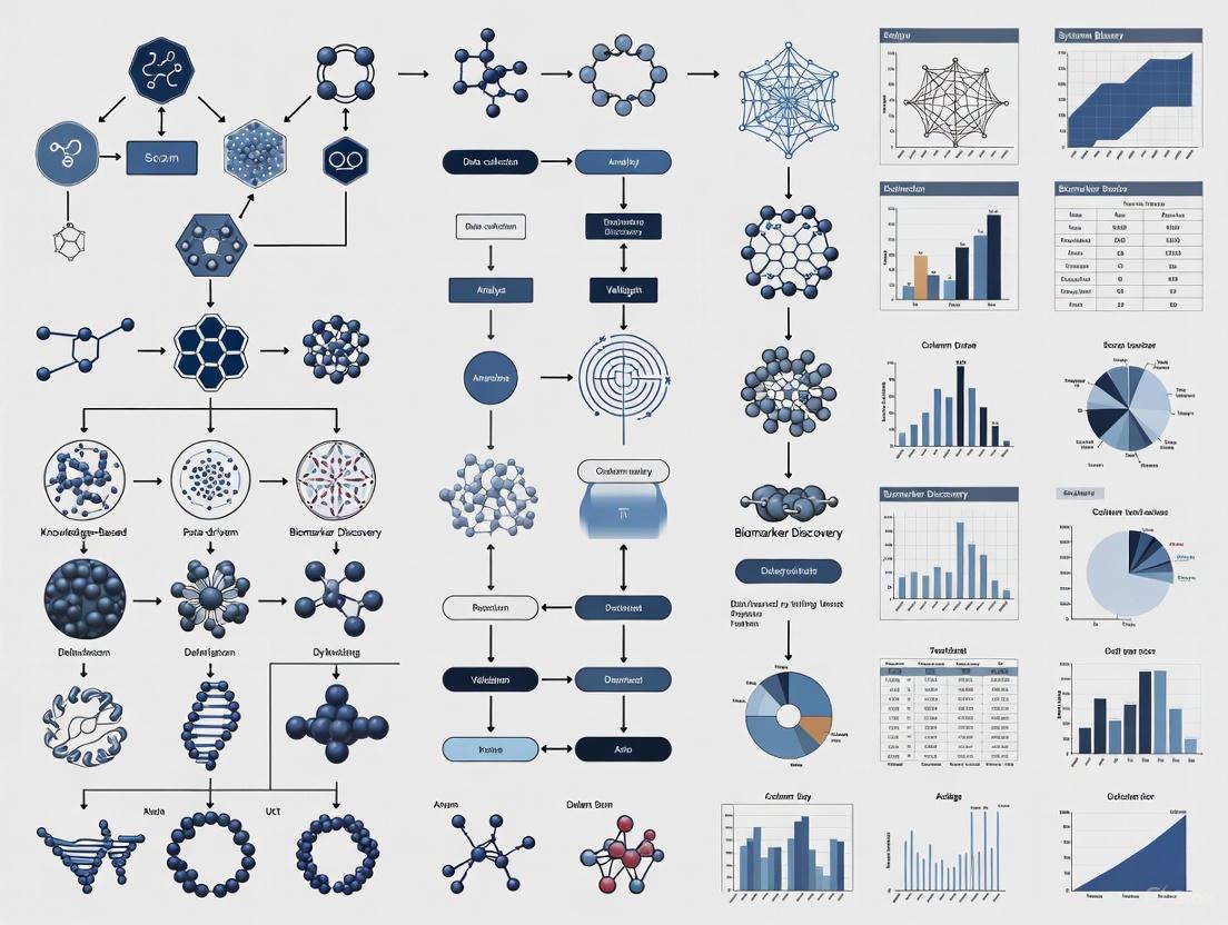
Diagram 1: Biomarker discovery workflow from sample to clinical application.
The Scientist's Toolkit: Essential Research Reagent Solutions
Table 3: Essential Research Reagents and Platforms for Biomarker Research
| Category | Specific Product/Platform | Primary Function | Key Applications |
|---|---|---|---|
| Sample Preparation | Omni LH 96 Homogenizer | Tissue disruption and homogenization | Standardized nucleic acid and protein extraction from tissue samples |
| Automation Systems | AquaMax 4000 Microplate Washer | Automated plate washing | High-throughput immunoassays in 96- or 384-well formats |
| Detection Instruments | SpectraMax ABS Plus Microplate Reader | Absorbance, fluorescence, and luminescence detection | Quantitative biomarker measurement in ELISA and other plate-based assays |
| Validated Assay Kits | Abcam SimpleStep ELISA Kits | Pre-optimized immunoassays with single-wash protocol | Rapid, reproducible quantification of specific protein biomarkers |
| Data Analysis Software | SoftMax Pro GxP Software | Curve fitting and data analysis | Compliance-ready data processing and reporting for biomarker assays |
| Next-Generation Sequencing | Comprehensive genomic profiling panels | Targeted sequencing of cancer-related genes | Mutation detection, fusion identification, and biomarker discovery |
| Liquid Biopsy Platforms | ctDNA extraction and analysis kits | Isolation and analysis of circulating tumor DNA | Non-invasive biomarker detection and monitoring |
Data-Driven Knowledge-Based Biomarker Discovery
The paradigm of biomarker discovery is shifting from traditional hypothesis-driven approaches toward hypothesis-free, data-driven strategies that leverage large-scale OMICS technologies and advanced computational analytics [5]. This approach systematically analyzes high-dimensional molecular data without preconceived notions of biological relevance, enabling the identification of novel biomarkers and associations that might be overlooked in targeted investigations.
Artificial intelligence is playing an increasingly transformative role in biomarker discovery. Machine learning algorithms, particularly deep learning networks, can identify complex patterns in multi-modal data that elude conventional statistical methods [9] [1]. The Predictive Biomarker Modeling Framework (PBMF) exemplifies this approach, using contrastive learning to systematically explore potential predictive biomarkers in an automated, unbiased manner [7]. When applied retrospectively to immuno-oncology trial data, this AI-driven framework has demonstrated the ability to identify biomarkers that could have improved patient selection for phase 3 trials, with identified patients showing a 15% improvement in survival risk compared to the original trial population [7].
The integration of dynamic prediction models (DPMs) represents another frontier in data-driven biomarker research. These models incorporate longitudinal biomarker measurements and time-dependent clinical events to continuously update prognostic predictions as new data becomes available during patient follow-up [8]. Joint models, which simultaneously analyze longitudinal biomarker data and time-to-event outcomes, are particularly valuable for capturing the evolving nature of cancer and its response to therapy [8].
Diagram 2: Data-driven knowledge-based biomarker discovery framework integrating multi-modal data and AI.
The field of oncology biomarkers is evolving toward increasingly sophisticated approaches that integrate multiple data modalities and leverage advanced computational methods. The future will see greater emphasis on multi-omics integration, combining genomic, transcriptomic, proteomic, epigenomic, and metabolomic data to develop comprehensive biomarker signatures that more accurately reflect the complexity of cancer biology [1] [5].
Artificial intelligence will play an expanding role in biomarker discovery and validation, with algorithms capable of identifying subtle patterns in high-dimensional data that escape human detection [9] [7]. The application of contrastive learning and other self-supervised approaches will enable more efficient identification of predictive biomarkers from complex clinicogenomic datasets [7]. Furthermore, the development of dynamic prediction models that incorporate longitudinal biomarker data will provide continuously updated prognostic assessments that better reflect the evolving nature of cancer and its response to therapy [8].
As biomarker science advances, addressing challenges related to data standardization, model generalizability across diverse populations, clinical implementation pathways, and regulatory alignment will be critical for translating these innovations into improved patient outcomes [1]. Through continued innovation in both molecular technologies and analytical approaches, biomarkers will increasingly fulfill their potential as essential tools for guiding personalized cancer care across the entire disease continuum.
The field of biomarker discovery is undergoing a profound transformation, moving from traditional hypothesis-driven approaches to data-driven strategies powered by artificial intelligence (AI). Traditional biomarker discovery methods, which often focus on single molecular features, face significant challenges including limited reproducibility, high false-positive rates, and inadequate predictive accuracy due to the inherent complexity and biological heterogeneity of diseases [10]. Machine learning (ML) and deep learning (DL) address these limitations by analyzing large, complex multi-omics datasets to identify more reliable and clinically useful biomarkers that capture the multifaceted biological networks underlying disease mechanisms [10]. This revolution enables the identification of patterns and relationships in high-dimensional data that far exceed human observational capacity and analytical capabilities [9] [11].
The integration of AI into biomarker research represents a fundamental shift toward proactive health management, transitioning from traditional disease diagnosis and treatment models to approaches based on prediction and prevention [1]. This transformation is grounded in the integration of diverse data types—including genomics, transcriptomics, proteomics, metabolomics, imaging data, and clinical records—providing comprehensive molecular profiles that facilitate the identification of highly predictive biomarkers across various disease areas [10] [1]. AI-driven biomarker discovery now spans oncology, infectious diseases, neurodegenerative disorders, and chronic inflammatory diseases, illustrating the versatility of these methodologies [10].
Table 1: Core AI Technologies in Biomarker Discovery
| AI Technology | Primary Applications in Biomarker Discovery | Key Advantages |
|---|---|---|
| Convolutional Neural Networks (CNNs) | Analysis of histopathology images, radiology scans, and spatial biology data [10] [9] | Identifies spatial patterns and features invisible to human observation |
| Recurrent Neural Networks (RNNs) | Processing sequential data, temporal biomarker patterns, and longitudinal monitoring data [10] | Captures time-dependent patterns in disease progression |
| Transformers & Large Language Models | Multi-omics data integration, literature mining, and clinical note analysis [10] [12] | Identifies complex non-linear associations across disparate data types |
| Random Forests & XGBoost | Feature selection, biomarker classification, and handling high-dimensional omics data [10] [13] | Robust against noise and overfitting; provides feature importance metrics |
| Generative Adversarial Networks | Molecular generation techniques, creating novel drug molecules [14] [15] | Generates novel molecular structures with desired biological properties |
AI Applications Across Data Modalities
Multi-Omics Data Integration
AI approaches have demonstrated remarkable capabilities in analyzing large-scale multi-omics datasets, enabling the identification of intricate patterns and interactions among various molecular features that were previously unrecognized [10]. By integrating genomic, epigenomic, transcriptomic, and proteomic data, ML models can develop comprehensive molecular disease maps that identify complex marker combinations traditional methods might overlook [1]. For instance, AI-driven analysis of multi-omics data has improved early Alzheimer's disease diagnosis specificity by 32%, providing a crucial intervention window [1]. The integration of multi-omics data reveals novel insights into the molecular basis of diseases and drug responses, identifying new biomarkers and therapeutic targets that predict and optimize individualized treatments [11].
Digital Biomarkers and Real-World Data
Digital biomarkers derived from wearables, smartphones, and connected medical devices are becoming invaluable tools that offer continuous, objective insights into a patient's health in real-world settings [16]. Unlike traditional clinical outcome assessments that rely on intermittent and sometimes subjective clinic-based measurements, digital biomarkers enable a richer, more dynamic understanding of disease progression and treatment response [16]. In oncology, wearable devices monitoring heart rate variability, sleep quality, and activity levels have reshaped how clinicians assess treatment tolerance and functional status [16]. When combined with electronic patient-reported outcome tools, these approaches capture daily symptom fluctuations—moving beyond static clinic visits and providing a real-world perspective of each patient's experience [16].
Spatial Biology and Imaging Data
Spatial biology techniques represent one of the most significant advances in biomarker discovery, revealing the spatial context of dozens of markers within a single tissue specimen [11]. Unlike traditional approaches, spatial transcriptomics and multiplex immunohistochemistry allow researchers to study gene and protein expression in situ without altering the spatial relationships or interactions between cells [11]. This provides critical information about physical distance between cells, cellular organization, and tissue architecture that is essential for understanding the complex tumor microenvironment [11]. Deep learning models, particularly CNNs, can extract hidden prognostic and predictive information directly from routine histological images, significantly enhancing traditional pathology workflows [10] [9].
Diagram 1: AI-Powered Spatial Biomarker Workflow (76 chars)
Experimental Protocols and Methodologies
Protocol 1: Plasma Proteomics-Based Biomarker Panel Discovery
Objective: To identify and validate a plasma protein signature for disease diagnosis and prognosis using machine learning approaches, as demonstrated in amyotrophic lateral sclerosis (ALS) research [17].
Materials and Reagents:
- Olink Explore 3072 platform or similar high-throughput proteomics system
- Plasma samples from patients and matched controls
- EDTA or heparin blood collection tubes
- Quality control materials for proteomic assays
- ELISA validation kits for candidate biomarkers
Methodology:
- Sample Preparation and Quality Control: Collect plasma samples following standardized protocols. Implement rigorous quality control measures, noting that the Olink platform typically demonstrates median intra-assay and inter-assay coefficients of variation of 9.9% and 22.3%, respectively [17].
Proteomic Profiling: Utilize the Olink Explore 3072 platform or equivalent to measure 2,886+ plasma proteins. Ensure adequate sample sizes (e.g., 183 ALS cases vs. 309 controls in discovery cohort) for statistical power [17].
Differential Abundance Analysis: Conduct proteome-wide association testing using generalized linear regression adjusted for age, sex, collection tube type, and population stratification factors. Apply false discovery rate (FDR) correction (e.g., FDR P < 0.05) to identify significantly differentially abundant proteins [17].
Machine Learning Model Development:
- Feature Selection: Include significantly differentially abundant proteins (e.g., 33 in ALS study) along with clinical parameters (age, sex) as initial features [17].
- Data Partitioning: Randomly designate 80% of samples as Discovery/Training Set and 20% as Replication/Test Set [17].
- Model Training: Employ supervised machine learning algorithms (e.g., random forests, XGBoost) to create binary classification models distinguishing cases from controls [17].
- Validation: Assess model performance using the independent replication cohort, with high-accuracy models achieving area under the curve (AUC) values up to 98.3% [17].
Pathway Analysis: Conduct enrichment analysis of significantly altered proteins to identify associated biological processes and pathways (e.g., skeletal muscle development, energy metabolism, neuronal function) [17].
Implementation Considerations: For studies aiming to predict disease onset in pre-symptomatic individuals, analyze plasma samples collected before symptom emergence to estimate the age of clinical onset and understand prodromal phase biology [17].
Protocol 2: Predictive Biomarker Discovery Using Network-Based Approaches
Objective: To identify predictive biomarkers for targeted cancer therapies by integrating network motifs and protein disorder information using the MarkerPredict framework [13].
Materials and Resources:
- Network data from Human Cancer Signaling Network (CSN), SIGNOR, and ReactomeFI databases
- Protein disorder data from DisProt, AlphaFold (pLLDT<50), and IUPred (score > 0.5) databases
- Biomarker annotations from CIViCmine text-mining database
- Machine learning environment with Random Forest and XGBoost implementations
Methodology:
- Network Motif Identification:
- Extract three-nodal motifs (triangles) from cancer signaling networks using FANMOD or similar tools
- Focus on triangles containing both intrinsically disordered proteins (IDPs) and known drug targets
- Note the significant overrepresentation of IDP-target triangles compared to random chance [13]
Training Dataset Construction:
- Positive Controls: Compile neighbor-target pairs where the neighbor is an established predictive biomarker for drugs targeting its pair partner (e.g., 332 of 4550 pairs in original study) [13]
- Negative Controls: Curate neighbor-target pairs where neighbors are not predictive biomarkers in CIViCmine database, supplemented with random pairs [13]
Machine Learning Framework:
- Feature Engineering: Incorporate topological network properties, protein disorder metrics, and functional annotations
- Model Training: Develop 32 different models using Random Forest and XGBoost algorithms on network-specific and combined data across three IDP databases [13]
- Hyperparameter Tuning: Utilize competitive random halving for parameter optimization [13]
Validation and Scoring:
- Employ leave-one-out-cross-validation (LOOCV), k-fold cross-validation, and train-test splits (70:30)
- Expect performance metrics in the range of 0.7-0.96 LOOCV accuracy for well-trained models [13]
- Calculate Biomarker Probability Score (BPS) as a normalized summative rank of model predictions to harmonize probability values [13]
Implementation Considerations: This approach identified 2084 potential predictive biomarkers for targeted cancer therapeutics, with 426 classified as biomarkers by all four calculations in the original study [13].
Table 2: Research Reagent Solutions for AI-Driven Biomarker Discovery
| Research Reagent | Function in Biomarker Discovery | Application Context |
|---|---|---|
| Olink Explore 3072 Platform | High-throughput proteomic profiling of 3,072 proteins from minimal sample volumes [17] | Plasma proteomic biomarker discovery for neurological and other diseases |
| Spatial Transcriptomics Platforms | Gene expression analysis with tissue spatial context preservation [11] | Tumor microenvironment characterization, spatial biomarker identification |
| Human Cancer Signaling Network Database | Provides curated cancer signaling pathways for network-based biomarker discovery [13] | Predictive biomarker identification for targeted therapies |
| DisProt Database | Centralized resource of experimentally characterized intrinsically disordered proteins [13] | Identification of disordered proteins as potential biomarkers |
| Organoid & Humanized Models | Physiologically relevant systems for functional biomarker validation [11] | Biomarker screening, target validation, resistance mechanism exploration |
Implementation Considerations and Challenges
Data Quality and Standardization
The performance of AI models in biomarker discovery heavily depends on data quality and standardization. Challenges include limited sample sizes, noise, batch effects, and biological heterogeneity that can severely impact model performance, leading to issues such as overfitting and reduced generalizability [10] [1]. Differences in sensor calibration, environmental factors, and user behavior can introduce variability or measurement errors in digital biomarker data [16]. Successful implementation requires robust data governance frameworks, including encryption, anonymization, and adherence to regulatory standards such as GDPR and HIPAA to protect patient confidentiality and build trust [16].
Model Interpretability and Validation
The interpretability of ML models remains a significant hurdle, as many advanced algorithms function as "black boxes," making it difficult to elucidate how specific predictions are derived [10]. This lack of interpretability poses practical barriers to clinical adoption, where transparency and trust in predictive models are essential [10]. Explainable AI techniques such as SHAP analysis can help demonstrate feature impact explanations, increasing transparency for biomarker-driven modeling [12]. Furthermore, biomarkers identified through computational methods must undergo stringent validation using independent cohorts and experimental methods to ensure reproducibility and clinical reliability [10] [17].
Regulatory and Ethical Considerations
The deployment of ML-derived biomarkers into clinical practice requires compliance with rigorous standards set by regulatory bodies such as the FDA and EMA [10] [9]. The dynamic nature of ML-driven biomarker discovery, where models continuously evolve with new data, presents particular challenges for regulatory oversight and demands adaptive yet strict validation and approval frameworks [10]. Algorithmic bias and generalizability also pose potential risks, as many digital biomarker algorithms are trained on limited demographic groups, potentially reducing accuracy in underrepresented populations [16]. Including diverse participants during algorithm development is essential to mitigate these biases [16].
Diagram 2: Biomarker Validation Pipeline (76 chars)
The future of AI-driven biomarker discovery lies in several promising directions. Expanding predictive models to rare diseases, incorporating dynamic health indicators, strengthening integrative multi-omics approaches, conducting longitudinal cohort studies, and leveraging edge computing solutions for low-resource settings emerge as critical areas requiring innovation and exploration [1]. Future research should focus on directly linking genomic data to functional outcomes, particularly with biosynthetic gene clusters and non-coding RNAs [10]. The recognition of the microbiome's impact on health has also spurred the application of ML to identify microbial pathways involved in metabolite production, revealing potential therapeutic targets and expanding the biomarker landscape beyond the human genome to include the broader holobiont [10].
The integration of AI biomarker analysis into early research and development will not happen in isolation—it requires collaboration across the entire ecosystem [9]. Pharmaceutical and biotech companies must invest in data infrastructure and AI partnerships, academic researchers should provide translational insights that bridge preclinical and clinical biology, and regulators need to evolve frameworks that foster innovation while ensuring patient safety [9]. As these partnerships mature and technologies advance, AI-driven biomarker discovery will continue to enhance our ability to develop personalized treatment strategies that improve patient outcomes, ultimately realizing the promise of precision medicine through patterns uncovered beyond human capability.
Multi-omics integration has emerged as a transformative approach in biomedical research, moving beyond the limitations of single-omics analyses to provide a comprehensive understanding of complex biological systems. By combining data from genomics, transcriptomics, proteomics, and metabolomics, researchers can now capture the full spectrum of molecular interactions that underlie health and disease states. This integrated framework is particularly powerful for biomarker discovery, offering unprecedented opportunities to identify robust diagnostic, prognostic, and predictive signatures across various disease areas, including oncology, tissue repair, and beyond. The implementation of multi-omics strategies requires sophisticated computational tools for data integration and visualization, coupled with rigorous experimental protocols. As the field advances, emerging technologies such as single-cell multi-omics, spatial omics, and artificial intelligence are further enhancing our ability to decipher disease mechanisms and develop personalized therapeutic strategies, ultimately driving the evolution of precision medicine.
Multi-omics represents a paradigm shift in biological research, integrating data from multiple molecular levels to construct comprehensive models of biological systems. This approach recognizes that biological functions emerge from complex interactions between various molecular layers—from genetic blueprint to metabolic activity. Where single-omics studies (examining only genomics, transcriptomics, proteomics, or metabolomics in isolation) provide limited insights, multi-omics integration reveals how alterations at one molecular level propagate through the system to influence phenotype and function [18]. This holistic perspective is particularly valuable for understanding complex diseases like cancer, where heterogeneity and dynamic changes across molecular layers drive pathogenesis and treatment response [19] [20].
The fundamental premise of multi-omics is that each molecular layer provides complementary information: genomics reveals inherited and acquired genetic variants; transcriptomics captures gene expression patterns; proteomics identifies protein abundance and modifications; and metabolomics profiles the end-products of cellular processes that most closely reflect phenotypic states [18] [20]. By integrating these layers, researchers can bridge the gap between genotype and phenotype, uncovering causal relationships and regulatory mechanisms that would remain invisible in single-omics approaches.
In the context of biomarker discovery, multi-omics strategies have demonstrated particular promise for identifying molecular signatures with greater specificity and clinical utility than single-omics biomarkers. The integration of multiple data types helps distinguish driver alterations from passenger events, identifies compensatory pathways that may mediate treatment resistance, and reveals biomarkers that accurately stratify patient populations for targeted therapies [19] [21] [20]. Furthermore, as pharmaceutical research increasingly focuses on personalized medicine, multi-omics approaches provide the comprehensive molecular profiling necessary to match patients with optimal treatments based on their unique molecular profiles [18] [22].
Methodological Framework for Multi-Omics Integration
Data Integration Strategies
Successful multi-omics integration requires methodological frameworks that can handle the complexity, high dimensionality, and heterogeneity of omics data. Several computational strategies have been developed, each with distinct strengths and applications in biomarker discovery research.
Table 1: Multi-Omics Data Integration Approaches
| Integration Method | Key Characteristics | Applications in Biomarker Discovery | Example Tools/Pipelines |
|---|---|---|---|
| Conceptual Integration | Links omics data through shared biological concepts or entities | Hypothesis generation; exploring associations between omics sets; functional annotation | STATegra, OmicsON, Gene Ontology, pathway databases |
| Statistical Integration | Uses quantitative measures to combine or compare datasets | Identifying co-expressed molecules across omics layers; clustering patients based on molecular profiles | Correlation analysis, regression models, clustering algorithms |
| Model-Based Integration | Employs mathematical models to simulate system behavior | Understanding dynamics and regulation of biological systems; predicting drug responses | Network models, PK/PD models, systems pharmacology |
| Network and Pathway Integration | Uses biological pathways to represent system structure and function | Visualizing interactions between molecules; identifying dysregulated pathways in disease | Protein-protein interaction networks, metabolic pathways |
Conceptual integration leverages existing biological knowledge from curated databases to link different omics datasets through shared entities such as genes, proteins, or pathways [18]. This approach is particularly valuable in the early stages of biomarker discovery, as it allows researchers to contextualize molecular findings within established biological processes. For example, identifying that differentially expressed genes, proteins, and metabolites from a multi-omics dataset all map to the same metabolic pathway significantly strengthens the case for that pathway's involvement in the disease process.
Statistical integration employs quantitative methods to identify patterns across omics datasets without requiring extensive prior biological knowledge [18]. These methods include correlation analysis to identify co-expressed genes or proteins across different molecular layers, and clustering techniques to group patients based on integrated molecular profiles. Such approaches can reveal novel biomarker combinations that would not be identified when analyzing each omics layer separately. For instance, statistical integration might identify that a specific genetic variant only leads to protein-level changes when accompanied by particular metabolic conditions—a finding with important implications for patient stratification.
Model-based integration uses mathematical and computational models to simulate biological system behavior, creating predictive frameworks that can inform biomarker discovery [18]. Network models can represent interactions between biomolecules across different omics layers, while pharmacokinetic/pharmacodynamic (PK/PD) models can describe how drugs are processed in the body and how they affect multiple molecular systems. These models are particularly valuable for predicting how interventions might affect biomarker levels and how biomarker combinations might predict treatment response.
Network and pathway-based integration provides a biological context for multi-omics data by mapping molecular measurements onto established pathways or interaction networks [18]. This approach helps researchers interpret complex multi-omics datasets in terms of disrupted biological processes, which is essential for understanding the functional implications of potential biomarkers. For example, mapping genomic, transcriptomic, and proteomic data onto protein-protein interaction networks can identify key hub proteins that might serve as biomarkers or therapeutic targets.
Workflow for Multi-Omics Biomarker Discovery
The typical workflow for multi-omics biomarker discovery involves several standardized steps:
Experimental Design: Careful planning of sample collection, storage, and processing to minimize technical variation across different omics platforms. This includes determining appropriate sample sizes, considering batch effects, and ensuring ethical compliance.
Data Generation: Simultaneous or sequential generation of data from multiple omics platforms, potentially including whole-genome sequencing, RNA sequencing, mass spectrometry-based proteomics, and NMR or LC-MS-based metabolomics.
Quality Control and Preprocessing: Rigorous quality assessment for each dataset, followed by normalization, missing value imputation, and data transformation to make datasets comparable.
Data Integration: Application of integration methods (as described in Table 1) to combine information from multiple omics layers.
Biomarker Identification: Use of statistical and machine learning approaches to identify molecular patterns associated with diseases, outcomes, or treatment responses.
Validation: Experimental validation of candidate biomarkers using independent samples and different analytical techniques.
A critical consideration throughout this workflow is the handling of data heterogeneity. Multi-omics data varies in scale, distribution, and noise characteristics, requiring specialized normalization and transformation approaches before integration [18]. Additionally, the high dimensionality of multi-omics data (with far more features than samples) necessitates appropriate statistical corrections to avoid false discoveries.
Experimental Protocols and Applications
Protocol 1: Multi-Omics Biomarker Discovery for Cancer Diagnostics
Objective: To identify integrated molecular signatures for cancer diagnosis, prognosis, and treatment selection using horizontally and vertically integrated multi-omics approaches.
Background: In oncology, multi-omics approaches have proven particularly valuable for addressing tumor heterogeneity and understanding the complex interplay between cancer cells and their microenvironment [19] [20]. Horizontal integration combines data from the same omics layer (e.g., multiple transcriptomic datasets), while vertical integration connects different biological layers (e.g., genomics to transcriptomics to proteomics) [20].
Table 2: Experimental Steps for Cancer Multi-Omics Biomarker Discovery
| Step | Procedure | Key Parameters | Quality Controls |
|---|---|---|---|
| Sample Collection | Collect tumor and matched normal tissues; record clinical metadata | Snap-freeze in liquid N₂; maintain cold chain | Assess tissue viability; document ischemia time |
| Nucleic Acid Extraction | Isolate DNA and RNA using commercial kits | Quantity and quality assessment | Bioanalyzer RNA Integrity Number (RIN) > 7.0 |
| Genomics (WES/WGS) | Library preparation and sequencing | Minimum 80x coverage for WGS; 100x for WES | Check coverage uniformity; mapping rates >90% |
| Transcriptomics | RNA-seq library prep (poly-A selection) | Minimum 30 million reads per sample | Check rRNA contamination; alignment rates |
| Proteomics | Protein extraction, digestion, LC-MS/MS | TMT or label-free quantification | Include QC reference samples; monitor retention time |
| Data Integration | Apply computational integration methods | Choice of horizontal vs. vertical integration | Assess batch effects; implement correction |
Detailed Procedures:
Sample Preparation and Quality Control:
- Obtain fresh-frozen tissue sections (5-10 mg) and allocate portions for each omics analysis.
- For nucleic acid extraction, use validated commercial kits with DNase/RNase-free conditions.
- Assess DNA/RNA quality using Agilent Bioanalyzer or TapeStation systems (RNA Integrity Number > 7.0 required).
- For proteomics, homogenize tissue in lysis buffer (e.g., 8M urea, 2M thiourea) with protease and phosphatase inhibitors.
Genomics Workflow:
- Perform library preparation using Illumina TruSeq DNA PCR-Free Library Prep Kit.
- Sequence on Illumina NovaSeq platform (150bp paired-end reads).
- Process data through bioinformatics pipeline: quality trimming (FastQC), alignment to reference genome (BWA-MEM), variant calling (GATK), and annotation (ANNOVAR).
Transcriptomics Workflow:
- Isolate mRNA using poly-A selection (Illumina Stranded mRNA Prep).
- Sequence on Illumina platform (minimum 30 million reads per sample).
- Process data: quality control (FastQC), alignment (STAR), quantitation (featureCounts), and differential expression analysis (DESeq2).
Proteomics Workflow:
- Digest proteins with trypsin (filter-aided sample preparation protocol).
- Desalt peptides using C18 solid-phase extraction.
- Analyze by LC-MS/MS on Orbitrap Eclipse Tribrid mass spectrometer.
- Process data: peak detection (MaxQuant), database searching (UniProt), and quantitation (LFQ or TMT-based).
Data Integration and Analysis:
- For horizontal integration: Combine scRNA-seq with spatial transcriptomics using Seurat v5 or Cell2location to map cell types to tissue locations [20].
- For vertical integration: Link genomic variants to transcriptomic and proteomic changes using iCluster or Multi-Omics Factor Analysis [20].
- Identify candidate biomarkers showing consistent alterations across multiple omics layers.
Applications: This protocol has been successfully applied in lung cancer research, revealing biomarkers such as KRT8+ alveolar intermediate cells that represent transitional states during early tumorigenesis [20]. The integrated approach has also identified TIM-3+ immune cells with impaired antigen presentation capacity, providing both diagnostic and therapeutic insights [20].
Protocol 2: Multi-Omics Data Visualization for Pathway Analysis
Objective: To simultaneously visualize multiple omics datasets on biological pathway diagrams to identify dysregulated pathways and network interactions.
Background: Visual integration of multi-omics data enables researchers to interpret complex molecular relationships in the context of biological pathways. Several tools are available for this purpose, each with unique capabilities for visualizing different omics data types simultaneously [23] [24] [25].
Detailed Procedures:
Data Preparation for PathVisio:
- Create a single data file containing all omics measurements with appropriate database identifiers.
- Format columns to include: Identifier, System Code (e.g., "L" for Entrez Gene, "S" for UniProt, "Ce" for ChEBI), quantitative values (e.g., log2FC), p-values, and Type (e.g., "Transcriptomics," "Proteomics") [23].
- Example data format:
PathVisio Visualization Workflow:
- Open PathVisio and load the pathway of interest.
- Ensure appropriate identifier mapping databases are loaded (species-specific gene and metabolite mapping).
- Import expression data through Data > Import Expression Data.
- In the import wizard, select the appropriate system code column for identifier mapping.
- Create advanced visualization through Data > Visualization Options:
- Select "Text label" and "Expression as color" > "Advanced."
- For log2FC values, create a color gradient from blue (down-regulated) to red (up-regulated).
- For data types, add rule-based coloring with distinct colors for each omics type [23].
OmicCircos Circular Visualization:
- Install OmicCircos in R:
BiocManager::install("OmicCircos") - Prepare genomic data in appropriate formats for different tracks (e.g., chromosome ideogram, gene expression, copy number variations).
- Use core functions:
sim.circos()to create simulated input data for practice.segAnglePo()to transform linear data into angular coordinates.circos()to create circular plots with multiple track types [24].
- Customize plots by adding scatterplots, lines, heatmaps, and curves to represent different omics data types and their relationships.
- Install OmicCircos in R:
PTools Cellular Overview Multi-Omics Visualization:
- Load up to four omics datasets simultaneously, assigning each to a different visual channel:
- Reaction edge color (e.g., for transcriptomics data)
- Reaction edge thickness (e.g., for proteomics data)
- Metabolite node color (e.g., for metabolomics data)
- Metabolite node thickness (e.g., for additional metabolic data) [25].
- Use interactive controls to adjust color and thickness mappings for optimal data representation.
- Utilize semantic zooming to reveal additional details at higher magnification levels.
- For time-series data, employ animation features to visualize dynamic changes across molecular layers.
- Load up to four omics datasets simultaneously, assigning each to a different visual channel:
Applications: These visualization approaches have been used to map relationships between human papillomavirus (HPV) genome and human genes in cervical cancer, and to display multi-omics profiles of breast cancer subtypes from The Cancer Genome Atlas data [24]. The simultaneous visualization of multiple data types helps identify coordinated changes across molecular layers that might be missed when examining individual datasets separately.
Visualization Approaches for Multi-Omics Data
Effective visualization is crucial for interpreting complex multi-omics datasets. Several specialized tools have been developed to represent multiple molecular layers simultaneously in biologically meaningful contexts.
Multi-Omics Visualization Tool Ecosystem
Table 3: Comparison of Multi-Omics Visualization Tools
| Tool | Diagram Type | Simultaneous Omics Types | Key Features | Best Applications |
|---|---|---|---|---|
| PTools Cellular Overview | Automated pathway-specific layout | 4 | Semantic zooming, animation, organism-specific diagrams | Metabolic pathway analysis, time-series multi-omics |
| PathVisio | Manually curated pathways | 3 | Rule-based visualization, multiple identifier systems | Pathway-centric biomarker validation |
| OmicCircos | Circular genomic plots | Multiple tracks | Genomic coordinate mapping, multiple track types | Genome-wide association studies, copy number variation |
| KEGG Mapper | Manual uber pathways | 2 | Standardized pathway diagrams, wide pathway coverage | Cross-species comparison, communication |
| PaintOmics | Manually drawn pathways | 3 | Web-based interface, no installation required | Collaborative projects, quick visualization |
The PTools Cellular Overview represents one of the most advanced multi-omics visualization systems, supporting the simultaneous display of four different omics datasets through distinct visual channels [25]. This tool uses automated layout algorithms to generate organism-specific metabolic network diagrams, ensuring that visualizations accurately reflect the specific metabolic capabilities of the organism being studied. A key advantage is the support for semantic zooming, which reveals different levels of detail as users zoom in and out of the diagram. Additionally, the animation capabilities enable researchers to visualize dynamic processes and time-series multi-omics data, revealing how molecular relationships evolve over time or in response to perturbations.
PathVisio offers powerful capabilities for creating intuitive visualizations of multiple omics data types on pathway diagrams [23]. The software supports rule-based visualization, allowing researchers to define custom display rules based on statistical thresholds, fold-changes, or data types. This flexibility is particularly valuable in biomarker discovery, where visualizing the same dataset with different thresholds can reveal meaningful patterns. PathVisio's support for multiple database identifier systems facilitates the integration of diverse omics data types that may use different naming conventions (e.g., Entrez Gene IDs for transcriptomics, UniProt IDs for proteomics, and ChEBI IDs for metabolomics).
OmicCircos specializes in circular plots for genomic data, enabling researchers to visualize multiple types of genomic information in coordinated tracks [24]. This approach is particularly useful for displaying relationships between genomic position and various molecular measurements, such as showing gene expression, copy number variations, and mutation status simultaneously across all chromosomes. The circular format facilitates identification of chromosomal patterns and hotspots that might be associated with disease processes or treatment responses.
Successful implementation of multi-omics approaches requires both wet-lab reagents for data generation and computational tools for data integration and analysis. This section outlines essential resources for a comprehensive multi-omics biomarker discovery pipeline.
Table 4: Essential Research Reagent Solutions for Multi-Omics Studies
| Category | Specific Products/Technologies | Key Applications | Considerations |
|---|---|---|---|
| Nucleic Acid Extraction | Qiagen AllPrep, TRIzol, magnetic bead-based systems | Simultaneous DNA/RNA extraction | Maintain RNA integrity, avoid cross-contamination |
| Sequencing Library Prep | Illumina TruSeq, NEBNext, SMARTer kits | WGS, WES, RNA-seq | Compatibility with downstream analysis |
| Proteomics Sample Prep | Filter-aided sample preparation (FASP), S-Trap kits | Protein digestion, cleanup | Efficiency, reproducibility, compatibility with MS |
| Mass Spectrometry | Orbitrap (Thermo), TIMS-TOF (Bruker) systems | Proteomics, metabolomics | Resolution, sensitivity, quantitative accuracy |
| Metabolomics | Biocrates kits, IROA technologies | Targeted metabolomics | Coverage, standardization, quantification |
| Single-Cell Technologies | 10x Genomics, BD Rhapsody | scRNA-seq, single-cell multi-omics | Cell viability, capture efficiency, cost |
Computational Resources and Tools:
Data Integration Platforms:
- STATegra: Provides a comprehensive framework for multi-omics data integration, with particular strength in time-series analyses [18].
- iCluster: Bayesian method for integrative clustering of multiple omics data types, useful for patient stratification [20].
- Multi-Omics Factor Analysis (MOFA): Discovers principal sources of variation across multiple omics layers [20].
Visualization Tools:
- PathVisio: Creates pathway-based visualizations with support for multiple omics data types and custom visualization rules [23].
- OmicCircos: Generates circular plots for genomic data, with capabilities for displaying multiple data tracks simultaneously [24].
- PTools Cellular Overview: Provides organism-specific metabolic network diagrams with multi-omics visualization capabilities [25].
Specialized Databases:
- Gene Ontology: Provides standardized vocabulary for gene function annotation, essential for conceptual integration [18].
- Pathway Databases: KEGG, Reactome, and WikiPathways offer curated pathway information for biological context [18] [25].
- Protein-Protein Interaction Networks: STRING and BioGRID provide interaction data for network-based integration [18].
The selection of appropriate reagents and computational tools should be guided by the specific research question, sample types, and available infrastructure. As multi-omics technologies continue to evolve, staying current with emerging platforms and methodologies is essential for maintaining cutting-edge biomarker discovery capabilities.
The field of multi-omics research is rapidly evolving, driven by technological advancements and increasingly sophisticated computational methods. Several emerging trends are poised to further transform biomarker discovery and precision medicine.
Emerging Technologies: Single-cell multi-omics technologies are revealing unprecedented insights into cellular heterogeneity within tissues and tumors [19] [20]. These approaches allow researchers to measure multiple molecular layers simultaneously from the same cell, providing direct evidence of how genomic variation influences transcriptomic and epigenomic states at the single-cell level. Spatial omics technologies add another dimension by preserving the architectural context of cells within tissues, enabling researchers to map molecular relationships within the tissue microenvironment [19]. These technologies are particularly valuable for understanding cell-cell interactions and spatial organization patterns that drive disease processes.
Artificial Intelligence Integration: Machine learning and deep learning approaches are becoming increasingly integral to multi-omics data analysis [19] [26]. These methods can identify complex, non-linear patterns across omics layers that might escape conventional statistical approaches. AI-based integration methods are particularly promising for predictive biomarker discovery, where they can integrate diverse molecular data to forecast disease progression, treatment response, or adverse events [19] [26]. As these methods mature, they are expected to enhance the precision and personalization of clinical interventions.
Clinical Translation Challenges: Despite the exciting potential of multi-omics approaches, several challenges remain for widespread clinical implementation. Standardization of protocols across laboratories, establishment of quality control metrics, and development of regulatory frameworks for multi-omics-based diagnostics are active areas of development [19] [18]. Additionally, the cost and computational complexity of multi-omics analyses present barriers to routine clinical application. However, as technologies continue to advance and costs decrease, multi-omics approaches are likely to become increasingly accessible for clinical biomarker discovery and implementation.
Conclusion: Multi-omics integration represents a powerful framework for biomarker discovery, providing a more comprehensive understanding of biological systems and disease processes than single-omics approaches. By simultaneously considering multiple molecular layers, researchers can identify robust biomarker signatures with greater predictive power and clinical utility. The successful implementation of multi-omics strategies requires careful experimental design, appropriate computational integration methods, and effective visualization approaches. As technologies continue to advance and computational methods become more sophisticated, multi-omics approaches are poised to drive significant advances in precision medicine, enabling more accurate diagnosis, prognosis, and treatment selection across a wide range of diseases.
The paradigm of biomarker discovery is shifting from traditional, hypothesis-driven approaches to data-driven, knowledge-based research. This transition is fueled by the convergence of high-throughput biotechnology, which can generate millions of clinicogenomic measurements per individual [7], and advanced artificial intelligence (AI). However, two significant challenges impede progress: the data accessibility problem, where sensitive clinical and genomic data cannot be centralized for analysis due to privacy and regulatory concerns, and the trust deficit, where the "black-box" nature of complex AI models hinders their adoption in clinical practice [27] [28].
In response, two key technological drivers have emerged as foundational to modern biomarker research: Federated Learning (FL) for secure, collaborative data analysis and Explainable AI (XAI) for building clinical trust. FL enables the training of machine learning models across multiple decentralized data sources without moving or sharing the underlying raw data [29]. Simultaneously, XAI provides transparent, interpretable results that allow clinicians and researchers to understand the reasoning behind an AI's output, fostering trust and facilitating clinical actionability [27] [28]. This Application Note details the protocols and methodologies for integrating these technologies into a robust framework for biomarker discovery.
Federated Learning for Secure, Collaborative Biomarker Discovery
Federated Learning is a distributed machine learning approach that is revolutionizing how researchers leverage real-world data from multiple institutions. It is particularly vital for biomarker discovery in oncology and rare diseases, where sample sizes from single centers are often insufficient for robust model development.
Core Protocol: Federated Model Training for a Predictive Biomarker Signature
The following protocol outlines the key steps for training a predictive biomarker model using a federated approach across multiple hospital sites.
Objective: To collaboratively train a predictive biomarker model for immunotherapy response in non-small cell lung cancer (NSCLC) using distributed clinicogenomic datasets without centralizing patient data. Data Type: Multi-modal data including genomic sequencing (e.g., from Comprehensive Genome Profiling panels), transcriptomics, and structured clinical data from Electronic Health Records (EHRs) [30] [29].
| Step | Procedure | Key Considerations & Parameters |
|---|---|---|
| 1. Initialization | A central server initializes a global machine learning model (e.g., a neural network or gradient boosting model). | Model architecture must be agreed upon by all participating sites. The PBMF framework is an example of a neural network suitable for this task [7]. |
| 2. Client Selection | The server selects a subset of available client sites (e.g., hospital servers) to participate in a training round. | Selection can be random or based on specific criteria like dataset size or computational availability. |
| 3. Client Download | Selected clients download the current global model weights from the central server. | Communication must be secured via encryption (e.g., HTTPS/TLS). |
| 4. Local Training | Each client trains the model on its local data. Training is performed for a predetermined number of local epochs. | Critical: Local data never leaves the hospital firewall. Algorithms like Stochastic Gradient Descent (SGD) are typically used. |
| 5. Model Export | Each client generates updated model parameters (e.g., weight updates, gradients) from the locally trained model. | Only the model parameters, not the training data, are exported. Differential privacy techniques can be added to further obscure the contribution of any single data point. |
| 6. Secure Aggregation | The clients send their model updates to the central server. The server aggregates these updates to improve the global model. | Aggregation algorithms like Federated Averaging (FedAvg) are standard. The formula for FedAvg is: ( w{t+1} \leftarrow \sum{k=1}^{K} \frac{nk}{n} w{t+1}^k ), where ( w ) are the weights, ( K ) is the number of clients, and ( n_k/n ) is the fraction of data on client ( k ). |
| 7. Model Update | The server updates the global model with the aggregated parameters. | The process repeats from Step 2 for a set number of rounds or until model performance converges. |
| 8. Validation | The final global model is evaluated on a held-out test set from each client or a centralized public dataset to assess its performance and generalizability. | Performance metrics (e.g., AUC, C-index) are reported for each site to check for consistency and identify potential biases [29]. |
Workflow Visualization
The diagram below illustrates the iterative cycle of federated model training.
Explainable AI for Clinical Trust and Actionable Insights
The development of a high-performance biomarker model is insufficient for clinical translation. Clinicians must understand the model's reasoning to trust and appropriately use its predictions. XAI techniques are essential for transforming a black-box prediction into an interpretable and clinically actionable insight [27] [31].
Core Protocol: Implementing XAI in a Biomarker-Driven Clinical Decision Support System (CDSS)
This protocol describes how to integrate XAI into a CDSS that provides biomarker-based treatment recommendations.
Objective: To explain an AI model's prediction of positive response to immunotherapy in a specific NSCLC patient, highlighting the key genomic and clinical features driving the decision. Model: A trained machine learning model (e.g., Random Forest, XGBoost, or Neural Network) for predicting treatment response. XAI Techniques: SHAP (SHapley Additive exPlanations) and LIME (Local Interpretable Model-agnostic Explanations) [27] [28].
| Step | Procedure | Key Considerations & Parameters |
|---|---|---|
| 1. Model & Data Setup | Deploy the trained predictive model and prepare the individual patient's data for explanation. | Data must be preprocessed identically to the training data (e.g., normalized, encoded). |
| 2. Global Explainability (SHAP) | Calculate SHAP values for the entire validation dataset to understand the model's overall behavior. | SHAP values quantify the marginal contribution of each feature to the model's prediction. Use shap.Explainer() and shap.summary_plot() to visualize global feature importance. |
| 3. Local Explainability (SHAP/LIME) | Generate an explanation for a single patient's prediction. | For SHAP: Use shap.force_plot() or shap.waterfall_plot() to show how each feature pushed the model's output from the base value to the final prediction. For LIME: Create a local surrogate model (e.g., linear model) that approximates the black-box model's behavior for that specific instance. Use lime_tabular.LimeTabularExplainer(). |
| 4. Explanation Presentation | Integrate the explanation into the CDSS user interface in a clear, concise manner for the clinician. | Present the top 5-10 features driving the prediction. Use natural language (e.g., "This patient is predicted to respond due to high PD-L1 expression and low tumor mutational burden"). Visual aids like bar charts or waterfall plots are highly effective [31]. |
| 5. Clinical Validation & Feedback | The clinician uses the explanation to contextualize the AI's recommendation against their own expertise. | This human-in-the-loop step is critical. Discrepancies between the explanation and clinical knowledge can reveal model biases or data quality issues, creating a feedback loop for model improvement [32] [31]. |
Workflow Visualization
The diagram below illustrates the pathway from a black-box model to a clinically trusted decision.
The Scientist's Toolkit: Essential Research Reagents & Computational Solutions
The following table details key resources and tools required for implementing the federated and explainable biomarker discovery workflows described in this note.
| Category | Item / Solution | Function & Application Note |
|---|---|---|
| Data & Standards | Real-World Clinicogenomic Data | Multi-modal data from EHRs, NGS, and transcriptomics. Must be harmonized using standards like OMOP CDM for federated analysis [30] [1]. |
| Biomarker Definitions | BEST (Biomarkers, EndpointS, and other Tools) Resource guidelines for defining and validating biomarker types (prognostic vs. predictive) [30]. | |
| Computational Frameworks | Federated Learning Platform | Software like Lifebit, NVIDIA FLARE, or Flower that orchestrates the federated training cycle across distributed data nodes [29]. |
| Predictive Biomarker Modeling Framework (PBMF) | A specific AI framework based on contrastive learning, designed to systematically discover predictive (not just prognostic) biomarkers from clinicogenomic data [7]. | |
| XAI Libraries | SHAP (SHapley Additive exPlanations) | A unified game theory-based library for explaining the output of any machine learning model. Provides both global and local interpretability [27] [28]. |
| LIME (Local Interpretable Model-agnostic Explanations) | Creates local, surrogate models to explain individual predictions. Useful for validating SHAP findings or for models where SHAP is computationally expensive [28]. | |
| Validation & Evaluation | Independent Clinical Cohorts | Held-out datasets from different geographical or demographic populations are essential for assessing model generalizability and preventing overfitting [1]. |
| Performance Metrics | Standard metrics (AUC, Accuracy) and clinical utility metrics (e.g., Net Reclassification Index) to evaluate the biomarker's impact on decision-making [29] [7]. |
The integration of Federated Learning and Explainable AI represents a foundational shift in data-driven biomarker discovery. FL directly addresses the critical constraints of data privacy and accessibility, enabling the pooling of statistical power from globally distributed datasets. Concurrently, XAI addresses the "trust gap" by making model outputs interpretable, which is a non-negotiable requirement for clinical adoption [31] [28]. The protocols outlined herein provide a actionable roadmap for researchers to build more robust, generalizable, and clinically trustworthy biomarker models. By adopting this integrated framework, the field can accelerate the translation of complex data into meaningful knowledge, ultimately advancing the goals of personalized medicine.
Building the Pipeline: Technologies and Workflows for Next-Generation Biomarker Identification
The discovery of robust, clinically relevant biomarkers is a cornerstone of modern precision medicine, yet the process remains notoriously challenging, expensive, and time-consuming. The integration of Artificial Intelligence (AI) and machine learning (ML) creates a powerful, data-driven paradigm shift, moving from a hypothesis-limited approach to a holistic, systems-level understanding of disease biology [33]. This document details the application notes and protocols for implementing an AI-powered discovery pipeline, designed to accelerate biomarker identification and validation within knowledge-based research frameworks. By systematically orchestrating the flow of data from heterogeneous sources through to model deployment, this pipeline ensures scalable, reproducible, and actionable insights that can fundamentally enhance drug development.
Pipeline Architecture and Quantitative Impact
An AI data pipeline automates the end-to-end flow of data, from raw collection to model training and deployment. It is distinguished from traditional data pipelines by its incorporation of ML-specific processes such as feature engineering, model training, continuous learning, and real-time data processing capabilities [34]. For biomarker discovery, this translates to a structured framework that transforms multi-modal, high-volume data into validated, predictive models.
Table 1: Quantitative Impact of AI Pipelines on Discovery Timelines and Success Rates
| Stage | Traditional Timeline | Traditional Success Rate (Phase Transition) | AI-Accelerated Timeline (Estimate) | AI-Improved Success Rate (Hypothesis) | Key AI Interventions |
|---|---|---|---|---|---|
| Target ID & Validation | 2-3 years | N/A (Impacts downstream success) | < 1 year | N/A (Improves downstream success) | Genomic data mining, multi-omics analysis, literature NLP, pathway modeling [33] |
| Hit-to-Lead & Preclinical | 3-6 years | ~69% (Preclinical) | 1-3 years | >75% | AI-powered virtual screening, predictive ADMET & toxicology [33] |
| Phase II Clinical Trials | ~2 years | ~29% | 1-1.5 years | >50% (with stratification) | Biomarker discovery, precision patient stratification, digital twins [33] |
The strategic value is clear: AI pipelines can condense discovery timelines, in some cases reducing a 12-month target identification phase to under 5 months, while simultaneously improving the likelihood of clinical success through better target selection and patient stratification [33].
Core Components and Experimental Protocols
Data Ingestion and Preprocessing
The initial stage involves aggregating and preparing diverse data types foundational to biomarker research.
Protocol 3.1.1: Multi-Omics Data Ingestion and Harmonization
- Objective: To systematically collect and harmonize structured and unstructured data from disparate biological sources for downstream feature engineering.
- Materials:
- Data Sources: Genomic sequencers, proteomic and metabolomic platforms, electronic Health Records (EHRs), published biomedical literature [33].
- Tools: Data integration platforms with pre-built connectors (e.g., Airbyte) or custom APIs [35]. Cloud storage solutions (e.g., AWS, Google Cloud, Azure) [34].
- Method:
- Configuration: Establish connections to source systems using secure credentials and APIs. For unstructured text data (e.g., scientific literature), implement a document processing pipeline [36].
- Synchronization: Set replication frequency (batch or real-time) and sync modes (e.g., Incremental - Append + Deduped) to ensure data currency without overloading systems [35].
- Validation & Checksumming: Implement automated data quality checks upon ingestion. Compute checksums (e.g., SHA-256) for all files to prevent and detect duplicate processing, a critical step for reproducibility [36].
- Metadata Capture: Record critical metadata including source, timestamp, and file checksum in a dedicated metadata table [36].
Protocol 3.1.2: Automated Data Preprocessing and Feature Engineering
- Objective: To clean, normalize, and transform raw data into informative features (biomarker candidates) suitable for ML models.
- Materials: Python environment with libraries like Pandas, Scikit-learn, and specialized AI tools (e.g.,
teradatagenaifor entity recognition in text) [36]. - Method:
- Cleaning: Handle missing values (e.g., imputation, removal), correct errors, and remove outliers.
- Normalization: Standardize numerical data (e.g., Z-score normalization) to ensure features are on a comparable scale.
- Feature Engineering: Create derived features that may have higher predictive power. This can include generating interaction terms between omics data points or calculating specific ratios of analyte concentrations.
- Dimensionality Reduction: Apply techniques like Principal Component Analysis (PCA) or autoencoders to reduce the feature space, mitigating the "curse of dimensionality" common in omics data.
Model Training and Validation
This phase involves selecting, training, and rigorously evaluating models to identify a candidate biomarker signature.
Protocol 3.2.1: Building and Training a Predictive Model for Biomarker Stratification
- Objective: To develop a model that identifies a biomarker pattern predictive of a specific clinical outcome or treatment response.
- Materials: Curated and preprocessed feature set from Protocol 3.1.2. ML frameworks like TensorFlow, PyTorch, or Scikit-learn. Access to GPU-accelerated computing (e.g., NVIDIA DGX Cloud) for deep learning models [37].
- Method:
- Data Partitioning: Split the dataset into training, validation, and hold-out test sets (e.g., 70/15/15). Ensure splits preserve the distribution of the target variable.
- Model Selection: Experiment with various algorithms, from simpler models (Logistic Regression, Random Forests) to more complex architectures (Graph Neural Networks for molecular data, Transformers for sequence data) [38].
- Hyperparameter Tuning: Use automated tools like Optuna or Ray Tune to systematically search the hyperparameter space for optimal model performance [39].
- Training: Train the model on the training set. For deep learning models, leverage optimized frameworks like NVIDIA BioNeMo, which provide pre-trained models and training recipes that can be fine-tuned with proprietary data [37].
- Cross-Validation: Perform k-fold cross-validation on the training/validation sets to obtain a robust estimate of model performance and mitigate overfitting.
Protocol 3.2.2: Rigorous Model Validation and Explainability Analysis
- Objective: To assess the model's generalizability and understand the contribution of individual features (biomarkers) to the prediction.
- Materials: Held-out test set. Model explainability tools (e.g., SHAP, LIME).
- Method:
- Performance Assessment: Evaluate the final model on the unseen test set using domain-relevant metrics: Area Under the Receiver Operating Characteristic Curve (AUC-ROC), precision, recall, F1-score.
- Explainability: Apply explainable AI (XAI) techniques. For instance, use SHAP (SHapley Additive exPlanations) values to quantify the impact of each feature on the model's output for a given patient, providing biological interpretability to the biomarker signature [38].
- Bias and Fairness Audit: Check for unintended biases in model performance across different demographic subgroups (e.g., age, sex, ancestry) to ensure equitable application of the biomarker.
Model Deployment and Continuous Monitoring
A trained model only provides value when integrated into a research or clinical workflow.
Protocol 3.3.1: Deployment via API Microservices
- Objective: To integrate the validated model into a production environment where it can score new patient data.
- Materials: Trained model artifact. Containerization technology (Docker, Kubernetes). API framework (e.g., FastAPI). Microservice platform (e.g., NVIDIA NIM) [37].
- Method:
- Containerization: Package the model, its dependencies, and a lightweight inference server into a Docker container. This guarantees consistency across different computing environments [39].
- API Development: Create a RESTful API with endpoints for model inference (e.g.,
POST /predict). The API should accept patient data in a predefined JSON format and return the prediction and confidence score. - Orchestration: Deploy the container as a microservice within a Kubernetes cluster or a specialized platform like NVIDIA NIM for scalable, managed inference [37].
- Integration: The API endpoint can now be called directly from electronic data capture (EDC) systems, clinical trial platforms, or other research applications.
Protocol 3.3.2: Continuous Performance Monitoring and Retraining
- Objective: To detect model degradation (e.g., data drift) and establish a feedback loop for continuous improvement.
- Materials: Monitoring and observability platform (e.g., Galileo, Prometheus, Grafana). Pipeline orchestration tool (e.g., Apache Airflow, Kubeflow) [39].
- Method:
- Monitoring Setup: Track key metrics in real-time: inference latency, data drift (changes in the distribution of input features), concept drift (changes in the relationship between features and target), and model accuracy over time [39] [40].
- Alerting: Configure automated alerts to trigger when performance metrics deviate from established baselines.
- Retraining Pipeline: Use an orchestration tool like Apache Airflow to create a automated workflow that, upon trigger, collects new ground-truth data, retrains the model, validates its performance, and if improved, deploys the new version (potentially using canary deployment strategies) [39].
Visualization of the AI-Powered Discovery Pipeline
The following diagram illustrates the complete, integrated workflow of the AI-powered biomarker discovery pipeline, highlighting the flow of data and iterative feedback loops.
Diagram Title: End-to-End AI-Powered Biomarker Discovery Pipeline
The Scientist's Toolkit: Key Research Reagent Solutions
Table 2: Essential Tools and Platforms for Implementing an AI Discovery Pipeline
| Category | Tool/Platform | Primary Function | Relevance to Biomarker Discovery |
|---|---|---|---|
| Data Integration & Orchestration | Airbyte [35] | Data ingestion from 600+ connectors (APIs, databases). | Streamlines collection of heterogeneous data from labs, EHRs, and public repositories. |
| Apache Airflow / Kubeflow [39] | Workflow orchestration and pipeline automation. | Manages complex, multi-step biomarker analysis workflows, ensuring reproducibility. | |
| AI/ML Frameworks & Platforms | NVIDIA BioNeMo [37] | Domain-specific framework for biomolecular AI. | Provides pre-trained models for genomics, proteomics, and chemistry for target & biomarker ID. |
| TensorFlow / PyTorch [38] | Open-source libraries for building deep learning models. | Core platforms for developing custom biomarker classification and stratification models. | |
| Feature Store & Data Management | Vector Databases (e.g., Pinecone) [35] | Stores high-dimensional data (e.g., embeddings). | Enables similarity search across molecular structures or patient profiles for novel biomarker finding. |
Teradata VantageCloud / teradatagenai [36] |
Cloud data analytics platform with built-in AI functions. | Securely processes and analyzes large-scale clinical and omics data within a governed environment. | |
| Monitoring & Observability | Galileo [39] | MLOps platform for model and data drift detection. | Critical for monitoring the performance of deployed biomarker models in clinical trials. |
| Prometheus / Grafana [39] | Infrastructure and application monitoring. | Tracks the health and performance of the entire pipeline infrastructure. |
The field of biomarker discovery is undergoing a revolutionary transformation, driven by technological advances that enable unprecedented resolution and real-time monitoring capabilities. Spatial transcriptomics, single-cell analysis, and liquid biopsies represent three interconnected technological pillars that are reshaping our approach to understanding disease heterogeneity, progression, and therapeutic response. These methodologies form the foundation of data-driven knowledge-based biomarker research, allowing scientists to move beyond bulk tissue analysis to capture the complex spatial, cellular, and temporal dimensions of biological systems [41] [42].
The integration of these technologies provides complementary insights into cancer biology and other complex diseases. Single-cell RNA sequencing (scRNA-seq) reveals cellular heterogeneity and identifies rare cell populations, while spatial transcriptomics preserves the architectural context of these cells within tissues. Liquid biopsies offer a minimally invasive window into disease dynamics, enabling real-time monitoring of treatment response and disease evolution through circulating biomarkers [43] [44]. Together, these approaches are accelerating the discovery of biomarkers with clinical utility for early detection, prognosis, and therapeutic selection.
Table 1: Multi-Omics Integration Strategies for Biomarker Discovery
| Integration Type | Description | Key Technologies | Biomarker Applications |
|---|---|---|---|
| Horizontal Integration | Combines same data type across multiple samples | Seurat, SC3, ASAP | Identification of consistent biomarkers across patient cohorts |
| Vertical Integration | Combines different molecular layers from same sample | Machine learning, Deep learning | Multi-omics biomarker panels for complex disease stratification |
| Single-cell Multi-omics | Simultaneous measurement of multiple molecules at single-cell level | CITE-seq, SPECTRACE | Cellular subtype classification and rare cell population identification |
| Spatial Multi-omics | Combines spatial information with molecular profiling | CosMx SMI, Visium, MERFISH | Tissue structure-associated biomarkers and cellular interaction networks |
Spatial Transcriptomics: Mapping Biomarkers in Context
Spatial transcriptomics encompasses a suite of technologies that enable comprehensive mapping of gene expression patterns within the context of intact tissue architecture. These methods bridge the critical gap between conventional molecular profiling and histopathological analysis by providing precise spatial localization of transcriptional activity [42]. The technological landscape includes sequencing-based approaches (e.g., 10x Visium, Slide-seqV2) that capture RNA from tissue sections using spatial barcodes, and imaging-based methods (e.g., MERFISH, seqFISH+, CosMx SMI) that detect transcripts through sequential hybridization and imaging [45] [42].
These platforms maintain the spatial organization of cells while quantifying dozens to thousands of RNA species simultaneously, enabling researchers to correlate gene expression patterns with specific tissue microenvironments. This spatial context is particularly valuable for understanding complex biological processes such as tumor-immune interactions, tissue development, and pathological alterations in disease states [42] [46]. The preservation of architectural information allows for the identification of spatially restricted biomarkers and neighborhood-specific cellular states that would be obscured in dissociated cell analyses.
Key Applications in Biomarker Discovery
Spatial transcriptomics has demonstrated significant utility in identifying biomarkers with prognostic and predictive value across various cancer types. In breast cancer research, spatial profiling of invasive micropapillary carcinoma (IMPC) identified sterol regulatory element-binding protein 1 (SREBF1) and fatty acid synthase (FASN) as potential prognostic biomarkers, with overexpression associated with higher rates of lymph node metastasis and worse disease-free survival [46]. These biomarkers were linked to metabolic reprogramming in specific tumor regions, highlighting how spatial localization informs biological function.
In immunotherapy response prediction, spatial technologies have enabled the identification of the Tumor Inflammation Signature (TIS), an 18-gene signature that measures T-cell infiltration and predicts response to anti-PD-1/PD-L1 immunotherapy [45]. Furthermore, spatial analysis of tertiary lymphoid structures (TLS) has revealed gene signatures that correlate with immunotherapy response in triple-negative breast cancer (TNBC), with distinct spatial archetypes demonstrating relevance to clinical outcomes [46]. These applications underscore how spatial context provides critical insights into biomarker function and clinical utility.
Experimental Protocol: Spatial Transcriptomics Workflow
Sample Preparation and Processing
- Tissue Collection: Obtain fresh frozen or formalin-fixed paraffin-embedded (FFPE) tissue specimens sectioned at 5-10μm thickness
- Tissue Optimization: Perform hematoxylin and eosin (H&E) staining and RNA quality control to ensure sample integrity
- Library Preparation: For sequencing-based methods (e.g., 10x Visium): Permeabilize tissue to release RNA, which binds to spatially barcoded oligonucleotides on the capture surface. For imaging-based methods (e.g., CosMx SMI): Hybridize with target-specific probes conjugated to fluorescent barcodes [45]
- Sequencing/Imaging: For sequencing approaches: Generate libraries and sequence on high-throughput platforms (Illumina). For imaging approaches: Perform multiple rounds of hybridization and imaging to decode spatial positions [42]
Data Analysis Pipeline
- Image Processing: Align fluorescence images with tissue morphology and identify cell boundaries
- Transcript Alignment: Map sequence reads or fluorescence signals to spatial barcodes and reference genome
- Spatial Clustering: Identify regions with similar gene expression patterns using algorithms such as BayesSpace or SpaGCN
- Cell-Type Deconvolution: Integrate with single-cell reference data to resolve cellular composition within spatial spots
- Spatial Pattern Identification: Detect spatially variable genes and cell-cell communication patterns
Spatial Analysis Workflow
Single-Cell Analysis: Decoding Cellular Heterogeneity
Single-cell RNA sequencing (scRNA-seq) has transformed our ability to characterize cellular heterogeneity and identify cell-type-specific biomarkers at unprecedented resolution. Unlike bulk RNA sequencing, which averages expression across thousands of cells, scRNA-seq profiles individual cells, revealing rare cell populations, transitional states, and cellular dynamics that are masked in ensemble measurements [47] [48]. This resolution is particularly valuable for understanding complex tissues like tumors, which contain diverse malignant, stromal, and immune cells that collectively influence disease progression and treatment response.
The scRNA-seq workflow involves isolating single cells, reverse transcribing their RNA into cDNA, amplifying the genetic material, and sequencing the resulting libraries. Modern platforms employ droplet-based methods (e.g., 10x Genomics) that use microfluidics to encapsulate individual cells in oil droplets containing barcoded beads, enabling high-throughput processing of thousands of cells simultaneously [47]. Alternatively, well-based platforms (e.g., SMART-seq2) provide greater sequencing depth per cell but at lower throughput. These technological advances have made scRNA-seq increasingly accessible for biomarker discovery across various disease contexts.
Key Applications in Biomarker Discovery
scRNA-seq has enabled the identification of novel biomarkers by characterizing cell-type-specific expression patterns associated with disease states or clinical outcomes. In lung adenocarcinoma, researchers integrated single-cell and bulk RNA-sequencing data to establish a novel prognostic risk model based on a 10-gene signature (CCL20, CP, HLA-DRB5, RHOV, CYP4B1, BASP1, ACSL4, GNG7, CCDC50, and SPATS2) that achieved stable prediction efficiency across datasets from different platforms [48]. This approach demonstrates how single-cell resolution can enhance the specificity of prognostic biomarkers.
In osteosarcoma, scRNA-seq revealed autophagy-related 16 like 1 gene (ATG16L1) as a potential prognostic biomarker associated with poor outcomes, particularly in patients with metastases [48]. Analysis suggested this gene mediates its effects through CD8+ T cells, highlighting how single-cell approaches can uncover both biomarkers and their potential mechanisms of action. Similarly, in hepatocellular carcinoma (HCC), scRNA-seq identified novel markers such as neuropeptide W (NPW) and interferon alpha inducible protein 27 (IFI27) on specific HCC subclusters, with corresponding protein-level validation confirming their potential clinical relevance [44].
Experimental Protocol: Single-Cell Analysis Pipeline
Sample Preparation and Single-Cell Isolation
- Tissue Dissociation: Use enzymatic digestion (collagenase, trypsin) to create single-cell suspensions while preserving cell viability
- Quality Control: Assess cell viability (>80%) and count using automated cell counters or flow cytometry
- Single-Cell Partitioning: Load cells onto droplet-based (10x Genomics) or well-based (SMART-seq) platforms
- Library Preparation: Perform cell lysis, reverse transcription, cDNA amplification, and library construction with cell barcodes
Bioinformatic Analysis Using DIscBIO Pipeline
- Data Preprocessing: Filter cells based on quality metrics (genes/cell, mitochondrial percentage), normalize using "median of ratios" method, and select highly variable genes [47]
- Cellular Clustering: Perform dimensionality reduction (PCA, t-SNE) and cluster cells using k-means or model-based clustering; estimate optimal cluster number with gap statistics
- Differential Expression: Identify marker genes for each cluster using statistical tests (Wilcoxon rank-sum)
- Biomarker Identification: Apply decision tree algorithms (RWeka, rpart) to select minimal gene sets that accurately classify cell states or clinical outcomes [47]
- Network and Enrichment Analysis: Map biomarker genes to protein-protein interaction networks (STRING) and perform functional enrichment analysis (EnrichR)
Table 2: Single-Cell Biomarkers in Cancer
| Cancer Type | Biomarker | Function | Clinical Utility |
|---|---|---|---|
| Lung Adenocarcinoma | 10-gene signature (CCL20, CP, etc.) | Various cellular processes | Prognostic risk stratification [48] |
| Osteosarcoma | ATG16L1 | Autophagy, CD8+ T cell mediation | Prognosis, especially in metastatic disease [48] |
| Hepatocellular Carcinoma | NPW, IFI27 | Novel oncogenes | HCC subcluster identification [44] |
| Breast Cancer (CTC) | Golgi-related genes | Golgi apparatus organization | Circulating tumor cell characterization [47] |
Single-Cell Analysis Pipeline
Liquid Biopsies: Real-Time Monitoring Biomarkers
Liquid biopsy represents a minimally invasive approach for cancer detection and monitoring through the analysis of circulating biomarkers in blood and other bodily fluids. This methodology provides a dynamic snapshot of tumor heterogeneity and evolution, overcoming limitations of traditional tissue biopsies that capture only a spatial and temporal subset of the disease [43] [49]. The primary analytes in liquid biopsy include circulating tumor cells (CTCs), circulating tumor DNA (ctDNA), extracellular vesicles (EVs), and tumor-educated platelets (TEPs), each offering complementary biological information.
CTC analysis enables the study of intact cancer cells that have entered the circulation, providing insights into the metastatic process. ctDNA consists of fragmented DNA released from apoptotic and necrotic tumor cells, carrying tumor-specific genetic and epigenetic alterations. EVs contain proteins, nucleic acids, and lipids that reflect their cell of origin, while TEPs incorporate tumor-derived biomolecules during their circulation [43]. The short half-life of these analytes (ctDNA: ~2 hours; CTCs: ~1-2.5 hours) enables real-time monitoring of disease dynamics, making liquid biopsy particularly valuable for tracking treatment response and emergence of resistance [43].
Key Applications in Biomarker Discovery
Liquid biopsy has demonstrated clinical utility across multiple cancer types for various applications. In colorectal cancer, monitoring ctDNA mutations in genes such as APC, KRAS, TP53, and PIK3CA has enabled real-time assessment of tumor burden and treatment response [43]. The mutational profile of ctDNA can reveal heterogeneous resistance mechanisms across different metastatic sites, guiding combination treatment strategies. Similarly, in breast cancer, CTC enumeration using the FDA-approved CellSearch system provides prognostic information, with higher counts associated with reduced progression-free and overall survival [43].
Liquid biopsy also shows promise in predicting immunotherapy response and toxicity. Researchers have developed biomarker signatures to identify patients likely to benefit from immune checkpoint inhibitors, potentially preventing overtreatment of non-responders [50]. Additionally, liquid biopsy approaches are being explored for detecting immune-related adverse events associated with immunotherapy, helping to balance treatment efficacy and toxicity [50]. For early detection, multi-cancer early detection tests (e.g., Galleri test) that analyze methylation patterns in cell-free DNA are under clinical evaluation and could transform cancer screening strategies [41].
Experimental Protocol: Liquid Biopsy Workflow
Sample Collection and Processing
- Blood Collection: Draw peripheral blood (typically 10-20ml) into specialized collection tubes (e.g., Streck, EDTA) that preserve analyte integrity
- Plasma Separation: Perform centrifugation within 2-4 hours of collection to separate plasma from cellular components
- Analyte Extraction: Isolate ctDNA using silica membrane columns or magnetic beads; concentrate CTCs through size-based filtration, density gradient centrifugation, or immunomagnetic capture (e.g., CellSearch)
Downstream Analysis
- ctDNA Analysis:
- Targeted sequencing: Amplify and sequence specific genomic regions (ARMS-PCR, ddPCR, NGS) to detect mutations
- Genome-wide approaches: Perform whole-genome sequencing to assess copy number alterations or shallow whole-genome sequencing for methylation profiling
- CTC Analysis:
- Molecular characterization: Perform RNA or DNA analysis after whole-genome amplification to identify transcriptional profiles and genetic alterations
- Functional studies: Culture CTCs ex vivo for drug sensitivity testing
- Data Interpretation:
- Variant calling: Use bioinformatic tools (MuTect, VarScan) to distinguish somatic mutations from sequencing artifacts
- Quantification: Calculate variant allele frequency for ctDNA or enumerate CTCs per blood volume
- Clinical correlation: Associate molecular findings with clinical parameters
Table 3: Liquid Biopsy Biomarkers and Applications
| Analyte | Key Features | Detection Methods | Clinical Applications |
|---|---|---|---|
| Circulating Tumor DNA (ctDNA) | Short fragments (20-50 bp), low concentration (~0.1% of cfDNA) | ddPCR, NGS, BEAMing | Treatment response monitoring, MRD detection, identification of resistance mutations [43] |
| Circulating Tumor Cells (CTCs) | Rare cells (1 per 10^6 leukocytes), epithelial and mesenchymal markers | CellSearch, microfluidic devices, filtration | Prognostic assessment, metastasis research, pharmacodynamic studies [43] |
| Extracellular Vesicles (EVs) | Membrane-bound vesicles carrying proteins, nucleic acids | Ultracentrifugation, precipitation, immunoaffinity | Early detection, subtyping, therapy guidance |
| Tumor-Educated Platelets (TEPs) | Platelets containing tumor-derived RNA | RNA sequencing, PCR | Cancer diagnosis, therapy monitoring [50] |
Liquid Biopsy Workflow
The Scientist's Toolkit: Essential Research Reagents and Platforms
Table 4: Research Reagent Solutions for Emerging Technologies
| Technology | Essential Reagents/Platforms | Function | Key Features |
|---|---|---|---|
| Spatial Transcriptomics | 10x Visium Spatial Gene Expression | Captures whole transcriptome data from tissue sections | 55μm spot size, FFPE and fresh frozen compatibility [46] |
| CosMx SMI (NanoString) | Imaging-based spatial molecular profiling | 1000-plex RNA and 64-plex protein detection, subcellular resolution [45] | |
| GeoMx DSP (NanoString) | Digital spatial profiling of RNA and protein | Region of interest selection, whole transcriptome capability | |
| Single-Cell Analysis | 10x Genomics Chromium | Single-cell partitioning and barcoding | High-throughput, cell surface protein detection (CITE-seq) |
| DIscBIO R Package | Computational analysis pipeline | User-friendly interface, biomarker discovery with decision trees [47] | |
| Cellenics | Cloud-based scRNA-seq analysis | No programming required, designed for academic researchers [48] | |
| Liquid Biopsy | CellSearch System | CTC enumeration and isolation | FDA-cleared, immunomagnetic capture based on EpCAM [43] |
| Streck Cell-Free DNA Blood Collection Tubes | Blood sample stabilization | Preserves ctDNA for up to 14 days at room temperature | |
| QIAamp Circulating Nucleic Acid Kit | ctDNA extraction from plasma | High sensitivity, removal of contaminating genomic DNA |
Integrated Data Analysis Framework
The true power of these emerging technologies emerges through integrated analysis that combines spatial, single-cell, and liquid biopsy data within a unified computational framework. Such integration enables the construction of comprehensive models of disease biology that span molecular, cellular, spatial, and temporal dimensions. Multi-omics integration strategies can be categorized as horizontal (combining similar data types across samples) or vertical (combining different molecular layers from the same sample), with machine learning approaches increasingly employed to extract biologically meaningful patterns from these complex datasets [41].
Several computational tools and databases support these integrative approaches. The DIscBIO pipeline provides a user-friendly framework for analyzing scRNA-seq data from read counts through biomarker discovery, incorporating clustering, differential expression, decision trees, and network analysis [47]. For spatial data analysis, methods include BayesSpace for enhancing spatial resolution, SpaGCN for identifying spatial domains, and Giotto for detecting spatially variable genes. Multi-omics databases such as DriverDBv4, GliomaDB, and HCCDBv2 integrate genomic, epigenomic, transcriptomic, and proteomic data from large patient cohorts, enabling researchers to place their findings in the context of existing knowledge [41].
The integration of single-cell and spatial data is particularly powerful for understanding tissue organization and cellular interactions. For example, in hepatocellular carcinoma, combined single-cell and spatial analysis revealed how HCC subclusters shape the tumor ecosystem by manipulating tumor-associated macrophages, with M1-type macrophages exhibiting disturbed metabolism and impaired antigen-presentation capabilities despite their pro-inflammatory classification [44]. Similarly, in breast cancer, this integration has identified distinct spatial archetypes and cellular neighborhoods associated with clinical outcomes and treatment response [46]. These insights highlight how multimodal data integration can uncover biologically and clinically relevant patterns that would remain hidden when analyzing each data type in isolation.
The transition from biomarker discovery to clinical validation remains a major bottleneck in translational research. Data-driven approaches often identify dozens of candidate biomarkers, but their functional validation requires model systems that faithfully recapitulate human physiology and disease. Organoids and humanized mouse models have emerged as transformative platforms that bridge this critical gap, enabling researchers to move beyond correlative associations to establish causal relationships between biomarker expression and therapeutic response. These advanced model systems provide a physiological context for assessing biomarker function, mechanism of action, and clinical predictive value, thereby enhancing the reliability of biomarker signatures for precision medicine applications [11] [51].
Organoids—three-dimensional, stem cell-derived structures that mimic organ architecture and function—offer unprecedented opportunities for studying biomarker expression and function in a human-specific context. Their ability to preserve the genetic and phenotypic heterogeneity of patient tumors makes them particularly valuable for validating biomarkers across diverse patient populations [52] [53]. Complementing organoid systems, humanized mouse models provide an in vivo platform for studying biomarker function within the complexity of an intact immune system and circulatory system. By co-engrafting human tumors and immune components in immunodeficient mice, these models enable the evaluation of human-specific immunotherapies and their associated biomarkers in a physiological context that captures critical tumor-immune interactions [54]. Together, these platforms form a powerful toolkit for functional biomarker validation, each offering unique advantages that address different aspects of biomarker research and development.
Model System Characteristics and Applications
Technical Specifications and Comparative Analysis
The selection of an appropriate model system for biomarker validation requires careful consideration of technical specifications, capabilities, and limitations. Organoids and humanized mouse models offer complementary advantages that make them suitable for different stages of the biomarker validation pipeline.
Table 1: Comparative Analysis of Advanced Model Systems for Biomarker Validation
| Characteristic | Organoid Models | Humanized Mouse Models |
|---|---|---|
| Architecture | 3D microtissues preserving cellular diversity and organization of original tissue [52] [53] | Human tumors and immune components in immunodeficient mice (e.g., NSG, MISTRG) [54] |
| Source Materials | PSCs (ESCs/iPSCs) or ASCs from patient tissues [52] [55] | CD34+ hematopoietic stem cells or PBMCs co-engrafted with human tumors [54] |
| Key Advantages | Preserves tumor heterogeneity; suitable for high-throughput screening; genetically tractable [56] [57] | Functional human immune system; enables study of tumor-immune interactions; systemic context [54] [58] |
| Limitations | Lack vascularization, neural innervation, and full immune components; matrix-dependent variability [52] [57] | Restricted development of mature innate immune cells; limited HLA matching; high cost and technical complexity [54] |
| Biomarker Applications | Functional biomarker screening, drug sensitivity assays, resistance mechanism studies [11] [56] | Immunotherapy response biomarkers, immune-related adverse event predictors, pharmacokinetic biomarkers [54] [57] |
| Throughput | High-throughput capability for drug screening and biomarker validation [56] | Lower throughput; longer experimental timelines (months) [54] |
| Human Specificity | Fully human system; captures human-specific biology [53] | Human immune and tumor components in murine systemic environment [54] |
Research Reagent Solutions for Model Establishment
The successful implementation of organoid and humanized mouse models requires specialized reagents and materials that support the growth, maintenance, and experimental manipulation of these systems.
Table 2: Essential Research Reagents for Advanced Model Systems
| Reagent Category | Specific Examples | Function and Application |
|---|---|---|
| Extracellular Matrices | Matrigel, synthetic hydrogels (e.g., GelMA) [57] | Provides 3D scaffold for organoid growth; regulates cell signaling and differentiation |
| Stem Cell Niche Factors | EGF, R-spondin-1, Noggin, Wnt3A [52] [57] | Maintains stemness and promotes self-renewal in organoid cultures |
| Cytokines for Immune Development | Human SCF, FLT3-L, IL-3, IL-15, M-CSF [54] | Supports human hematopoietic stem cell differentiation and immune cell development in humanized mice |
| Immunodeficient Mouse Strains | NOD-scid IL2Rγnull (NSG), MISTRG, BRGS [54] | Enables engraftment of human immune cells and tumor tissues without rejection |
| Tissue Dissociation Reagents | Collagenase, Dispase, Accutase [57] | Digests patient tissues for organoid establishment and passage |
| Cell Sorting Markers | Anti-human CD34, CD45, CD3, CD19 antibodies [54] | Isolation of specific cell populations for model construction |
Experimental Protocols for Biomarker Validation
Protocol 1: Patient-Derived Organoid Generation for Biomarker Screening
Objective: Establish patient-derived organoid (PDO) biobanks from tumor specimens for functional biomarker validation and drug sensitivity testing.
Materials:
- Fresh tumor tissue from surgical resection or biopsy
- Advanced DMEM/F12 medium
- Dissociation reagents (Collagenase/Dispase solution)
- Matrigel or synthetic hydrogel
- Organoid culture medium with niche factors (EGF, Noggin, R-spondin, etc.)
- 24-well or 48-well cell culture plates
- Centrifuge tubes (15 mL and 50 mL)
Procedure:
- Tissue Processing: Mince fresh tumor tissue into approximately 1-2 mm³ fragments using sterile surgical blades. Transfer tissue fragments to digestion solution containing Collagenase IV (1-2 mg/mL) and Dispase (1 mg/mL) in Advanced DMEM/F12.
- Enzymatic Digestion: Incubate tissue fragments at 37°C for 30-60 minutes with gentle agitation. Monitor digestion progress visually and mechanically disrupt larger fragments by pipetting every 15 minutes.
- Cell Isolation: Neutralize digestion enzymes with complete medium containing FBS. Filter cell suspension through 70-100 μm cell strainers to remove undigested fragments. Centrifuge at 300 × g for 5 minutes and resuspend pellet in Advanced DMEM/F12.
- Matrix Embedding: Mix isolated cells with Matrigel or synthetic hydrogel on ice at a density of 1-2 × 10⁴ cells per 50 μL dome. Plate matrix-cell mixture as domes in pre-warmed culture plates and polymerize at 37°C for 20-30 minutes.
- Culture Maintenance: Overlay polymerized matrix domes with complete organoid medium containing appropriate niche factors. Culture at 37°C in 5% CO₂, replacing medium every 2-3 days.
- Passaging and Expansion: Passage organoids every 1-2 weeks by mechanical disruption and enzymatic dissociation using TrypLE Express. Re-embed dissociated cells in fresh matrix for continued expansion.
- Biobanking: Cryopreserve organoids in freezing medium containing 10% DMSO and 90% FBS using controlled-rate freezing containers at -80°C before transfer to liquid nitrogen storage.
Quality Control:
- Verify organoid morphology and growth characteristics relative to original tumor tissue
- Confirm retention of key genetic alterations found in original tumor via genomic analysis
- Assess viability and recovery post-cryopreservation
- Test for mycoplasma contamination regularly
Protocol 2: Humanized Mouse Model Generation for Immunotherapy Biomarker Validation
Objective: Generate immune-system humanized mice for validating biomarkers of response to immunotherapy.
Materials:
- Neonatal or adult NSG or MISTRG mice (6-12 weeks old)
- Human CD34+ hematopoietic stem cells from cord blood, bone marrow, or mobilized peripheral blood
- Patient-derived tumor organoids or tumor fragments
- Irradiation apparatus (for adult mice)
- Anti-human CD34 microbeads and magnetic separation columns
- HBSS with 2% FBS for cell washing
- Anesthetic reagents (isoflurane or ketamine/xylazine)
Procedure:
- HSC Isolation: Isolate CD34+ hematopoietic stem cells from human tissue sources using magnetic-activated cell sorting with anti-human CD34 microbeads according to manufacturer's protocol. Assess cell viability and purity by flow cytometry.
- Pre-conditioning: For adult mice, administer sublethal irradiation (1-3 Gy) 4-24 hours before transplantation to enhance engraftment. Neonatal mice (≤4 weeks old) do not require pre-conditioning.
- Cell Transplantation: Resuspend CD34+ cells (1-5 × 10⁵ cells/mouse) in cold HBSS and inject via intrafemoral, intravenous (retro-orbital), or intrasplenic route. For neonatal mice, inject intracardially or intrahepatically.
- Engraftment Monitoring: Assess human immune cell chimerism in peripheral blood at 8-12 weeks post-transplantation by flow cytometry using anti-human CD45 antibody. Successful engraftment typically shows >25% human CD45+ cells in peripheral blood.
- Tumor Engraftment: Once human immune reconstitution is established (typically 12-16 weeks), implant patient-derived tumor organoids (1-5 × 10⁶ cells) or tumor fragments (1-2 mm³) subcutaneously or orthotopically.
- Treatment and Biomarker Analysis: After tumor establishment (100-200 mm³), randomize mice into treatment groups and administer immunotherapies (immune checkpoint inhibitors, CAR-T cells, bispecific antibodies). Monitor tumor growth and collect serial blood samples for biomarker analysis.
Quality Control:
- Monitor human immune cell reconishment by flow cytometry at 8, 12, and 16 weeks post-engraftment
- Assess diversity of human immune populations (T cells, B cells, NK cells, monocytes)
- Monitor mice for signs of graft-versus-host disease (weight loss, hunched posture, poor coat condition)
- Verify tumor engraftment and growth characteristics
Protocol 3: Organoid-Immune Co-culture for Functional Biomarker Assessment
Objective: Establish organoid-immune cell co-culture systems to validate biomarkers of immune cell recruitment and activation.
Materials:
- Mature tumor organoids (10-14 days post-passage)
- Autologous or allogeneic human peripheral blood mononuclear cells (PBMCs)
- Immune cell isolation reagents (Ficoll-Paque, anti-CD3/CD28 beads)
- Cytokine cocktails for immune cell activation (IL-2, IL-15, etc.)
- 96-well U-bottom plates or transwell systems
- Flow cytometry antibodies for immune phenotyping
Procedure:
- Immune Cell Isolation: Isolate PBMCs from whole blood using Ficoll-Paque density gradient centrifugation. Activate T cells with anti-CD3/CD28 beads and IL-2 (100 IU/mL) for 3-5 days.
- Organoid Preparation: Harvest mature organoids from Matrigel using Cell Recovery Solution or mechanical disruption. Wash organoids with PBS and resuspend in co-culture medium.
- Co-culture Establishment: Seed organoids (10-50 organoids/well) in 96-well plates and add immune cells at various effector-to-target ratios (5:1 to 20:1). Include organoid-only and immune cell-only controls.
- Treatment Application: Add immunotherapeutic agents (immune checkpoint inhibitors, bispecific antibodies, etc.) at clinically relevant concentrations.
- Endpoint Analysis: After 3-7 days of co-culture, assess organoid viability using ATP-based assays, measure cytokine secretion in supernatants via multiplex ELISA, and analyze immune cell activation and exhaustion markers by flow cytometry.
- Biomarker Correlation: Correlate biomarker expression (e.g., PD-L1, MHC molecules, antigen presentation machinery) with functional outcomes (organoid killing, immune cell activation).
Figure 1: Integrated Workflow for Functional Biomarker Validation Using Advanced Model Systems
Data Integration and Biomarker Assessment
Multi-dimensional Biomarker Analysis
Functional biomarker validation requires integration of data across multiple dimensions to establish robust correlations between biomarker expression and treatment response.
Table 3: Multi-omics Approaches for Comprehensive Biomarker Assessment
| Analysis Dimension | Key Technologies | Biomarker Applications | Data Integration Insights |
|---|---|---|---|
| Genomic | Whole exome sequencing, SNP arrays, targeted NGS panels [1] | Somatic mutation profiles, copy number variations, tumor mutation burden | Correlate genetic alterations with drug sensitivity in organoids and treatment response in humanized mice |
| Transcriptomic | RNA sequencing, single-cell RNA-seq, spatial transcriptomics [11] [1] | Gene expression signatures, immune cell infiltration scores, pathway activation | Identify expression biomarkers predictive of therapy response across model systems |
| Proteomic | Mass spectrometry, multiplex IHC/IF, CyTOF, protein arrays [1] | Protein phosphorylation, signaling pathway activity, immune checkpoint expression | Validate protein-level biomarkers in spatial context and correlate with functional responses |
| Metabolomic | LC-MS/MS, GC-MS, NMR spectroscopy [1] | Metabolic pathway activities, oncometabolite levels, nutrient utilization | Identify metabolic biomarkers of treatment efficacy and resistance mechanisms |
| Digital Biomarkers | Automated image analysis, AI-based pattern recognition [11] [1] | Morphological features, growth kinetics, cell death patterns in organoids | Extract quantitative features from organoid imaging as surrogate biomarkers |
Data Integration Framework
The integration of multi-omics data with functional outcomes from organoid and humanized mouse models enables the identification of robust biomarker signatures with high predictive value for clinical translation.
Figure 2: Data-Driven Knowledge-Based Framework for Biomarker Discovery and Validation
Organoids and humanized mouse models represent complementary pillars in a robust framework for functional biomarker validation. By integrating these advanced model systems with multi-omics technologies and AI-driven analytics, researchers can bridge the critical gap between biomarker discovery and clinical application. The protocols outlined here provide a systematic approach for leveraging these tools to establish causal relationships between biomarker expression and therapeutic response, ultimately enhancing the predictive power of biomarker signatures for precision medicine. As these technologies continue to evolve—through improvements in organoid complexity, enhanced immune system development in humanized models, and more sophisticated data integration methods—their value in de-risking biomarker-driven clinical development will only increase, accelerating the delivery of effective personalized therapies to patients.
Osteoarthritis (OA) is a prevalent chronic degenerative joint disease and a leading cause of global disability, affecting over 500 million people worldwide [59] [60]. The disease manifests with significant heterogeneity in its clinical presentation, progression patterns, and underlying biological mechanisms, complicating effective treatment and prevention strategies [61]. This heterogeneity, combined with an absence of disease-modifying therapies, has created an urgent need for precise risk assessment tools and personalized intervention approaches [59].
The emergence of large-scale biobanks, particularly the UK Biobank with its deep multimodal phenotyping of over 500,000 participants, provides unprecedented opportunities to address OA heterogeneity through data-driven approaches [59] [62]. This case study examines how integrative analysis of clinical, lifestyle, and molecular data through advanced machine learning can identify distinct OA risk subgroups, characterize their predictive biomarkers, and illuminate underlying pathogenic mechanisms to enable personalized OA prevention.
Materials and Experimental Protocols
UK Biobank Cohort and OA Study Population
The foundational dataset for this research was derived from the UK Biobank, a population-based cohort study with extensive health information collected during participant recruitment (2006-2010) and through linkage to electronic health records [59].
Experimental Protocol: Cohort Identification
- Case Identification: 103,086 participants with OA diagnoses were identified from linked EHR data (~21% of UK Biobank participants)
- Data Sources: Diagnosis codes were extracted from both primary care (Read v2, CTV3/Read v3 codes) and secondary care (ICD-9, ICD-10) settings
- Control Selection: An equal number of control participants without OA diagnoses were randomly selected and date-matched with case diagnosis dates
- Temporal Filtering: Cases and controls were filtered to include only those with OA diagnosis/matched index date within 5 years after UK Biobank recruitment assessment
- Validation Cohort: An independent hold-out population was generated following the same procedure but with longer follow-up (5-11 years after assessment)
The final analytical cohort consisted of 19,120 OA cases and 19,252 controls, while the validation cohort included 7,341 cases and 5,999 controls [59]. Baseline characteristics confirmed that OA cases were generally older, had higher BMI, and included a higher proportion of females compared to controls.
Data Modalities and Feature Engineering
The study integrated diverse multimodal data sources to capture the complex etiology of OA:
Table 1: Multimodal Data Sources Utilized in OA Risk Modeling
| Data Category | Specific Data Types | Temporal Collection | Example Features |
|---|---|---|---|
| Clinical & Sociodemographic | Basic demographics, clinical measurements, medical history | Assessment center baseline | Age, sex, BMI, previous injuries |
| Longitudinal EHR | Diagnoses, medications, laboratory tests | 5 years pre-diagnosis/index date | NSAID prescriptions, blood/urine biomarkers |
| Lifestyle & Environmental | Physical activity, diet, smoking, alcohol use | Assessment center baseline | Exercise frequency, dietary patterns |
| Omics Data | Genomics, proteomics, metabolomics | Assessment center baseline (subsets) | GDF5 gene, CRTAC1 protein, metabolic profiles |
Longitudinal electronic health record data was captured in yearly bins during the 5-year period preceding OA diagnosis or index date, enabling analysis of temporal patterns in clinical biomarkers and medication use [59]. Missing data was systematically documented, and appropriate imputation strategies were applied.
Machine Learning Framework for Risk Prediction and Subgroup Identification
The analytical approach employed an interpretable machine learning framework to predict OA risk while enabling biomarker discovery at population and individual levels.
Experimental Protocol: Machine Learning Pipeline
- Model Selection: eXtreme Gradient Boosting (XGBoost) was implemented as the primary prediction algorithm
- Model Training: The model was trained to predict 5-year risk of OA diagnosis using retrospective multimodal data
- Validation Framework: Performance was evaluated using 5x5 cross-validation on the test set
- Benchmark Comparison: A baseline model using only age, sex, and BMI was developed for performance comparison
- Model Interpretation: SHAP (SHapley Additive exPlanations) values were calculated to quantify feature importance
- Subgroup Identification: Unsupervised clustering algorithms were applied to the latent representations learned by the model to identify distinct patient subgroups
Multiple model configurations were tested by incrementally adding data modalities (clinical, genetic, proteomic, metabolomic) to assess the contribution of each data type to predictive performance [59].
Results and Analytical Findings
Performance of OA Risk Prediction Models
The multimodal machine learning approach demonstrated robust performance in predicting 5-year OA risk:
Table 2: Predictive Performance of OA Risk Models
| Model Type | ROC-AUC (95% CI) | Key Predictors | Sensitivity | Specificity |
|---|---|---|---|---|
| Full Clinical Model | 0.72 (0.71-0.73) | Age, BMI, NSAIDs, clinical biomarkers | 70% | 60% |
| Baseline (Age, Sex, BMI) | 0.67 (0.67-0.68) | Age, BMI, sex | N/A | N/A |
| Joint-Specific Models | 0.67-0.73 | Joint-dependent risk profiles | Variable | Variable |
| Knee OA Specific | 0.73 | Weight-bearing joint factors | N/A | N/A |
| Hip OA Specific | 0.72 | Weight-bearing joint factors | N/A | N/A |
The full clinical model correctly identified 7 out of 10 individuals who would develop OA, with 66% of positive predictions being true OA cases [59]. The model demonstrated highest predictive accuracy for weight-bearing joints (knee and hip OA), suggesting distinct risk profiles for different joint types.
Identification and Characterization of OA Risk Subgroups
Unsupervised analysis of the OA risk population revealed 14 distinct subgroups with unique risk profiles [59]. These subgroups were validated in an independent patient set evaluating 11-year OA risk, with 88% of patients uniquely assigned to one subgroup.
Table 3: Characteristics of Representative OA Risk Subgroups
| Subgroup | Defining Characteristics | Key Biomarkers | Progression Pattern |
|---|---|---|---|
| Low Tissue Turnover | Low repair, minimal cartilage turnover | Low CRTAC1, COL9A1 | Highest proportion of non-progressors |
| Structural Damage | High bone formation/resorption, cartilage degradation | Elevated bone/cartilage biomarkers | Primarily structural progression |
| Systemic Inflammation | Joint tissue degradation, inflammation markers | Inflammatory cytokines, CRP | Sustained or progressive pain |
| Metabolic Profile | BMI-driven, metabolic syndrome features | Adipokines, insulin resistance | Weight-bearing joint progression |
| Genetic Predisposition | Family history, specific genetic variants | GDF5, TGF-β pathway genes | Early onset, multiple joints |
Personalized biomarker profiles characterized each subgroup, enabling precise risk attribution and highlighting potential intervention targets [59]. The validation of these subgroups across independent cohorts with extended follow-up demonstrates their robustness and clinical relevance.
Key Biomarkers and Pathway Analysis
Integrative omics analysis revealed significant biomarkers and biological pathways associated with OA risk:
Diagram 1: OA Risk Biomarkers & Pathways
The molecular characterization identified:
- Genetic Factors: GDF5 gene variants and TGF-β signaling pathway alterations [59]
- Protein Biomarkers: CRTAC1 and COL9A1 showed significant predictive value for OA risk [59]
- Pathway Alterations: Dysregulation in TGF-β signaling, inflammation, and extracellular matrix organization pathways [59] [63]
These biomarkers demonstrated complementary value when integrated with clinical predictors, enhancing both risk stratification and biological understanding of OA pathogenesis.
The Scientist's Toolkit: Research Reagent Solutions
Table 4: Essential Research Resources for OA Biomarker Discovery
| Resource Category | Specific Solution | Research Application |
|---|---|---|
| Cohort Data | UK Biobank multimodal data | Large-scale population analytics, model training |
| Biomarker Assays | Immunoassays for CRTAC1, COL9A1 | Protein biomarker quantification |
| Genotyping Arrays | GWAS panels, custom SNP chips | Genetic association studies |
| Omics Platforms | LC-MS/MS, NMR spectroscopy | Metabolomic and proteomic profiling |
| Machine Learning | XGBoost, SHAP, clustering algorithms | Predictive modeling, subgroup identification |
| Bioinformatics | STRING, KEGG, GO databases | Pathway analysis, functional enrichment |
Discussion and Research Implications
This case study demonstrates the significant potential of data-driven approaches for unraveling OA heterogeneity using multimodal data from the UK Biobank. The identification of 14 distinct OA risk subgroups provides a refined taxonomy for this clinically heterogeneous condition, moving beyond a one-size-fits-all approach to OA risk assessment.
The research methodology exemplifies several advances in biomarker discovery:
- Multimodal Integration: Combining clinical, lifestyle, and molecular data captures complementary aspects of OA pathophysiology
- Temporal Modeling: Analyzing longitudinal data preceding diagnosis identifies early warning signs
- Interpretable AI: Using SHAP values provides biological insight alongside predictive accuracy
- Validation Rigor: Demonstrating subgroup stability across independent cohorts ensures clinical relevance
These findings have important implications for both clinical practice and therapeutic development. The identified subgroups enable targeted prevention strategies for high-risk individuals, while the biomarker signatures offer potential endpoints for clinical trials of targeted therapies. Pharmaceutical developers can leverage these subgroups to enrich trial populations with patients most likely to respond to specific mechanism-based interventions.
Future research directions should include:
- Prospective validation of the subgroups in diverse populations
- Integration of additional omics layers (epigenomics, transcriptomics)
- Development of targeted interventions for specific subgroups
- Exploration of dynamic biomarker changes during disease progression
This case study establishes a robust framework for data-driven osteoarthritis subtyping using UK Biobank multimodal data. Through integrative machine learning analysis, we identified 14 distinct OA risk subgroups with unique biomarker profiles and progression patterns. These findings significantly advance our understanding of OA heterogeneity and provide a foundation for personalized prevention strategies and targeted therapeutic development. The methodologies demonstrated—including multimodal data integration, interpretable machine learning, and rigorous validation—provide a template for biomarker discovery in other complex chronic diseases characterized by significant clinical heterogeneity.
Navigating Implementation: Overcoming Data, Technical, and Translational Hurdles
The pursuit of biomarker discovery in oncology is increasingly reliant on the integration of diverse, high-dimensional data modalities. Technological advances now make it possible to study a patient from multiple angles, generating massive amounts of data ranging from molecular and histopathology to radiology and clinical records [64]. However, this wealth of information presents a fundamental challenge: data heterogeneity. This heterogeneity manifests across dimensions—varying dimensionality (e.g., 2D imaging vs. 3D volumes), format (structured to unstructured), scale, and distribution across institutions [65] [66]. Single-modality approaches often fail to capture the complex heterogeneity of diseases like cancer, limiting progress in personalized medicine [67]. Consequently, developing effective multimodal fusion strategies, supported by robust data governance standards, has become critical for unlocking the full potential of data-driven biomarker research. This document outlines practical strategies and experimental protocols to conquer data heterogeneity, enabling reliable and reproducible biomarker discovery.
Multi-Modal Data Fusion Strategies and Applications
Technical Approaches to Fusion
Data fusion techniques can be categorized by the stage at which integration occurs. The selection of an appropriate strategy is often determined by the specific data characteristics and research question.
Table 1: Multi-Modal Data Fusion Strategies
| Fusion Type | Description | Best-Suited Applications | Key Advantages | Key Limitations |
|---|---|---|---|---|
| Early Fusion | Integration of raw or low-level features from different modalities before model input. | Modalities with natural alignment and similar dimensionality. | Preserves all original information; can model complex cross-modal interactions early. | Highly sensitive to noise and missing data; requires modalities to be aligned. |
| Late Fusion | Separate models process each modality; decisions or high-level features are fused at the end. | Modalities with different characteristics or update frequencies. | Resilient to missing modalities; allows use of modality-specific models. | Cannot leverage low-level correlations between modalities. |
| Intermediate Fusion | Fusion occurs at intermediate processing layers, allowing interaction between modality-specific features. | Most complex scenarios, particularly with deep learning architectures. | Highly flexible; can capture complex non-linear interactions; balance of early and late benefits. | Computationally complex; requires careful architecture design. |
| Cross-Modal Learning | Emphasizes mapping, alignment, or translation between modalities rather than direct fusion [68]. | Tasks like generating reports from images or retrieving images based on textual queries. | Can work with loosely paired data; enables knowledge transfer from data-rich to data-poor modalities. | Does not create a unified representation for a single prediction task. |
Advanced deep learning architectures, particularly Transformers, have revolutionized intermediate fusion. Their self-attention mechanism excels at capturing long-range dependencies and complex interactions between different data modalities without the constraints of sequential processing [69] [68]. Innovations like domain-specific multi-scale attention and contrastive cross-modal alignment frameworks are now addressing the unique temporal hierarchies and semantic correspondence challenges in biomedical data [69].
Handling Heterogeneous Dimensionality with Projective Networks
A specific challenge in biomedical contexts is fusing data with inherently different dimensionalities, such as combining 3D volumetric MRI scans with 2D histopathology whole-slide images. Standard fusion methods are incompatible with this task.
Experimental Protocol: Fusion via Projective Networks
- Objective: To create a unified feature representation from modalities of heterogeneous dimensionality (e.g., 3D and 2D) for a localization task like tumor segmentation.
- Method Overview: A novel framework where features from different modalities are extracted and then projected into a common latent feature subspace before fusion and final prediction [65].
- Detailed Workflow:
- Input: Paired multi-modal data (e.g., 3D OCT scans and 2D fundus images for retinal analysis).
- Feature Extraction: Process each modality using separate, modality-specific encoders (e.g., a 3D CNN for volumetric data, a 2D CNN for image data).
- Feature Projection: Pass the extracted features through a projective network. This network learns a non-linear transformation that maps the features from each modality into a shared feature space of common dimensionality.
- Fusion & Prediction: Fuse the projected features (e.g., via concatenation or weighted summation). The fused feature vector is then processed by a final decoder network (e.g., a CNN or transformer) to generate the output, such as a segmentation mask.
- Validation: The method was validated on segmentation of geographic atrophy in multimodal retinal imaging, outperforming state-of-the-art monomodal methods by up to 3.10% in Dice score [65].
The following diagram illustrates the workflow of this projective network architecture.
Application in Distributed Learning with HeteroSync
Data heterogeneity is a critical bottleneck in distributed AI, such as Federated Learning (FL), where data across institutions varies in feature distribution, label distribution, and quantity [66]. The HeteroSync Learning (HSL) framework provides a privacy-preserving solution.
Experimental Protocol: HeteroSync Learning for Multi-Center Studies
- Objective: To train a robust, global model on distributed, heterogeneous medical imaging data (e.g., from multiple hospitals) without sharing raw data.
- Core Components:
- Shared Anchor Task (SAT): A homogeneous reference task derived from a public dataset (e.g., CIFAR-10, RSNA) that is uniformly distributed across all nodes. This task is used to align feature representations globally [66].
- Auxiliary Learning Architecture: A Multi-gate Mixture-of-Experts (MMoE) model that coordinates the co-optimization of the local primary task (e.g., cancer diagnosis on private data) and the global SAT [66].
- Workflow:
- Each node locally trains the MMoE model on its private data and the SAT dataset.
- Only the model parameters (not the data) are shared and aggregated across nodes.
- Steps are repeated until convergence. The SAT acts as a homogenizing force, aligning learning across heterogeneous nodes.
- Performance: In a real-world multi-center thyroid cancer study, HSL achieved an AUC of 0.846, outperforming 12 other federated learning methods by 5.1-28.2% and matching the performance of a model trained on centralized data [66].
The logical relationship and workflow of the HSL framework is shown below.
Standardized Data Governance for Biomarker Research
Effective data fusion is impossible without a foundation of robust data governance. Governance standards are the formal rules that define how data is created, classified, protected, shared, and retired, ensuring data is AI-ready and compliant [70].
Table 2: Core Components of Data Governance Standards for Research
| Component | Description | Application in Biomarker Discovery |
|---|---|---|
| Data Quality Standards | Defines thresholds for accuracy, completeness, and timeliness. | Ensures genomic variants, lab values, and image-derived features are reliable for model training. |
| Metadata Management | Manages descriptive, structural, and administrative information about data. | Critical for tracking sample provenance, sequencing protocols, and image acquisition parameters. |
| Security & Privacy Standards | Implements RBAC/ABAC, encryption, and data masking to protect sensitive information. | Enables privacy-preserving analysis of patient genomic and health data in line with HIPAA/GDPR. |
| Lifecycle Management | Defines data retention, archival, and disposal policies. | Manages the vast volume of interim data generated during model training and analysis. |
| Interoperability Standards | Employs common data models, schemas, and APIs for integration. | Allows fusion of EHR data, genomic files, and radiology images from disparate hospital systems. |
Implementation Protocol: Establishing Data Governance
- Assess Current State: Audit all data assets, uses, and existing management practices. Identify gaps against business goals and compliance requirements (e.g., FDA guidelines for AI/ML-based software) [70] [67].
- Define Roles: Assign a Chief Data Officer (CDO) for strategic direction, Data Owners for domain-specific standards, and Data Stewards for implementation and monitoring [70].
- Define and Implement Standards: Translate policies into clear procedures for data access, quality checks, and classification. Implement tools like data catalogs and lineage tools to automate governance [70].
- Communicate and Train: Ensure all researchers and data users understand their roles and responsibilities in maintaining data integrity [70].
Table 3: Essential Research Reagent Solutions for Multi-Modal Studies
| Item / Resource | Function / Description | Example Use Case |
|---|---|---|
| Vision Transformers (ViTs) | Deep learning model for image recognition that uses self-attention mechanisms. | Feature extraction from whole-slide histopathology images or radiographic scans [68]. |
| Transformer Architecture | Neural network architecture based on self-attention, capable of processing multiple data types. | Core engine for multi-modal fusion models, handling long-range dependencies in heterogeneous data [69]. |
| Multi-gate Mixture-of-Experts (MMoE) | A neural network layer designed to model task relationships for multi-task learning. | Used in HeteroSync Learning to coordinate Shared Anchor Tasks with local primary tasks [66]. |
| Data Catalogs | Tools for inventorying, classifying, and organizing data assets with business context. | Creating a single source of truth for available multi-omics, imaging, and clinical data within a research institution [70]. |
| Federated Learning Platforms | Frameworks that enable model training across decentralized data sources without data sharing. | Privacy-preserving collaboration between multiple hospitals to train a cancer classification model [66]. |
| Canonical Correlation Analysis (CCA) | A statistical method for finding relationships between two sets of variables. | A conventional fusion technique for identifying correlations between gene expression and radiographic features [68]. |
In data-driven knowledge-based biomarker discovery, the reliability of predictive models hinges on the quality and comparability of input data. Biological variance among samples from different cohorts, however, presents a significant challenge for the long-term validation of developed models [71]. Data-driven normalization methods offer promising tools for mitigating inter-sample biological and technical variance, which, if unaddressed, can obscure true biological signals and lead to inaccurate findings [71] [72]. This application note provides a comparative analysis of various normalization techniques, detailing their experimental protocols and efficacy in minimizing cohort discrepancies to enhance the robustness of biomarker research. The content is structured to serve researchers, scientists, and drug development professionals by providing actionable methodologies and critical evaluations.
Core Concepts: Normalization in Biomarker Research
The Critical Need for Normalization
Biomarker discovery utilizes high-throughput technologies such as mass spectrometry, single-cell RNA-sequencing (scRNA-seq), and proteomic platforms to generate vast, complex datasets. These datasets are inherently affected by multiple sources of variation. Technical variability arises from discrepancies in sample preparation, instrumental analysis, and sequencing protocols [72] [73]. Biological variability, including factors like age, gender, and external conditions, can also lead to experimental-condition-associated molecular profile variances that overshadow those attributed to individual subjects [71].
The primary purpose of normalization is to maximize the discovery of meaningful biological differences by reducing these non-biological, systematic variations [72]. Effective normalization is particularly crucial for multi-omics integration and cross-study investigations, where inconsistencies can prevent the merging of datasets from different cohorts and times, ultimately hindering the discovery of reproducible biomarkers [71] [74].
Classification of Normalization Methods
Normalization methods can be broadly categorized based on their underlying assumptions and correction approaches. The following diagram outlines the primary categories and their relationships.
Comparative Analysis of Normalization Methods
Performance Evaluation Across Omics Types
The effectiveness of normalization methods varies significantly across different omics data types and experimental designs. Recent comparative studies provide quantitative metrics for evaluating method performance.
Table 1: Comparative Performance of Normalization Methods in Metabolomics and Lipidomics [71] [72]
| Normalization Method | Class | Key Assumption | Reported Performance (AUC/Sensitivity/Specificity) | Best Suited For |
|---|---|---|---|---|
| Variance Stabilizing Normalization (VSN) | Transformation | Feature variance depends on its mean | 86% sensitivity, 77% specificity [71] | Large-scale, cross-study metabolomic investigations |
| Probabilistic Quotient Normalization (PQN) | Global Scaling | Majority of metabolites unaffected by extreme changes | Optimal for metabolomics & lipidomics in temporal studies [72] | Datasets with stable majority of metabolites |
| Median Ratio Normalization (MRN) | Global Scaling | Geometric averages of concentrations are stable | High diagnostic quality in metabolomics [71] | General metabolomic applications |
| Trimmed Mean of M-values (TMM) | Scaling | Balanced differential abundance | Consistent performance in microbiome data [75] | Microbiome data with population heterogeneity |
| Locally Estimated Scatterplot Smoothing (LOESS) | Linear Model | Balanced up/down-regulated features | Optimal for lipidomics and proteomics in temporal studies [72] | Time-course data with technical variation |
| Systematic Error Removal using Random Forest (SERRF) | Machine Learning | Correlated compounds in QC samples correct systematic errors | Outperformed others but sometimes masked biological variance [72] | Datasets with extensive QC samples |
Table 2: Performance of Normalization Methods in Microbiome and Single-Cell Transcriptomic Studies [75] [73]
| Normalization Method | Class | Key Assumption | Reported Performance | Limitations |
|---|---|---|---|---|
| Batch Correction Methods (BMC, Limma) | Between-sample | Batch effects can be modeled and removed | Consistently outperformed other approaches in microbiome data [75] | Requires careful parameter tuning |
| Transformation Methods (Blom, NPN) | Transformation | Data should follow normal distribution | Demonstrated promise in capturing complex associations [75] | May not preserve all biological variance |
| Global Scaling Methods (TSS, CSS) | Scaling | Total signal intensity is constant across samples | Rapid performance decline with increased population effects [75] | Sensitive to abundant features |
| Quantile Normalization | Between-sample | Overall distribution of feature intensities is similar | Distorted biological variation in microbiome data [75] | Over-correction in heterogeneous datasets |
Advanced and Novel Normalization Approaches
Recent methodological advances have addressed specific limitations of traditional approaches. Local Neighbor Normalization (LNN) represents a significant innovation by correcting for dilution effects while preserving the intrinsic variability of metabolomics data [76]. Unlike global methods like PQN and CSN, which assume invariant statistics across all samples, LNN identifies a neighbor set for each sample based on similarity metrics and normalizes each sample against a tailored reference spectrum derived from these neighbors [76]. This approach is particularly valuable for datasets with over 50% differential metabolites, where traditional methods fail.
In proteomics, Adaptive Normalization by Maximum Likelihood (ANML) normalizes measurements to a healthy reference population, enabling the combination of data from different times and sources without requiring bridging samples [74]. This method reduced the median coefficient of variation (CV) on raw SomaScan data from 22.4% to 5.3% after application, demonstrating substantial improvement in data quality [74].
Experimental Protocols
General Workflow for Normalization Evaluation
Implementing and evaluating normalization methods requires a systematic approach. The following workflow outlines the key steps for assessing normalization performance in biomarker discovery studies.
Detailed Protocol: Evaluating Normalization for Metabolomic Data
This protocol adapts methodologies from recent studies on hypoxic-ischemic encephalopathy (HIE) in rats and multi-omics time-course experiments [71] [72].
Sample Preparation and Data Acquisition
- Sample Collection: Obtain biological samples (e.g., dried blood spots, tissue biopsies, cell cultures) from both control and case groups. Ensure appropriate sample size (typically n≥10 per group) to achieve statistical power.
- Quality Control (QC) Samples: Prepare pooled QC samples by combining equal aliquots from all experimental samples. These will be used to monitor technical variability throughout the data acquisition process.
- Randomization: Randomize the injection order of samples to avoid confounding batch effects with biological conditions.
- Data Generation: Perform quantitative analysis using appropriate platforms (e.g., LC-MS for metabolomics, LC-MS/MS for proteomics, sequencing for transcriptomics). The study by Lu et al. utilized quantitative analysis of 57 metabolites in rat blood spots [71].
Data Preprocessing Steps
- Peak Detection and Alignment: Use software specific to your analytical platform (e.g., Compound Discoverer for metabolomics, Proteome Discoverer for proteomics) to identify and align features across samples.
- Missing Value Imputation: Address missing values using appropriate methods (e.g., k-nearest neighbor, minimum value imputation). Filter out features with excessive missing values (e.g., >20%).
- Data Filtering: Remove low-quality features based on QC criteria (e.g., relative standard deviation >20% in QC samples).
Application of Normalization Methods
- Data Partitioning: Split the dataset into training and validation sets. The study by Kulakov et al. used samples from one experiment as a training dataset and samples from another experiment as a test dataset [71].
- Method Implementation: Apply multiple normalization methods to the training set. Key methods include:
- Probabilistic Quotient Normalization (PQN): Calculate correction factors using the median relative signal intensity compared to a reference sample (usually the mean/median of all samples) [71] [72].
- Variance Stabilizing Normalization (VSN): Determine optimal parameters for generalized log (glog) transformation that reduce signal intensity variation relative to mean signal intensity [71].
- Local Neighbor Normalization (LNN): For each sample, identify k-nearest neighbors based on similarity metrics, derive a reference spectrum from these neighbors, and compute sample-specific normalization factors [76].
- Test Set Normalization: Apply the parameters learned from the training set to the test set. For methods like PQN and VSN, this involves using the reference spectrum or transformation parameters derived from the training set [71].
Performance Assessment
- Multivariate Modeling: Build Orthogonal Partial Least Squares (OPLS) or similar multivariate models on normalized training data.
- Model Validation: Apply the model to the normalized test set and calculate performance metrics including sensitivity, specificity, accuracy, and area under the curve (AUC).
- Variance Assessment: Evaluate the proportion of variance explained by biological factors versus technical factors using Principal Component Analysis (PCA) or similar methods.
- Biomarker Identification: Identify potential biomarkers using Variable Importance in Projection (VIP) scores and compare consistency across normalization methods.
The Scientist's Toolkit
Research Reagent Solutions
Table 3: Essential Research Reagents and Tools for Normalization Experiments
| Item | Function/Application | Example Use Cases |
|---|---|---|
| Pooled QC Samples | Monitor technical variability throughout data acquisition; used as reference in many normalization methods | Essential for PQN, LOESS QC, and SERRF normalization [72] |
| External RNA Control Consortium (ERCC) Spike-ins | Create standard baseline measurement for counting and normalization in transcriptomic studies | scRNA-seq normalization [73] |
| Stable Isotope-Labeled Internal Standards | Enable absolute quantification and assay standardization in mass spectrometry-based workflows | Critical for validation phase in targeted MS [77] |
| Reference Population Samples | Enable normalization to an external standard, facilitating data combination across different cohorts and times | ANML normalization in proteomics [74] |
Software and Computational Tools
Table 4: Key Software Packages for Implementing Normalization Methods
| Software/Package | Primary Function | Supported Methods |
|---|---|---|
| R preprocessCore | Data normalization for high-throughput biological data | Quantile normalization [71] |
| R vsn package | Variance stabilization and calibration for omics data | Variance Stabilizing Normalization (VSN) [71] |
| R limma package | Differential expression analysis | LOESS, Median, and Quantile normalization [72] |
| R edgeR package | Differential expression analysis of digital gene expression data | Trimmed Mean of M-values (TMM) normalization [71] |
| Rcpm package | General data preprocessing methods for omics data | Probabilistic Quotient Normalization (PQN) [71] |
| Custom LNN scripts | Advanced normalization preserving local data structures | Local Neighbor Normalization [76] |
The critical role of data normalization in minimizing cohort discrepancies cannot be overstated in biomarker discovery research. This analysis demonstrates that method selection must be tailored to specific data types, experimental designs, and research questions. While PQN and LOESS excel in temporal metabolomics and lipidomics studies [72], VSN shows superior performance for cross-study metabolomic investigations [71]. For microbiome data, batch correction methods and specific transformations outperform traditional scaling methods in predictive tasks [75].
Emerging methods like LNN offer promising approaches for preserving biological heterogeneity while removing technical artifacts [76]. Regardless of the method chosen, rigorous validation using independent cohorts and biological experiments remains essential. By implementing the protocols and considerations outlined in this application note, researchers can significantly enhance the reliability, reproducibility, and clinical translatability of their biomarker discoveries.
In the field of data-driven biomarker discovery, researchers increasingly face the "high-dimensional, small-sample-size" (HD-SSS) problem, where datasets contain a vast number of potential features (p) relative to a limited number of patient samples (n). This scenario is particularly prevalent in 'omics' research (genomics, proteomics, metabolomics) where thousands of molecular features can be measured from individual patient specimens that are often difficult and expensive to collect [78] [1]. The confluence of high dimensionality and limited samples creates a perfect storm for overfitting, where models learn noise and spurious correlations specific to the training data rather than biologically meaningful signals, ultimately failing to generalize to new patient populations [79] [80].
The stakes for addressing these challenges are immense. Cardiovascular diseases alone remain the world's leading cause of death, yet the biomarker validation bottleneck means only 0-2 new protein biomarkers achieve FDA approval annually across all diseases [78]. Beyond statistical challenges, HD-SSS problems introduce significant ethical concerns through bias amplification, where overfitted models may exacerbate existing biases in sparse datasets, leading to unfair outcomes across demographic groups and potentially jeopardizing patient care through incorrect diagnoses or treatment recommendations [81]. This protocol provides a comprehensive framework to navigate these challenges and build robust, generalizable predictive models for biomarker discovery.
Quantitative Analysis of Feature Selection Methods
Feature selection serves as a critical defense against overfitting in HD-SSS contexts by reducing model complexity and eliminating irrelevant variables [79]. Recent research has evaluated several hybrid feature selection algorithms on high-dimensional biological datasets, with performance metrics providing crucial guidance for method selection.
Table 1: Performance Comparison of Feature Selection Algorithms on High-Dimensional Biological Datasets
| Algorithm | Full Name | Key Mechanism | Average Accuracy | Features Selected | Key Advantage |
|---|---|---|---|---|---|
| TMGWO | Two-phase Mutation Grey Wolf Optimization | Two-phase mutation strategy for exploration/exploitation balance [79] | 96.0% [79] | 4 [79] | Superior accuracy with minimal features |
| ISSA | Improved Salp Swarm Algorithm | Adaptive inertia weights and local search techniques [79] | 94.2% [79] | 6-8 [79] | Enhanced convergence accuracy |
| BBPSO | Binary Black Particle Swarm Optimization | Velocity-free mechanism with chaotic search [79] | 93.5% [79] | 5-7 [79] | Computational efficiency |
| Transformer-based (FS-BERT) | Feature Selection using BERT | Attention mechanisms for feature importance [79] | 95.3% [79] | Varies | Native handling of feature interactions |
| Transformer-based (TabNet) | Tabular Network | Sequential attention for feature selection [79] | 94.7% [79] | Varies | Interpretable feature selection |
The TMGWO algorithm has demonstrated particular efficacy, achieving 96% classification accuracy on the Wisconsin Breast Cancer Diagnostic dataset using only 4 features, outperforming both traditional methods and recent Transformer-based approaches [79]. This performance highlights the value of hybrid optimization strategies that maintain a balance between exploring new feature subsets and exploiting known high-performing combinations.
Experimental Protocols for Robust Biomarker Discovery
Protocol 1: Hybrid Feature Selection with TMGWO
Principle: Leverage metaheuristic optimization to identify minimal feature subsets that maximize predictive performance while maintaining biological interpretability [79].
Procedure:
- Data Preparation:
Algorithm Initialization:
- Initialize grey wolf population representing potential feature subsets
- Set mutation parameters for the two-phase exploration-exploitation balance
- Define fitness function combining classification accuracy and feature parsimony
Iterative Optimization:
- Execute exploration phase with higher mutation rates for diversity
- Transition to exploitation phase with focused local search
- Evaluate candidate solutions using 10-fold cross-validation [79]
- Apply elitism to preserve top-performing feature subsets
Validation:
- Hold out 20-30% of data as validation set from initial partitioning
- Compare selected features with known biological pathways
- Perform statistical significance testing against null models
Protocol 2: Stacking Ensemble Framework with Maximal Information Coefficient
Principle: Combine diverse base models with low correlation to enhance generalization while mitigating overfitting through strategic model diversity [82].
Procedure:
- Base Model Selection:
Ensemble Construction:
- Implement stacking architecture with base models as first layer
- Train meta-learner (often linear model or simple neural network) on base model predictions
- Apply regularization constraints to meta-learner to prevent overfitting
Performance Validation:
- Utilize nested cross-validation to avoid data leakage [80]
- Compare ensemble performance against individual base models
- Conduct calibration assessment to evaluate prediction confidence
Protocol 3: Case-Only Analysis for Rare Event Biomarkers
Principle: Address limited sample size challenges in predictive biomarker validation through specialized statistical designs that focus on informative patient subsets [83].
Procedure:
- Study Design:
- Identify cases (patients who experienced event of interest) from cohort
- Apply inclusion criteria: randomized treatment assignment and rare event rate (<20%) [83]
- Ensure biomarker measurement quality from archived specimens
Statistical Analysis:
- Implement logistic regression with Firth correction to reduce small-sample bias [83]
- Calculate confidence intervals using profile likelihood methods
- Include interaction term between biomarker and treatment
Bias Assessment:
- Verify independence between biomarker status and treatment assignment
- Evaluate sensitivity to censoring assumptions
- Compare results with full cohort analysis when sample size permits
Visualization of Experimental Workflows
Biomarker Discovery and Validation Pipeline
Overfitting Mitigation Strategy Framework
The Scientist's Toolkit: Research Reagent Solutions
Table 2: Essential Computational Tools for Robust Biomarker Discovery
| Tool Category | Specific Solutions | Function in Biomarker Discovery | Key Features |
|---|---|---|---|
| Feature Selection Algorithms | TMGWO, ISSA, BBPSO [79] | Identify minimal feature subsets with maximal predictive power | Handles high dimensionality, maintains biological interpretability |
| Ensemble Learning Frameworks | Stacking with MIC [82] | Combines diverse models to improve generalization | Reduces variance, mitigates overfitting through model diversity |
| Data Preprocessing Tools | MICE, CatBoost, SMOTE [82] | Handles missing data, creates features, balances datasets | Robust to data quality issues, captures interaction effects |
| Statistical Analysis Platforms | Firth Correction, Profile Likelihood [83] | Addresses small-sample bias in biomarker validation | Redoves systematic overestimation, provides accurate confidence intervals |
| Validation Frameworks | Nested Cross-Validation [80] | Provides unbiased performance estimation | Prevents data leakage, generates realistic performance metrics |
| Multi-Omics Integration Platforms | Systems Biology Approaches [1] | Combines data from genomics, proteomics, metabolomics | Provides comprehensive biological insight, enhances biomarker specificity |
Discussion and Implementation Guidelines
The protocols outlined herein address the critical challenges in HD-SSS biomarker discovery through a multi-layered approach. The integration of hybrid feature selection methods like TMGWO with sophisticated validation frameworks represents a paradigm shift from traditional single-algorithm approaches [79]. Furthermore, the emergence of specialized statistical designs such as case-only analysis provides powerful alternatives when traditional cohort studies are infeasible due to sample size constraints [83].
Implementation of these protocols requires careful attention to domain-specific considerations. In oncology applications, where biomarker-driven personalized treatment has demonstrated significant success, ensuring adequate representation of molecular subtypes is essential for generalizability [2] [1]. For neurological disorders, digital biomarkers derived from wearables and smartphones offer continuous monitoring capabilities that traditional snapshot biomarkers cannot provide, but introduce new dimensionality challenges that must be addressed through the feature selection methods described in Protocol 1 [78] [16].
Future directions in HD-SSS biomarker research will likely focus on the integration of artificial intelligence explainability (XAI) frameworks to enhance model interpretability without sacrificing performance [78]. Additionally, federated learning approaches that enable model training across distributed datasets while maintaining data privacy show promise for addressing sample size limitations while preserving patient confidentiality [78]. As regulatory frameworks like ICH E6(R3) evolve to accommodate digital biomarkers and novel trial designs, the rigorous validation approaches outlined in these protocols will become increasingly essential for regulatory approval and clinical adoption [16].
The implementation of the European Union's In Vitro Diagnostic Regulation (IVDR, EU 2017/746) represents one of the most significant regulatory shifts for diagnostic medical devices in recent decades [84]. Replacing the previous In Vitro Diagnostic Directive (IVDD), the IVDR introduces substantially stringent requirements for clinical evidence, performance evaluation, and post-market surveillance [85]. This regulatory transformation has profound implications for biomarker translation, potentially creating substantial hurdles in the pathway from discovery to clinical application.
A fundamental challenge under IVDR is the dramatically expanded scope of regulated devices. Where approximately 10% of IVDs required notified body involvement under IVDD, now 80-90% fall under stricter classification [86]. For biomarker developers, this means assays used for patient selection, treatment allocation, or safety monitoring in clinical trials now face more rigorous evidentiary requirements [85]. The regulation introduces a risk-based classification system (Class A-D) that dictates the conformity assessment pathway, with companion diagnostics (CDx) automatically classified as Class C [85] [87].
Table: IVDR Device Classification and Examples Relevant to Biomarker Development
| Class | Risk Level | Description | Examples |
|---|---|---|---|
| Class A | Low individual and low public health risk | Self-declared conformity | Specimen containers [86] |
| Class B | Moderate individual and/or low public health risk | Notified body assessment | Fertility, cholesterol tests [86] |
| Class C | High individual and/or moderate public health risk | Notified body assessment with stricter scrutiny | Genetic testing, cancer staging, companion diagnostics [85] [86] |
| Class D | High individual and high public health risk | Notified body assessment plus EU reference laboratory review | Blood transfusion testing, life-threatening disease detection [86] |
IVDR's Impact on Biomarker Translation Workflow
The IVDR fundamentally alters the development pathway for biomarker-based tests, requiring integrated regulatory planning from earliest discovery phases. The diagram below illustrates the key decision points and workflows for IVDR-compliant biomarker translation.
Figure 1. IVDR Compliance Workflow for Biomarker Translation. The pathway begins with defining intended use, which determines regulatory classification and subsequent evidence requirements. Class C and D devices face the most stringent assessment pathways.
Critical Regulatory Definitions
Understanding several key definitions is essential for navigating IVDR compliance:
In Vitro Diagnostic (IVD) Medical Device: Any medical device which is a reagent, reagent product, calibrator, control material, kit, instrument, apparatus, piece of equipment, software or system, used alone or in combination, intended by the manufacturer to be used in vitro for the examination of specimens, including blood and tissue donations, derived from the human body [85].
Companion Diagnostic (CDx): A device which is essential for the safe and effective use of a corresponding medicinal product to identify, before and/or during treatment, patients who are most likely to benefit from the corresponding medicinal product or identify patients likely to be at increased risk of serious adverse reactions [87].
Performance Study: A study undertaken to establish or confirm the analytical or clinical performance of a device [87].
In-House Devices (IH-IVD): Tests manufactured and used within the same health institution, which are exempt from most IVDR requirements but must meet specific conditions [86].
Experimental Protocols for IVDR-Compliant Biomarker Validation
Protocol: Analytical Validation for IVDR Compliance
This protocol establishes minimum requirements for analytical validation of biomarker assays under IVDR, focusing on Class C companion diagnostics.
3.1.1 Materials and Reagents
Table: Research Reagent Solutions for IVDR-Compliant Biomarker Validation
| Reagent/Category | Specific Examples | Function/Application | IVDR Consideration |
|---|---|---|---|
| Reference Standards | Certified reference materials (NIST, IRMM), cell line derivatives | Establish metrological traceability, calibrate assays | Required for analytical performance claims [85] |
| Control Materials | Commercial controls, pooled patient samples, synthetic controls | Monitor assay precision, accuracy, reproducibility | Must be commutable to patient samples [85] |
| Sample Collection Kits | CE-marked blood collection tubes, preservatives, stabilizers | Standardize pre-analytical variables | Must be validated as part of total test system [85] |
| Assay Components | Antibodies, primers, probes, enzymes, NGS panels | Detect and measure biomarkers | Documentation of source, characterization, and quality controls required [88] |
| Data Analysis Tools | Algorithm software, bioinformatics pipelines | Convert raw data to clinical results | Software must be validated as medical device software [87] |
3.1.2 Procedure
Precision Testing: Evaluate repeatability (within-run) and reproducibility (between-run, between-operator, between-lot, between-instrument) using at least two levels of controls (normal and pathological) over minimum 5 days with duplicate measurements [85] [89].
Analytical Sensitivity (Limit of Detection): Prepare serial dilutions of sample with known analyte concentration. Determine the lowest concentration detectable with 95% confidence (n≥20 replicates per concentration) [85].
Analytical Specificity:
- Interference Testing: Spike potential interferents (hemoglobin, lipids, bilirubin, common medications) at pathological concentrations and measure recovery (<10% deviation acceptable).
- Cross-reactivity: Test structurally similar compounds, related biomarkers, and common endogenous substances [85].
Reportable Range: Establish measuring interval through serial dilution of high-concentration sample. Verify linearity through polynomial regression (second-order coefficient not significantly different from zero) [89].
Sample Stability: Evaluate stability under various conditions (freeze-thaw cycles, ambient temperature, refrigerated storage) using clinically relevant concentrations [85].
3.1.3 Data Analysis and Acceptance Criteria
All validation data must include point estimates with 95% confidence intervals. Performance specifications should be established prior to validation and compared against state-of-the-art (SoA) performance where available. Documentation must include all raw data, statistical analyses, and justification for acceptance criteria [85] [89].
Protocol: Clinical Performance Study for Biomarker Validation
Clinical performance studies under IVDR require demonstration of scientific validity, analytical performance, and clinical performance [85] [87].
3.2.1 Study Design Considerations
Intended Use and Target Population: Precisely define the clinical indication, target population, and clinical claims [85].
Comparator Methods: Select appropriate comparator methods (gold standard, reference method) with justification [89].
Sample Size Calculation: Calculate sample size based on pre-specified performance goals (sensitivity, specificity) with adequate precision (confidence interval width) [89].
Sample Selection: Implement consecutive enrollment or random sampling to avoid spectrum bias. Include adequate representation of borderline cases, differential diagnoses, and interfering conditions [89].
3.2.2 Statistical Analysis Plan
The analysis plan, finalized before study initiation, must include:
- Sensitivity and Specificity: Calculate with 95% confidence intervals using exact binomial methods [89].
- Receiver Operating Characteristic (ROC) Analysis: For continuous biomarkers, plot ROC curve and calculate area under curve (AUC) with confidence interval [89].
- Positive/Negative Predictive Values: Report with prevalence-based interpretation [89].
- Subgroup Analyses: Pre-specify important subgroups (age, sex, disease severity, comorbidities) [89].
Operational Infrastructure for Clinical Workflow Integration
Integrated Clinical Trial Design for Combined Assessment
Combined trials evaluating both medicinal products and companion diagnostics face significant operational challenges due to separate regulatory frameworks [87]. The diagram below illustrates the complex parallel processes required.
Figure 2. Parallel Regulatory Pathways for Combined Trials. The EU CTR and IVDR have distinct application and assessment procedures that must be carefully coordinated, creating operational complexity for combined medicinal product and companion diagnostic trials.
4.1.1 Country-Specific Implementation Challenges
The implementation of IVDR performance studies varies significantly across EU member states, creating substantial operational hurdles:
- Sequential vs. Parallel Reviews: Some countries (Czech Republic, Finland, Germany, Hungary, Italy, Poland, Romania) require sequential ethical review before regulatory assessment, while others conduct parallel reviews [87].
- National Application Forms: Several countries (Belgium, France, Lithuania) have implemented their own application forms despite MDCG template availability [87].
- Additional Documentation: Countries including France and Portugal require additional documents beyond MDCG guidelines [87].
4.1.2 Coordinated Application Strategy: The Danish Model
Denmark has pioneered a coordinated application process that aligns assessment timelines for combined trials [87]:
- Timeline Synchronization: IVDR application submitted no more than 5 days before EU CTR application
- Simultaneous Validation: Requests for information (RFIs) issued simultaneously with coordinated response deadlines
- Parallel Assessment: Regulatory and ethical reviews conducted concurrently
- Coordinated Approval: Decisions issued simultaneously around day 45
Documentation and Translation Infrastructure
IVDR mandates comprehensive documentation translated into all official languages of countries where devices are marketed [90] [91].
Table: IVDR Documentation and Translation Requirements
| Document Category | Translation Requirement | Key Considerations | Deadlines |
|---|---|---|---|
| Instructions for Use (IFU) | All official languages of marketing countries | Must be accurate, understandable to non-professionals | Prior to market entry [91] |
| Labeling | All official languages of marketing countries | Includes primary packaging, outer packaging | Prior to market entry [91] |
| EU Declaration of Conformity | Required languages of countries where device available | Must be translated in full | Prior to market entry [91] |
| Field Safety Corrective Actions | All official languages of affected countries | Includes field safety notices | Immediately upon identification of risk [91] |
| Technical Documentation | Upon request by competent authorities | Must be readily available for review | Within specified timeframe upon request [91] |
Emerging Technologies and Future Directions
Advanced Technologies in Biomarker Discovery
Emerging technologies are reshaping biomarker discovery but introduce additional validation considerations under IVDR:
- Spatial Biology: Techniques including spatial transcriptomics and multiplex immunohistochemistry preserve tissue architecture context, revealing biomarker distribution patterns with prognostic significance [11].
- Artificial Intelligence/Machine Learning: AI algorithms can identify subtle patterns in high-dimensional data (multi-omics, digital pathology) beyond human capability [9]. However, these require rigorous validation as medical device software under IVDR [9].
- Multi-omic Integration: Combined analysis of genomic, epigenomic, proteomic, and metabolomic data provides comprehensive biological signatures but increases validation complexity [11].
- Advanced Models: Organoids and humanized systems better recapitulate human biology for functional biomarker validation but require standardization for regulatory acceptance [11].
Strategic Implementation Framework
Successfully navigating IVDR requires proactive strategic planning throughout the biomarker development lifecycle:
- Early Regulatory Planning: Integrate IVDR requirements during protocol design phase to minimize remediation needs [84].
- Gap Analysis: Conduct thorough assessment of existing assays against IVDR requirements, identifying additional validation needs [84].
- Expert Partnerships: Leverage specialized expertise in IVDR compliance, particularly for companion diagnostic co-development [84] [85].
- Post-Market Planning: Implement robust post-market surveillance and performance follow-up systems at study inception [85] [86].
The IVDR represents both a challenge and opportunity for biomarker translation. While compliance requires more rigorous evidence generation and operational complexity, successful navigation can accelerate biomarker adoption and improve patient access to personalized medicine approaches. Strategic implementation that addresses regulatory, operational, and technological dimensions is essential for bridging the clinical translation gap in the evolving regulatory landscape.
From Candidate to Clinic: Robust Validation, Stability Assessment, and Benchmarking
The transition from biomarker discovery to clinical application represents a major challenge in modern precision medicine. Despite decades of discovery-driven research, only a limited number of biomarkers successfully translate into routine clinical practice [92]. This high attrition rate often stems from inadequate validation frameworks that fail to adequately demonstrate analytical robustness, clinical relevance, and practical utility. A comprehensive validation strategy is therefore essential for establishing biomarkers as trustworthy tools for clinical decision-making.
The complexity of contemporary biomarker research, particularly within data-driven paradigms that integrate multi-omics data and artificial intelligence (AI), necessitates equally sophisticated validation approaches [1] [51]. This document outlines a structured, three-phase validation framework—encompassing analytical, clinical, and utility validation—to ensure that novel biomarkers are not only scientifically sound but also clinically actionable and beneficial to patient care.
Foundational Principles: V3 and V3+ Frameworks
A robust foundation for biomarker validation can be built upon established frameworks for evaluating medical technologies. The V3 framework provides a foundational structure comprising Verification, Analytical Validation, and Clinical Validation [93]. This framework is specifically adapted for Biometric Monitoring Technologies (BioMeTs) and digital tools, recognizing that validation must confirm both technical correctness and clinical relevance.
An extension, the V3+ framework, introduces greater modularity and includes an additional critical component: Usability Validation [94]. This is particularly important for tools that require interaction from healthcare professionals or patients, ensuring that human factors do not compromise performance. The modular nature of V3+ allows for targeted re-evaluation of specific components as the technology evolves, without necessitating a full re-validation [94].
Table 1: Core Components of the V3 and V3+ Validation Frameworks
| Framework Component | Definition | Primary Objective |
|---|---|---|
| Verification | Confirmation through objective evidence that specified requirements have been fulfilled [93]. | Ensure the technology meets its predefined design specifications. |
| Usability Validation (V3+) | Evaluation of how well users can interact with the technology [94]. | Ensure the tool is intuitive and minimizes use-related risks. |
| Analytical Validation | Assessing the technology's ability to accurately and reliably measure the intended analyte [93] [94]. | Establish that the test is accurate, precise, and reproducible. |
| Clinical Validation | Establishing the correlation between the tool's output and the clinical condition of interest [93] [94]. | Confirm that the test identifies or predicts a clinical state or experience. |
The following diagram illustrates the sequential yet interconnected nature of this validation pathway.
Phase 1: Analytical Validation
Objectives and Key Parameters
Analytical validation establishes that a biomarker test or tool accurately, reliably, and reproducibly measures the intended analyte. It answers the fundamental question: "Does the test measure what it claims to measure?" This phase focuses on the technical performance of the assay itself, independent of its clinical meaning [93] [94].
Key parameters for analytical validation are detailed in the table below.
Table 2: Key Parameters and Metrics for Analytical Validation
| Parameter | Definition | Exemplary Metrics & Targets |
|---|---|---|
| Accuracy | The closeness of agreement between a test result and the accepted reference value. | % within range (e.g., ±5%); Correlation coefficient vs. gold standard. |
| Precision | The closeness of agreement between independent test results obtained under stipulated conditions. | % Coefficient of Variation (CV); Intra-assay, inter-assay, inter-operator. |
| Reliability | The failure rate of the technology or assay under normal operating conditions. | Failure rate < 0.1% [94]. |
| Specificity | The ability of the assay to unequivocally assess the analyte in the presence of interfering components. | % Specificity; Demonstrated lack of cross-reactivity. |
| Sensitivity | The lowest amount of analyte in a sample that can be consistently detected. | Limit of Detection (LOD); Limit of Quantification (LOQ). |
| Reportable Range | The range of analyte values that can be reliably measured. | Defined upper and lower limits with linearity demonstrated. |
Experimental Protocol for Analytical Validation
This protocol outlines a generalized workflow for validating a quantitative biomarker assay, such as an ELISA for a protein biomarker or a qPCR assay for a miRNA signature.
Protocol Title: Analytical Validation of a Quantitative Biomarker Assay
1. Objective: To establish the accuracy, precision, sensitivity, and specificity of the [Assay Name] for measuring [Biomarker Name] in [Matrix, e.g., human plasma].
2. Materials and Reagents:
- Samples: Purified analyte (for standard curve), pooled matrix (e.g., plasma/serum) for precision and recovery studies, and clinical samples for comparison studies.
- Reference Standard: Internationally recognized standard or well-characterized in-house primary reference.
- Assay Kit/Components: [List key components, e.g., capture antibody, detection antibody, substrate, plates].
- Equipment: [List key equipment, e.g., microplate reader, pipettes, incubator].
3. Procedure: 1. Accuracy and Linearity: * Prepare a dilution series of the reference standard in the appropriate matrix across the expected reportable range. * Analyze each concentration in replicate (n≥5). * Plot observed concentration vs. expected concentration and perform linear regression analysis. The coefficient of determination (R²) should be >0.98. 2. Precision: * Repeatability (Intra-assay): Analyze three quality control (QC) samples (low, mid, high concentration) multiple times (n≥10) in a single run. Calculate the mean, standard deviation (SD), and %CV for each. Acceptable %CV is typically <10-15%. * Intermediate Precision (Inter-assay): Analyze the same three QC samples in duplicate over at least 10 separate runs (e.g., different days, different operators). Calculate the overall mean, SD, and %CV. Acceptable %CV is typically <15-20%. 3. Limit of Blank (LoB), LOD, and LOQ: * Analyze at least 20 replicates of a blank sample (matrix without analyte). LoB = Meanblank + 1.645(SDblank). * Analyze diluted samples near the expected LoB. LOD = LoB + 1.645(SDlow concentration sample). * LOQ is the lowest concentration measured with acceptable precision (e.g., %CV <20%) and accuracy (e.g., ±20% of nominal value). 4. Specificity/Interference: * Spike the analyte at a mid-level concentration into individual samples containing potentially interfering substances (e.g., hemoglobin, lipids, bilirubin, related proteins). * Compare recovery to a control sample without interferents. Recovery should be within 85-115%.
4. Data Analysis: All data should be analyzed using predefined statistical criteria. The results should be compiled into a formal Analytical Validation Report.
Phase 2: Clinical Validation
Establishing Clinical Relevance
Clinical validation moves beyond technical performance to answer the critical question: "Is the biomarker measurement associated with a clinically relevant state or outcome?" [93] This phase establishes that the biomarker is a reliable indicator of a specific disease, prognosis, or prediction of treatment response within a defined target population [92] [94].
The core aspects of clinical validation include:
- Clinical Sensitivity: The proportion of subjects with the clinical condition who test positive.
- Clinical Specificity: The proportion of subjects without the clinical condition who test negative.
- Predictive Value: The probability that the test result correctly predicts the presence (Positive Predictive Value) or absence (Negative Predictive Value) of the condition.
- Clinical Utility (Early Assessment): Initial assessment of how the biomarker could be used to improve patient care, which is more deeply explored in Utility Validation.
Experimental Protocol for Clinical Validation
This protocol describes a case-control or prospective cohort study designed to validate a prognostic circulating miRNA signature for colorectal cancer (CRC), as exemplified in the search results [51].
Protocol Title: Clinical Validation of a Prognostic miRNA Signature in Colorectal Cancer
1. Objective: To validate that an 11-miRNA plasma signature is associated with overall survival in patients with stage III colorectal cancer.
2. Study Design: Prospective, multi-center, observational cohort study.
3. Patient Population:
- Inclusion Criteria: Patients with histologically confirmed stage III CRC after curative resection; ECOG performance status 0-2; adequate organ function.
- Exclusion Criteria: Prior chemotherapy for metastatic disease; inability to provide informed consent.
- Sample Size: Power calculation to detect a significant hazard ratio (e.g., ≥2.0) for overall survival, requiring approximately [Number] patients per prognostic group.
4. Materials and Reagents:
- Sample Collection: K3EDTA blood collection tubes, centrifuge, -80°C freezer.
- RNA Isolation: Commercially available miRNA isolation kit (e.g., MirVana PARIS kit [51]).
- miRNA Profiling: Platform for quantitative analysis (e.g., RT-qPCR with TaqMan assays, OpenArray platform [51]).
- Data Analysis Software: Statistical software (e.g., R, SPSS) for survival analysis.
5. Procedure: 1. Sample Acquisition and Processing: * Collect blood samples at baseline (post-resection, pre-chemotherapy). * Process plasma within 30 minutes of collection (centrifuge at 2500 × g for 20 min) and store at -80°C. * Assess samples for hemolysis (e.g., via spectrophotometry or miR-16 levels) and exclude hemolyzed samples [51]. 2. Laboratory Analysis: * Isolate total RNA from plasma according to the manufacturer's protocol. * Perform reverse transcription and quantitative PCR (RT-qPCR) for the 11 target miRNAs and reference genes (e.g., miR-16-5p for normalization in plasma). * All samples should be run in duplicate with appropriate non-template controls. 3. Data Preprocessing: * Calculate Cq values. Perform quantile normalization across samples. * Impute any missing data using a robust method (e.g., k-nearest neighbors [51]). * Calculate normalized expression levels (e.g., ΔCq) for each target miRNA. 4. Statistical Analysis: * Apply a pre-specified algorithm or model to classify patients into "High-Risk" or "Low-Risk" groups based on the miRNA signature. * The primary endpoint is Overall Survival (OS), defined as time from surgery to death from any cause. * Use Kaplan-Meier curves to visualize survival and the log-rank test to compare "High-Risk" vs. "Low-Risk" groups. * Calculate the hazard ratio (HR) and its 95% confidence interval using a Cox proportional-hazards model, adjusting for key clinical variables (e.g., age, sex, T/N stage) to demonstrate independent prognostic value.
6. Deliverables: A clinical validation report detailing the association between the miRNA signature and patient outcome, including estimates of clinical sensitivity, specificity, and the HR for the primary endpoint.
Phase 3: Utility Validation
Demonstrating Value in Clinical Practice
Utility validation, sometimes framed within Health Technology Assessment (HTA), addresses the ultimate question: "Does using this biomarker in clinical practice lead to improved patient outcomes, and is it a worthwhile investment?" [92] A biomarker can be analytically and clinically valid but still lack utility if it does not inform decisions that meaningfully improve patient care, quality of life, or healthcare efficiency.
Key aspects of utility validation include:
- Clinical Impact: Demonstrating that biomarker-guided decisions lead to better health outcomes (e.g., improved survival, reduced recurrence, fewer side effects).
- Economic Feasibility: Evidence of cost-effectiveness compared to standard of care [94].
- Usability and Workflow Integration: Confirmation that the test can be seamlessly integrated into clinical workflows without undue burden [94].
- Patient-Centered Value: Assessment of the biomarker's value from the patient's perspective, often achieved through direct patient involvement in the research process [92].
Framework for Assessing Utility
A robust utility validation often requires a randomized controlled trial (RCT) where patients are assigned to receive either biomarker-guided therapy or standard therapy. The following diagram illustrates the decision-impact logic that underpins such a trial.
Table 3: Key Components of a Utility Validation Study
| Component | Description | Evidence Generated |
|---|---|---|
| Trial Design | Randomized Controlled Trial (RCT) comparing biomarker-guided care vs. standard care. | Causal evidence of the biomarker's impact on patient management and outcomes. |
| Patient Involvement | Close involvement of patients and patient associations in study set-up, conduct, and dissemination [92]. | Ensures the research addresses patient needs and that outcomes are meaningful. |
| Early Health Technology Assessment (HTA) | Evaluation of clinical effectiveness, cost-effectiveness, and organizational impact alongside clinical validation [92]. | Provides comprehensive data for payers and health systems to support adoption. |
| Economic Analysis | Detailed analysis of healthcare costs, cost-effectiveness, and budget impact. | Demonstrates the financial value proposition of the biomarker. |
The Scientist's Toolkit: Essential Research Reagent Solutions
The following table catalogues critical reagents and materials frequently employed in data-driven biomarker discovery and validation research, as evidenced in the search results.
Table 4: Essential Research Reagent Solutions for Biomarker Research
| Reagent / Material | Function / Application | Specific Examples / Notes |
|---|---|---|
| Specialized Blood Collection Tubes | Preservation of labile biomarkers (e.g., RNA, proteins) in plasma/serum. | K3EDTA tubes for plasma; tubes with RNA stabilizers [51]. |
| Nucleic Acid Isolation Kits | Extraction of high-quality DNA/RNA from complex biological fluids (liquid biopsies). | MirVana PARIS kit for miRNA isolation from plasma [51]. |
| Hemolysis Detection Tools | Quality control of plasma/serum samples; hemolysis can drastically alter miRNA and protein profiles. | Spectrophotometry (free hemoglobin); RT-qPCR for erythrocyte-derived miRNAs (e.g., miR-16) [51]. |
| High-Throughput Profiling Platforms | Unbiased discovery and validation of molecular signatures (e.g., miRNA, protein, metabolomic). | OpenArray platform for miRNA [51]; Mass spectrometry for proteomics/metabolomics [1]. |
| Reference Standards & Controls | Calibration of assays and monitoring of inter-assay performance during analytical validation. | International standards (WHO); well-characterized in-house primary references. |
| Pre-characterized Biobank Samples | Access to well-annotated sample cohorts for discovery and initial validation studies. | Samples with linked clinical data (e.g., survival, treatment response) [95] [51]. |
| Bioinformatic & AI/ML Tools | Data preprocessing, multi-omics integration, and development of predictive algorithms. | Multi-objective optimization software [51]; Deep learning algorithms for pattern recognition [9] [1]. |
The transition from biomarker discovery to clinical application is fraught with challenges, primarily concerning the stability and reproducibility of the proposed markers. In the context of data-driven, knowledge-based biomarker discovery research, robustness—the ability of a biomarker to maintain its performance characteristics despite variations in sample handling and technical measurement—is a critical gatekeeper for translational success [1]. Biomarkers are objectively measurable indicators of biological processes, but their measurements can be significantly influenced by pre-analytical variables and technical noise [1]. This application note provides detailed protocols and frameworks for systematically evaluating biomarker robustness, with a particular focus on circulating microRNAs (miRNAs) as a case study due to their emerging prominence and documented stability profiles [96].
Biomarker measurements are susceptible to multiple sources of variability that can compromise their clinical utility. Understanding these sources is essential for designing appropriate robustness assessment protocols.
Batch Effects and Measurement Error
In large-scale studies, samples are often processed in separate batches under different conditions, leading to batch-specific measurement errors [97]. Traditional measurement error models often assume additive, normally distributed errors, but these assumptions are frequently unrealistic in practice. A more robust approach considers batch/experiment specific errors where measurements within the same batch are rank-preserving, even if the absolute values shift between batches [97]. This structure is characterized by the model: $$W_{bi} = h(X_{bi}, \etab)$$ where $W_{bi}$ is the observed measurement of the true biomarker value $X_{bi}$ for the $i$-th observation in batch $b$, and $\etab$ represents the batch-specific error conditions. The function $h$ is assumed to be monotonic in $X_{bi}$ for fixed $\eta_b$, preserving within-batch rankings [97].
Pre-analytical Variables
Pre-analytical variables encompass all factors from sample collection to analysis that can alter biomarker measurements. For circulating biomarkers, these include:
- Time and temperature conditions between sample collection and processing
- Sample type (serum vs. plasma) and anticoagulant used
- Hemolysis levels that can release cellular biomarkers into circulation
- Freeze-thaw cycles during storage
Experimental Protocols for Assessing Biomarker Robustness
Protocol 1: Stability Assessment of Circulating miRNAs
Purpose
To verify the stability of circulating miRNA profiles in plasma and serum under different processing and storage conditions to inform standardized protocols for biomarker studies [96].
Materials and Reagents
Table 1: Essential Research Reagents for miRNA Stability Studies
| Reagent/Equipment | Specification | Function/Purpose |
|---|---|---|
| Blood collection tubes | K2EDTA tubes (plasma), clotting tubes (serum) | Sample collection with different anticoagulants |
| RNA isolation kit | miRNeasy Serum/Plasma Kit | Extraction of high-quality miRNA from biofluids |
| Reverse transcription kit | High-Capacity RNA-to-cDNA kit | cDNA synthesis for downstream analysis |
| qPCR assays | TaqMan MicroRNA Assays | Specific quantification of target miRNAs |
| Thermal cycler | CFX96 Real-Time System | Amplification and detection of miRNA targets |
| Small RNA-seq platform | Illumina or similar | Untargeted profiling of miRNAome |
Experimental Workflow
- Sample Collection: Collect blood from healthy volunteers or patients with appropriate consent following IRB-approved protocols [96].
- Sample Processing:
- For plasma: Collect in K2EDTA tubes, invert immediately, centrifuge at 1200×g for 10 minutes at room temperature
- For serum: Collect in clotting tubes, allow to clot for 30 minutes at room temperature, then centrifuge at 1200×g for 10 minutes
- Stability Conditions:
- Aliquot processed samples and expose to different conditions:
- Time delays: 0, 2, 6, 24 hours at room temperature or on ice
- Storage temperatures: -80°C (long-term), 4°C (short-term), room temperature
- Freeze-thaw cycles: 1, 3, 5 cycles
- Aliquot processed samples and expose to different conditions:
- RNA Isolation: Extract miRNA using validated kits with minor adjustments to manufacturer's protocol (e.g., increased elution volume and centrifugation time) [96].
- Quality Assessment:
- Assess hemolysis by visual inspection (pink discoloration) and/or miR-16 levels
- Exclude hemolyzed samples from analysis
- Analysis Methods:
- Targeted analysis: RT-qPCR for specific miRNAs (e.g., miR-15b, miR-16, miR-21, miR-24, miR-223)
- Untargeted analysis: Small RNA sequencing for comprehensive miRNAome profiling
Figure 1: Experimental workflow for assessing miRNA stability under various pre-analytical conditions
Data Analysis and Interpretation
- Calculate mean Cq values for target miRNAs across conditions
- Compare variance components using ANOVA models
- For small RNA-seq data, assess profile consistency (>99% miRNA signals unchanged indicates high stability) [96]
- Establish acceptance criteria for stability (e.g., <1 Cq value shift across conditions)
Protocol 2: Batch Effect Assessment and Correction
Purpose
To evaluate and mitigate batch effects in biomarker measurements that can compromise data integrity and reproducibility.
Experimental Design
- Sample Design:
- Include replicate samples across batches
- Use quality control pools with known characteristics
- Balance case/control status across batches
- Data Collection: Process samples in multiple batches reflecting typical laboratory conditions
- Statistical Analysis:
- Principal Component Analysis (PCA) to visualize batch separation
- Linear mixed models to quantify batch variance components
- Intra-class correlation coefficients (ICC) to assess reproducibility
Batch Correction Methods
- Standardization: Subtract batch-specific means from each measurement [97]
- Advanced Methods: ComBat, percentile normalization, or ratio-based approaches
- Rank-Based Approaches: Leverage within-batch rank preservation property [97]
Data Analysis Framework for Robustness Assessment
Statistical Methods for Robust Inference
When biomarkers are measured with batch-specific errors, standard statistical methods may yield biased results. Robust alternatives include:
Rank-Preserving Transformation
For observations within batch $b$, define transformed variables: $$Z{bi}^{*} = I(W{bi} \leq \hat{\xi}b)$$ where $\hat{\xi}b$ is the sample percentile of $W$ in the $b$-th batch. As sample size increases, $Z{bi}^{*}$ converges in probability to $Z{bi} = I(X{bi} \leq \xiX)$, where $\xi_X$ is the true percentile in the population [97]. This transformation enables valid inference without assumptions about the specific structure of the measurement error.
Association Analysis
To assess the association between an outcome $Y$ and a biomarker $X$ measured with error, fit the model: $$g(\mu{bi}) = \alpha1 + \gamma Z{bi}^{*}$$ where $g$ is an appropriate link function. The maximum likelihood estimate $\hat{\gamma}$ consistently estimates the true parameter $\gammaT$ when batch sizes are large [97].
Figure 2: Analytical framework for robust biomarker assessment accommodating batch effects and measurement errors
Quantitative Assessment Metrics
Table 2: Key Metrics for Biomarker Robustness Assessment
| Metric Category | Specific Metrics | Acceptance Criteria | Interpretation |
|---|---|---|---|
| Technical Precision | Intra-batch CV (<15%), Inter-batch CV (<20%) | CV < 20% generally acceptable | Lower CV indicates better precision |
| Stability | Mean Cq shift in RT-qPCR, % miRNA signals unchanged in sequencing | <1 Cq value shift, >99% signals unchanged | Minimal change indicates high stability |
| Reproducibility | Intra-class correlation coefficient (ICC), Pearson/Spearman correlation | ICC > 0.7 good, > 0.9 excellent | Higher values indicate better reproducibility |
| Batch Effects | Variance explained by batch in ANOVA models | <10% variance from batch effects | Lower values indicate minimal batch effects |
Case Study: Circulating miRNA Stability Evidence
Recent research demonstrates the remarkable stability of circulating miRNAs, making them promising biomarker candidates:
Stability Findings
- Time and Temperature: Specific miRNAs (miR-15b, miR-16, miR-21, miR-24, miR-223) show consistent mean Cq values between 0-24 hours when serum and plasma are stored on ice [96]
- Processing Delays: Small RNA sequencing detected approximately 650 different miRNA signals in plasma, with over 99% of the miRNA profile unchanged even when blood draw tubes were left at room temperature for 6 hours prior to processing [96]
- Clinical Applications: The stability of miRNAs enables their investigation as biomarkers for various conditions including cancer, autoimmune, metabolic, and cardiovascular diseases [96]
Integration in Data-Driven Discovery
In colorectal cancer research, a multi-objective optimization framework integrating data-driven approaches with knowledge from miRNA-mediated regulatory networks identified a robust prognostic signature comprising 11 circulating miRNAs [51]. This approach effectively adjusted for conflicting biomarker objectives and incorporated heterogeneous information, facilitating systems approaches to biomarker discovery.
Implementation Considerations for Robust Biomarker Development
Standardization Protocols
Based on stability evidence, implement standardized protocols:
- Specify maximum time intervals between sample collection and processing
- Define optimal storage conditions and acceptable temperature ranges
- Establish quality control measures including hemolysis assessment
- Implement batch correction methods for large studies
Validation Framework
- Analytical Validation: Assess precision, accuracy, sensitivity, and specificity
- Clinical Validation: Evaluate diagnostic/prognostic performance in independent cohorts
- Robustness Validation: Verify performance stability across pre-analytical variables and measurement platforms
Assessing biomarker robustness to sample variation and technical noise is an essential component of the biomarker development pipeline. The protocols and frameworks presented here provide a systematic approach to evaluating stability and reproducibility, with circulating miRNAs serving as an exemplary case due to their documented stability profiles. By implementing these rigorous assessment protocols within a data-driven, knowledge-based biomarker discovery framework, researchers can enhance the translational potential of proposed biomarkers and advance their application in precision medicine.
The advancement of high-throughput molecular profiling technologies has generated vast amounts of omics data, creating unprecedented opportunities for biomarker discovery in precision medicine. A significant challenge in this domain is the "curse of dimensionality," where the number of features (e.g., genes, proteins, single nucleotide polymorphisms) vastly exceeds the number of samples [98]. This imbalance complicates the development of robust and generalizable predictive models. Feature selection has therefore emerged as a critical preprocessing step to identify the most informative biomarkers, remove irrelevant and redundant features, enhance model performance, and improve the interpretability of results [98] [99]. This application note provides a structured comparative analysis of various feature selection methodologies, evaluating them on predictive performance and biological consistency to guide researchers and drug development professionals in data-driven, knowledge-based biomarker discovery.
Comparative Analysis of Feature Selection Techniques
Feature selection methods can be broadly categorized into filter, wrapper, embedded, and hybrid approaches. The table below summarizes the key characteristics, advantages, and limitations of these methodologies.
Table 1: Categories of Feature Selection Methods
| Method Category | Core Principle | Key Advantages | Potential Limitations |
|---|---|---|---|
| Filter Methods [100] [101] | Selects features based on statistical measures (e.g., correlation, chi-square) independent of a classifier. | Computationally efficient; scalable to high-dimensional data; less prone to overfitting. | Ignores feature dependencies and interactions with the classifier. |
| Wrapper Methods [99] [102] | Uses the performance of a specific predictive model to evaluate feature subsets. | Captures feature dependencies; generally provides high predictive performance. | Computationally intensive; higher risk of overfitting. |
| Embedded Methods [10] | Performs feature selection as part of the model training process (e.g., LASSO, Random Forest). | Balances efficiency and performance; considers feature interactions during model building. | Model-specific; the selected feature set is tied to the learning algorithm. |
| Hybrid Methods [102] | Combines filter and wrapper methods to leverage their respective strengths. | Improves computational efficiency while maintaining high predictive performance. | Design and implementation can be complex. |
Quantitative Performance Comparison
The predictive performance of feature selection methods can vary significantly based on the dataset and the number of features selected. The following table synthesizes findings from comparative studies.
Table 2: Predictive Performance of Different Feature Selection Approaches
| Study Context | Top-Performing Methods | Key Performance Metrics | Comparative Findings |
|---|---|---|---|
| Drug Response Prediction [103] | Transcription Factor Activities (Feature Transformation), Ridge Regression | Pearson's Correlation Coefficient (PCC) | TF activities outperformed other knowledge-based (OncoKB, Pathway genes) and data-driven (PCA, Autoencoder) methods on tumor data. |
| Medical Diagnosis (Gastric Cancer) [101] | Causal-based Selection (for few features), Univariate Selection (for more features) | Sensitivity (at Fixed Specificity of 0.9) | With 3 biomarkers: Causal methods (0.240 sensitivity) > Univariate + ML (0.240) > Logistic Regression (0.000). With 10 biomarkers: Univariate + ML (0.520) > Causal methods > Logistic Regression (0.040). |
| Multi-Objective Biomarker Discovery [104] | Genetic Algorithms (NSGA2-CH/CHS) | Balanced Accuracy, Feature Set Size | Genetic algorithms effectively balanced classification performance with small, optimized feature set sizes (e.g., 2-7 features achieving ~0.8 balanced accuracy). |
| Biomarker Selection Stability [100] | Multivariate Rankers | Stability (I-overlap), AUC | Different techniques selected different gene sets; stability was a significant issue, emphasizing the need for stability assessment in the evaluation protocol. |
Experimental Protocols for Evaluation
To ensure a comprehensive evaluation of feature selection techniques, the following protocol outlines a standardized workflow encompassing performance and biological consistency assessment.
Protocol 1: A Standardized Workflow for Evaluating Feature Selection Techniques
Objective: To systematically evaluate and compare the predictive performance, stability, and biological relevance of feature selection methods for biomarker discovery.
Inputs: A dataset ( D ) with ( Z ) samples and ( N ) molecular features (e.g., gene expression, SNPs) and a target outcome ( Y ) (categorical or survival).
Pre-processing:
- Quality Control: Perform standard QC, such as removing low-quality samples and features, imputing missing values, and normalizing data [98].
- Data Splitting: Split the dataset ( D ) into a training set ( D{train} ) (e.g., 2/3) and a hold-out test set ( D{test} ) (e.g., 1/3) [99]. ( D_{test} ) will be used only for the final evaluation of the selected model.
Procedure:
- Feature Selection on Training Data:
- Predictive Performance Evaluation (via Nested Cross-Validation):
- On ( D{train} ), perform K-fold cross-validation (e.g., K=5 or 10). Within each fold: a. Use the feature selector ( Ri ) on the training fold to get a feature subset. b. Train a predictive model (e.g., Ridge Regression, Random Forest) using only the selected features. c. Validate the model on the left-out validation fold.
- Use performance metrics such as AUC, accuracy, or Pearson's Correlation, and average them across all folds [98] [103].
- Stability Assessment:
- Generate ( P ) perturbed datasets ( Dk ) ( ( k = 1, 2, ..., P ) ) by randomly subsampling (e.g., 80%) from ( D{train} ) [100].
- Apply each feature selector ( Ri ) to each ( Dk ) to get a feature subset ( S_{ik} ).
- Calculate the average pairwise similarity (e.g., using Kuncheva index or I-overlap) between all subsets ( { S{i1}, S{i2}, ..., S{iP} } ) for each ( Ri ). A higher average similarity indicates a more stable method [100].
- Biological Consistency Analysis:
- Gene Overlap: For the final selected feature sets ( Si ) from different methods, compute the pairwise overlap ( |Si \cap Sj| ) normalized by ( |Si \cup Sj| ) [100].
- Functional Similarity: For each gene set ( Si ), extract associated Gene Ontology (GO) terms or KEGG pathways. Use semantic similarity measures to compare the functional profiles of the gene sets, even if their gene-level overlap is low [100] [105].
- Final Model Selection and Testing:
- Select the best-performing and most stable feature-selector and model combination based on the cross-validation results.
- Re-train this final model on the entire ( D{train} ) using the selected features.
- Report the final predictive performance on the held-out test set ( D{test} ) [99].
Outputs:
- Ranked list of feature selection methods based on predictive performance and stability.
- A set of robust biomarker candidates.
- An assessment of the biological consistency across different methods.
Visualization of Workflows and Relationships
The following diagram illustrates the logical workflow of the standardized evaluation protocol, showing the sequence of steps from data input to final evaluation.
Evaluation Workflow for Feature Selection
The relationship between different feature selection categories and their core characteristics can be summarized as follows:
Feature Selection Categories and Characteristics
The Scientist's Toolkit: Research Reagent Solutions
The following table details key computational tools and resources essential for conducting rigorous feature selection analysis in biomarker discovery.
Table 3: Essential Research Reagents and Computational Tools
| Tool/Resource Name | Type/Category | Primary Function in Biomarker Discovery |
|---|---|---|
| BioDiscML [99] | Automated Machine Learning Software | Automates the entire ML pipeline, including feature selection, model selection, and validation, for both classification and regression problems from high-dimensional omics data. |
| Recursive Feature Elimination with Cross-Validation (RFECV) [106] | Feature Selection Algorithm | Recursively removes the least important features and uses cross-validation to identify the optimal number of features for a given estimator. |
| Weka [99] | Machine Learning Workbench | Provides a comprehensive collection of machine learning algorithms for data preprocessing, feature selection, classification, and regression. |
| Gene Ontology (GO) Database [100] | Biological Knowledge Base | Provides a controlled vocabulary of terms for describing gene product function, used for functional enrichment analysis of selected biomarker sets. |
| Iterative Bayesian Model Averaging (IBMA) [102] | Wrapper Feature Selection Method | A sophisticated wrapper method that considers multiple models and their probabilities to select influential genes, often used in survival analysis. |
| Knowledge-Based Feature Sets [103] | Prior Biological Knowledge | Pre-defined gene sets (e.g., Landmark genes, Drug pathway genes, OncoKB genes) used to constrain feature selection to biologically meaningful pathways. |
Leveraging Real-World Evidence and Longitudinal Cohort Studies for Enhanced Biomarker Performance Assessment
The advancement of precision medicine is intrinsically linked to the robust assessment of biomarker performance. Moving beyond the controlled environment of traditional Randomized Controlled Trials (RCTs), which often exclude patients with poorer functional status or comorbidities, a new paradigm is emerging [107]. This framework synergistically integrates Real-World Evidence (RWE) and Longitudinal Cohort Studies to capture the complex reality of disease progression and treatment response across diverse populations [1] [108]. This approach is foundational to data-driven, knowledge-based biomarker discovery, enabling researchers to understand how biomarkers perform in the heterogeneous patient populations encountered in routine clinical practice.
The core strength of this integration lies in addressing the complementary aspects of validity. While RCTs excel in internal validity through randomization, RWE derived from longitudinal data offers superior external validity, ensuring findings are generalizable to broader patient populations, including those typically underrepresented in clinical trials [107] [108]. The longitudinal setup is uniquely powerful because it tracks within-individual changes over time, capturing dynamic biomarker trajectories that are more informative than single, cross-sectional measurements [109]. This is critical for understanding the temporal patterns of biomarker expression and their relationship with disease onset, progression, and therapeutic intervention.
This protocol outlines a structured methodology for leveraging these data sources to enhance the assessment of biomarker clinical utility, reliability, and applicability, thereby strengthening the pipeline from biomarker discovery to clinical validation.
Core Conceptual Pillars
The proposed framework is built on three interconnected pillars derived from current research:
- Multi-Modal Data Fusion: Integrating diverse data sources, from multi-omics profiles (genomics, proteomics, metabolomics) to clinical electronic health records (EHRs) and patient-generated health data, to create comprehensive biomarker signatures [1] [110].
- Standardized Governance Protocols: Implementing rigorous data quality controls and standardized processing pipelines to ensure RWD is fit-for-purpose for regulatory-grade evidence generation [111] [110].
- Interpretability Enhancement: Utilizing explainable artificial intelligence (AI) models to ensure that biomarker-disease associations are transparent and clinically actionable [1].
Key Advantages and inherent Challenges
A critical step in study design is understanding the distinct value proposition and limitations of both RWE and longitudinal studies compared to traditional RCTs.
Table 1: Strengths and Limitations of Real-World Evidence (RWE) vs. Randomized Controlled Trials (RCTs)
| Aspect | Randomized Controlled Trials (RCTs) | Real-World Evidence (RWE) |
|---|---|---|
| Patient Population | Carefully selected, homogenous, often excludes complex patients [111] [107] | Diverse, heterogeneous, reflects everyday clinical practice [111] [108] |
| Setting & Data Collection | Controlled, protocol-driven [108] | Routine clinical care settings; data from EHRs, claims, registries, wearables [111] [108] |
| Primary Strength (Validity) | High internal validity due to randomization, providing a strong estimate of efficacy [107] | High external validity/generalizability to broader patient populations [107] [108] |
| Key Limitations | Limited generalizability (external validity); high cost and slow recruitment [111] [107] | Susceptibility to bias and confounding; variable data quality requiring extensive curation [111] [107] |
| Best Use Case | Establishing causal efficacy of a new intervention [107] | Understanding effectiveness in routine practice, long-term safety, and outcomes in rare/underrepresented populations [107] |
Table 2: Opportunities and Challenges of Longitudinal Cohort Studies
| Aspect | Opportunities | Challenges |
|---|---|---|
| Scientific Value | Unlocks individual developmental/aging trajectories; identifies deviant paths predictive of disease; establishes temporal sequences [109] | Requires non-ergodic statistical models as inter-individual variation often does not reflect intra-individual change [109] |
| Data & Methodology | Every individual acts as their own control; increases signal-to-noise ratio with multiple acquisitions [109] | Missing data (attrition); risk of bias if dropouts are systematic; aging technologies/methods over long study periods [109] [112] |
| Operational & Resource | Enables study of rare diseases and long-term outcomes [107] | High financial cost, significant time, and organizational effort [109] [112] |
Experimental Protocols
This section provides detailed methodologies for key experiments that leverage RWE and longitudinal data for biomarker assessment.
Protocol: Target Trial Emulation for Biomarker Validation
Objective: To estimate the causal effect of a biomarker-stratified treatment strategy on a clinical outcome (e.g., overall survival) using RWD, while minimizing confounding bias inherent in observational data [111] [108].
Workflow Overview:
Detailed Methodology:
Define a Target Trial Protocol: Explicitly specify the components of a hypothetical RCT that would answer the research question [108].
- Eligibility Criteria: Mirror inclusion/exclusion criteria for the biomarker-positive population.
- Treatment Strategies: Define the treatment and comparator arms (e.g., biomarker-guided therapy vs. standard of care).
- Outcomes: Primary (e.g., progression-free survival) and secondary endpoints.
- Follow-up: Start, end, and assessment intervals.
Create the Study Cohort from RWD Sources:
Address Confounding via Propensity Score Matching:
- Estimate a propensity score for each patient—the probability of being in the biomarker-guided therapy group based on all measured baseline confounders (e.g., age, comorbidities, disease stage) [111] [108].
- Match each patient in the treatment group with one or more patients in the comparator group with a similar propensity score. This creates a balanced pseudo-randomized cohort.
Estimate the Causal Effect:
- In the matched cohort, compare the outcome between the two groups using an appropriate statistical model (e.g., Cox proportional hazards model for time-to-event data).
- The hazard ratio represents the estimated effect of the biomarker-guided strategy.
Validation and Sensitivity Analysis:
- Conduct multiple sensitivity analyses to assess the robustness of findings to potential unmeasured confounding [107].
- Validate the biomarker's performance by comparing RWE results with existing RCT data, if available.
Protocol: Longitudinal Biomarker Trajectory Analysis for Early Risk Stratification
Objective: To identify dynamic patterns and critical inflection points in biomarker measurements over time that are predictive of future clinical events (e.g., disease conversion, relapse) [109].
Workflow Overview:
Detailed Methodology:
Data Preparation and Modeling:
- Utilize data from deeply phenotyped longitudinal cohorts with repeated biomarker assessments (e.g., neuroimaging, liquid biopsy, proteomic panels) [109].
- For each individual, model their biomarker trajectory over time using appropriate statistical techniques:
- Linear Mixed Effects (LME) Models: For linear trends.
- Generalized Additive Mixed Models (GAMM): For capturing non-linear, complex shapes of development and aging [109].
Trajectory Clustering and Pattern Identification:
- Use machine learning (ML) clustering algorithms (e.g., k-means, latent class mixed models) on the modeled trajectories to identify distinct subgroups of patients with similar biomarker progression patterns [1].
- These patterns may reflect different disease endotypes or variations in aging.
Linking Trajectories to Clinical Outcomes:
- Statistically test whether membership in a specific trajectory cluster predicts a future clinical outcome (e.g., using survival analysis).
- This identifies high-risk biomarker patterns.
Normative Modeling and Individual-Level Deviation Detection:
- Build a "normative" model of biomarker change using data from a healthy control population [109].
- For a new individual, compute their deviation from this normative trajectory. Significant deviations can serve as a personalized early warning signal.
The Scientist's Toolkit: Essential Research Reagent Solutions
Table 3: Key Technologies and Analytical Tools for RWE and Longitudinal Biomarker Research
| Tool Category | Specific Technology/Platform | Function in Biomarker Assessment |
|---|---|---|
| Multi-Omics Profiling | Single-cell RNA sequencing (e.g., 10x Genomics) [110] | Uncover cellular heterogeneity and identify rare cell populations driving disease. |
| Spatial Transcriptomics/Proteomics [110] | Map biomarker expression within the tissue microenvironment, preserving spatial context. | |
| High-throughput Proteomics (e.g., Sapient Biosciences) [110] | Profile thousands of proteins from a single sample to discover comprehensive biomarker signatures. | |
| Liquid Biopsy Technologies | Circulating Tumor DNA (ctDNA) Analysis [113] | Enables non-invasive, real-time monitoring of disease burden and genomic evolution. |
| Exosome Profiling [113] | Isolate and analyze exosomes for protein and nucleic acid biomarkers. | |
| Data Integration & AI | Natural Language Processing (NLP) [111] [108] | Extract critical biomarker information and clinical phenotypes from unstructured EHR notes. |
| Federated Learning Platforms (e.g., Lifebit) [111] [108] | Train AI models on data from multiple institutions without moving sensitive patient data, ensuring privacy. | |
| Explainable AI (XAI) [1] | Interpret complex AI model decisions to build trust and identify key biomarker features. | |
| Data Management & Standards | Common Data Models (e.g., OMOP CDM) [111] | Harmonize disparate RWD sources into a standard structure for large-scale analytics. |
| Trusted Research Environments (TREs) [111] | Provide secure, centralized or federated platforms for analyzing sensitive RWD with governed access. |
Conclusion
Data-driven, knowledge-based biomarker discovery represents a fundamental shift in biomedical research, moving us from a reactive to a proactive and precise approach to medicine. The integration of AI with multi-omics and advanced spatial technologies is no longer a futuristic concept but a present-day engine for uncovering complex, clinically actionable signatures. However, the journey from discovery to clinical impact hinges on systematically addressing key challenges: data heterogeneity through sophisticated normalization and fusion, model generalizability via robust validation frameworks, and clinical adoption through streamlined regulatory pathways and interoperable digital infrastructure. The future will be defined by the expansion into rare diseases, the dynamic monitoring of health through digital biomarkers, the strengthening of multi-omics integration, and the leveraging of edge computing for broader accessibility. For researchers and drug developers, success will depend on embracing this integrated, collaborative approach to turn the immense promise of data-driven biomarkers into tangible improvements in patient outcomes.
References
- 1. Applications and challenges of biomarker-based predictive ... [https://pmc.ncbi.nlm.nih.gov/articles/PMC12399543/]
- 2. What Are Biomarkers? Definition, Applications and ... [https://blog.omni-inc.com/blog/what-are-biomarkers-and-whats-new-in-2025]
- 3. Cancer Biomarkers: Reflection on Recent Progress, Emerging ... [https://pmc.ncbi.nlm.nih.gov/articles/PMC12468363/]
- 4. Precision cancer medicine 2025: some concerns - PMC [https://pmc.ncbi.nlm.nih.gov/articles/PMC12439214/]
- 5. Hypothesis-Free Biomarker Discovery in Oncology [https://blog.crownbio.com/hypothesis-free-biomarker-discovery-in-oncology]
- 6. Biomarker Discovery: How Automation and Integration are ... [https://www.moleculardevices.com/lab-notes/microplate-readers/biomarker-discovery-automation-integration]
- 7. AI-driven predictive biomarker discovery with contrastive ... [https://www.sciencedirect.com/science/article/pii/S1535610825001308]
- 8. Evaluating the application of dynamic prediction models in ... [https://www.nature.com/articles/s41698-025-01162-7]
- 9. AI and the future of biomarker analysis in early R&D [https://www.drugtargetreview.com/article/190428/ai-and-the-future-of-biomarker-analysis-in-early-rd/]
- 10. Machine Learning in Biomarker Discovery for Precision ... [https://esmed.org/machine-learning-in-biomarker-discovery-for-precision-medicine/]
- 11. Emerging Technologies in Biomarker Discovery You ... [https://blog.crownbio.com/emerging-technologies-in-biomarker-discovery-you-should-know-about]
- 12. Artificial Intelligence in Clinical and Translational Science [https://pmc.ncbi.nlm.nih.gov/articles/PMC12580936/]
- 13. MarkerPredict: predicting clinically relevant ... [https://www.nature.com/articles/s41540-025-00603-0]
- 14. Transformative Role of Artificial Intelligence in Drug ... [https://pmc.ncbi.nlm.nih.gov/articles/PMC12406033/]
- 15. The future of pharmaceuticals: Artificial intelligence in drug ... [https://www.sciencedirect.com/science/article/pii/S2095177925000656]
- 16. Unlocking the Power of Digital Biomarkers in Clinical Trials [https://acrpnet.org/2025/08/19/unlocking-the-power-of-digital-biomarkers-in-clinical-trials]
- 17. A plasma proteomics-based candidate biomarker panel ... [https://www.nature.com/articles/s41591-025-03890-6]
- 18. Multi-Omics Integration for the Design of Novel Therapies and ... [https://pmc.ncbi.nlm.nih.gov/articles/PMC10594525/]
- 19. Multi-omics strategies for biomarker discovery and ... [https://pubmed.ncbi.nlm.nih.gov/41269529/]
- 20. Harnessing multi-omics approaches to decipher tumor ... [https://biomarkerres.biomedcentral.com/articles/10.1186/s40364-025-00859-y]
- 21. The role of multi-omics in biomarker discovery, diagnosis ... [https://www.sciencedirect.com/science/article/pii/S2214031X25001196]
- 22. Exemplar uses of multi-omics in drug discovery [https://frontlinegenomics.com/exemplar-uses-of-multi-omics-in-drug-discovery/]
- 23. Tutorial: Multi-omics data visualization with PathVisio [https://pathvisio.org/tutorials/multi-omics-tutorial.html]
- 24. Using Circle Plots for Visualizing Multi-Omics Data - NCI [https://www.cancer.gov/about-nci/organization/cbiit/training/library/omnicircos]
- 25. Visual analysis of multi-omics data - PMC [https://pmc.ncbi.nlm.nih.gov/articles/PMC11420163/]
- 26. Emerging trends and hot topics in the application of multi ... [https://www.sciencedirect.com/science/article/pii/S1573412924000021]
- 27. Explainable AI in Clinical Decision Support Systems [https://pmc.ncbi.nlm.nih.gov/articles/PMC12427955/]
- 28. Fostering trust and interpretability: integrating explainable AI ... [https://diagnosticpathology.biomedcentral.com/articles/10.1186/s13000-025-01686-3]
- 29. AI-Powered Biomarker Discovery: 7 ... [https://lifebit.ai/blog/ai-powered-biomarker-discovery/]
- 30. A State-of-the-Art Roadmap for Biomarker-Driven Drug ... [https://pmc.ncbi.nlm.nih.gov/articles/PMC9143954/]
- 31. Trust in Artificial Intelligence–Based Clinical Decision ... [https://www.jmir.org/2025/1/e69678]
- 32. The human factor in explainable artificial intelligence [https://www.nature.com/articles/s41746-025-02023-0]
- 33. AI in Action: Accelerating the Drug Discovery Pipeline [https://www.drugpatentwatch.com/blog/ai-in-action-accelerating-the-drug-discovery-pipeline/?srsltid=AfmBOoorvWqxHf2-m6LQacrRRenSXDElY2BlfILrSqxKRNagKGeVBC1G]
- 34. AI Data Pipeline: Benefits, Features & Use Cases [https://rivery.io/data-learning-center/ai-data-pipeline/]
- 35. How to Build an AI Data Pipeline Using Airbyte [https://airbyte.com/data-engineering-resources/ai-data-pipeline]
- 36. AI beyond chatbots, AI powered data ingestion from ... [https://medium.com/teradata/ai-beyond-chatbots-ai-powered-data-ingestion-from-unstructured-sources-9217b1d6f32d]
- 37. BioNeMo for Biopharma | Drug Discovery with Generative AI [https://www.nvidia.com/en-us/industries/healthcare-life-sciences/biopharma/]
- 38. AI-Driven Drug Discovery: A Comprehensive Review - PMC [https://pmc.ncbi.nlm.nih.gov/articles/PMC12177741/]
- 39. How to Build Automated AI Pipeline Architectures [https://galileo.ai/blog/automated-ai-pipelines-architectures]
- 40. Optimize Your Data Pipeline with AI Techniques [https://www.acceldata.io/blog/optimize-your-data-pipeline-with-ai-techniques]
- 41. Multi-omics strategies for biomarker discovery and application ... [https://pmc.ncbi.nlm.nih.gov/articles/PMC12638490/]
- 42. Clinical and translational values of spatial transcriptomics [https://www.nature.com/articles/s41392-022-00960-w]
- 43. Liquid biopsy in cancer: current status, challenges and ... [https://www.nature.com/articles/s41392-024-02021-w]
- 44. Single cell-spatial transcriptomics and bulk multi-omics ... [https://www.nature.com/articles/s41698-024-00752-1]
- 45. Using Spatial Transcriptomics for Biomarker Discovery [https://nanostring.com/blog/using-spatial-transcriptomics-for-biomarker-discovery/]
- 46. Spatial omics in biomarker discovery in breast cancer [https://abs.amegroups.org/article/view/9441/html]
- 47. DIscBIO: A User-Friendly Pipeline for Biomarker Discovery in ... [https://pmc.ncbi.nlm.nih.gov/articles/PMC7866810/]
- 48. Biomarker discovery using scRNA-seq: what's the big deal? [https://www.biomage.net/blog/biomarker-discovery-using-scrnaseq]
- 49. Liquid biopsy in cancer management: Integrating ... [https://www.sciencedirect.com/science/article/pii/S2352551724000921]
- 50. Editorial: Liquid-biopsy-guided biomarker and drug discovery [https://pmc.ncbi.nlm.nih.gov/articles/PMC11286551/]
- 51. A data-driven, knowledge-based approach to biomarker ... [https://www.nature.com/articles/s41540-018-0056-1]
- 52. Organoids: development and applications in disease models ... [https://pmc.ncbi.nlm.nih.gov/articles/PMC11416091/]
- 53. Human organoids: model systems for human biology and ... [https://www.nature.com/articles/s41580-020-0259-3]
- 54. Humanized mouse models for immuno-oncology research | Nature Reviews Clinical Oncology [https://www.nature.com/articles/s41571-022-00721-2]
- 55. Recent progress on the organoids: Techniques ... [https://www.sciencedirect.com/science/article/pii/S0753332225001362]
- 56. Organoid models in oncology: advancing precision cancer therapy and vaccine development | Cancer Biology & Medicine [https://www.cancerbiomed.org/content/early/2025/07/24/j.issn.2095-3941.2025.0127]
- 57. Breakthroughs and challenges of organoid models for ... [https://www.nature.com/articles/s41420-025-02505-w]
- 58. Harnessing Humanized Mouse Models Part 4 [https://www.genetargeting.com/exclusive-newsletter-content/model-vs-model-humanized-mice-compared-to-organoids-chips-and-ai]
- 59. Data-driven identification of predictive risk biomarkers for ... [https://pmc.ncbi.nlm.nih.gov/articles/PMC10985086/]
- 60. Deep learning–based clustering for endotyping and post ... [https://www.sciencedirect.com/science/article/pii/S0003496725000573]
- 61. Osteoarthritis endotype discovery via clustering of ... [https://www.sciencedirect.com/science/article/pii/S0003496724080245]
- 62. The integration of multimodal data to accelerate biomarker ... [https://www.ukbiobank.ac.uk/projects/the-integration-of-multimodal-data-to-accelerate-biomarker-identification-risk-stratification-and-therapeutic-development-for-degenerative-musculoskeletal-diseases/]
- 63. Bioinformatics-Led Discovery of Osteoarthritis Biomarkers ... [https://www.frontiersin.org/journals/immunology/articles/10.3389/fimmu.2022.871008/full]
- 64. Multimodal data fusion for cancer biomarker discovery with ... [https://www.nature.com/articles/s42256-023-00633-5]
- 65. Deep Multimodal Fusion of Data with Heterogeneous ... [https://arxiv.org/abs/2402.01311]
- 66. Addressing data heterogeneity in distributed medical ... [https://www.nature.com/articles/s41467-025-64459-y]
- 67. Multimodal data fusion for cancer biomarker discovery with ... [https://pmc.ncbi.nlm.nih.gov/articles/PMC10484010/]
- 68. Multimodal fusion techniques: Review, data representation ... [https://www.sciencedirect.com/science/article/abs/pii/S0925231225014997]
- 69. Multi-source heterogeneous data fusion and intelligent ... [https://www.nature.com/articles/s41598-025-22752-2]
- 70. Data Governance Standards (2025): What to Adopt & How [https://atlan.com/data-governance-standards/]
- 71. A Comparative Analysis of Data Normalization Approaches ... [https://www.mdpi.com/2079-3197/12/7/137]
- 72. Evaluation of normalization strategies for mass spectrometry ... [https://pmc.ncbi.nlm.nih.gov/articles/PMC12214035/]
- 73. Data normalization for addressing the challenges in the ... [https://bmcgenomics.biomedcentral.com/articles/10.1186/s12864-024-10364-5]
- 74. Normalization to external reference for reduction of ... [https://somalogic.com/poster/normalization-to-external-reference-for-reduction-of-technical-variation/]
- 75. Comparison of the effectiveness of different normalization ... [https://www.nature.com/articles/s41598-024-57670-2]
- 76. Local neighbor Normalization: Reconciling accurate ... [https://www.sciencedirect.com/science/article/abs/pii/S0003267025008347]
- 77. Harnessing Mass Spectrometry for Biomarker Discovery in ... [https://blog.crownbio.com/biomarker-and-mass-spectrometry-aml]
- 78. Biomarker discovery pipeline: 3 Critical Steps 2025 [https://lifebit.ai/blog/biomarker-discovery-pipeline/]
- 79. Optimizing high dimensional data classification with a ... [https://www.nature.com/articles/s41598-025-08699-4]
- 80. The importance of choosing a proper validation strategy in ... [https://www.sciencedirect.com/science/article/pii/S0003267025012322]
- 81. Overfitting In Sparse Models [https://www.meegle.com/en_us/topics/overfitting/overfitting-in-sparse-models]
- 82. High-Dimensional, Small-Sample Product Quality ... [https://www.mdpi.com/2076-3417/12/1/23]
- 83. Case-only analysis in small studies of predictive biomarkers [https://www.nature.com/articles/s41598-025-96904-9]
- 84. Overcoming IVDR Challenges with a Trusted Partner | LabConnect [https://www.labconnect.com/cracking-the-ivdr-compliance-code-for-clinical-trial-success/]
- 85. Navigating IVDR and Its Impact on Your Clinical Trial Assays [https://cellcarta.com/science-hub/navigating-ivdr-and-its-impact-on-your-clinical-trial-assays/]
- 86. Critical Implications of IVDR for Innovation in Diagnostics [https://pmc.ncbi.nlm.nih.gov/articles/PMC9126521/]
- 87. Integrating Performance Studies of In-Vitro Diagnostics into ... [https://www.iconplc.com/insights/blog/2024/05/10/integrating-performance-studies-vitro-diagnostics-clinical-trials-complex]
- 88. Biomarker Discovery & Translational Research [https://www.foundationmedicine.com/service/discovery-and-translational-research]
- 89. Translating biomarkers to clinical practice - PMC [https://pmc.ncbi.nlm.nih.gov/articles/PMC3752672/]
- 90. Disclosing IVDRs Impact on Medical Device Localization and Translation [https://powerling.com/blog/disclosing-ivdr-s-impact-on-medical-device-localization-and-translation]
- 91. IVDR Translation Requirements- Language Scientific [https://www.languagescientific.com/complying-with-european-in-vitro-diagnostic-regulation-ivdr-language-translation-requirements/]
- 92. Call for Validation of biomarker candidates 2025 [https://www.kwf.nl/en/forresearchers/funding/biomarkers2025]
- 93. Verification, analytical validation, and clinical validation (V3) [https://pmc.ncbi.nlm.nih.gov/articles/PMC7156507/]
- 94. Your New Guide to Validate Clinical Trial DHTs [https://www.clinicaltrialvanguard.com/article/your-new-guide-to-validate-clinical-trial-dhts/]
- 95. Opportunities and Challenges for Biomarker Discovery ... [https://pmc.ncbi.nlm.nih.gov/articles/PMC10530198/]
- 96. Stability of microRNAs in serum and plasma reveal promise as ... [https://pmc.ncbi.nlm.nih.gov/articles/PMC12414832/]
- 97. Robust Statistical Methods for Analysis of Biomarkers ... [https://pmc.ncbi.nlm.nih.gov/articles/PMC3177604/]
- 98. A Review of Feature Selection Methods for Machine Learning ... [https://pmc.ncbi.nlm.nih.gov/articles/PMC9580915/]
- 99. Large-Scale Automatic Feature Selection for Biomarker ... [https://www.frontiersin.org/journals/genetics/articles/10.3389/fgene.2019.00452/full]
- 100. A Comparative Analysis of Biomarker Selection Techniques [https://pmc.ncbi.nlm.nih.gov/articles/PMC3842054/]
- 101. Machine learning driven biomarker selection for medical ... [https://journals.plos.org/plosone/article?id=10.1371/journal.pone.0322620]
- 102. Consistency and Stability in Feature Selection for High- ... [https://www.mdpi.com/2306-5729/10/2/26]
- 103. Comparative evaluation of feature reduction methods for ... [https://www.nature.com/articles/s41598-024-81866-1]
- 104. A Comprehensive Evaluation Framework for ... [https://pubmed.ncbi.nlm.nih.gov/39401114]
- 105. Machine learning integrated ensemble of feature selection ... [https://www.sciencedirect.com/science/article/abs/pii/S001048252100038X#!]
- 106. A supervised machine learning approach with feature ... [https://www.nature.com/articles/s41540-025-00523-z]
- 107. A Narrative Review of the Strengths and Limitations of Real ... [https://pmc.ncbi.nlm.nih.gov/articles/PMC12651554/]
- 108. Real World Data: 5 Powerful Benefits & Challenges in 2025 [https://lifebit.ai/blog/real-world-data/]
- 109. Unique opportunities and challenges of longitudinal ... [https://www.sciencedirect.com/science/article/abs/pii/S0896627325003125]
- 110. Insights from Biomarkers & Precision Medicine 2025 [https://www.signifyresearch.net/insights/biomarkers-billions-and-breakthroughs-insights-from-biomarkers-precision-medicine-2025/]
- 111. RWD for Clinical Evidence in Oncology: Top 2025 Gains [https://lifebit.ai/blog/real-world-data-for-clinical-evidence-generation-in-oncology/]
- 112. Large-scale cohort studies in mental health research [https://pubmed.ncbi.nlm.nih.gov/40390602/]
- 113. Challenges and Future Trends in Biomarker Analysis for ... [https://blog.crownbio.com/challenges-and-future-trends-in-biomarker-analysis-for-clinical-research-whats-ahead-in-2025]