Adjoint Sensitivity Analysis for Large Biological Models: Advanced Methods for Efficient Parameter Estimation and Uncertainty Quantification
This article provides a comprehensive guide to adjoint sensitivity analysis (ASA) for researchers, scientists, and drug development professionals working with large biological models.
Adjoint Sensitivity Analysis for Large Biological Models: Advanced Methods for Efficient Parameter Estimation and Uncertainty Quantification
Abstract
This article provides a comprehensive guide to adjoint sensitivity analysis (ASA) for researchers, scientists, and drug development professionals working with large biological models. We explore the foundational principles of ASA, a powerful mathematical framework that enables the efficient computation of gradients for systems governed by differential equations, with a cost independent of the number of parameters. The article details methodological implementations, including non-intrusive techniques and tools like PEtab.jl, and offers practical guidance on solver selection, troubleshooting, and optimization. Through validation frameworks, benchmarking studies, and real-world applications in areas like Alzheimer's disease research and systems biology, we demonstrate how ASA accelerates parameter estimation, uncertainty quantification, and optimal control, ultimately enhancing the reliability and predictive power of computational models in biomedical research.
Understanding Adjoint Sensitivity Analysis: Core Principles and Advantages for Biological Systems
The advancement of computational biology hinges on our ability to construct and parameterize accurate mathematical models of complex biological systems. Traditional approaches to model fitting and sensitivity analysis often encounter computational bottlenecks when applied to large-scale biological models with numerous parameters. This application note explores the mathematical foundation that connects the Lagrangian formalism, a cornerstone of theoretical physics, with modern adjoint sensitivity analysis methods, creating a powerful framework for addressing computational challenges in biological research. We demonstrate how this unified mathematical approach enables efficient parameter estimation and uncertainty quantification for large biological models, with direct applications in drug development and systems biology.
The synergy between these mathematical frameworks provides researchers with powerful tools for working with complex biological systems, from population dynamics to neuronal activity and tumor growth modeling. By establishing this mathematical connection and providing practical protocols, we aim to equip biological researchers with methodologies that significantly enhance computational efficiency in model parameterization and analysis.
Theoretical Foundation
Lagrangian Formalism in Biological Modeling
The Lagrangian formalism, with its foundation in the Principle of Stationary Action (PSA), has recently been adapted for biological applications after centuries of successful application in physics [1]. The action integral is defined as:
[ \mathcal{A}(x,ti,tf) = \int{ti}^{t_f} L[\dot{x}(t), x(t), t] dt ]
where (L[\dot{x}(t), x(t), t]) represents the Lagrangian function. The requirement that the action be stationary ((\delta \mathcal{A} = 0)) leads to the Euler-Lagrange equation:
[ \left[\frac{d}{dt}\left(\frac{\partial}{\partial \dot{x}}\right) - \frac{\partial}{\partial x}\right] L[\dot{x}(t), x(t), t] = 0 ]
which determines the system's evolution [1]. In biological contexts, three families of Lagrangians are particularly relevant:
Table 1: Families of Lagrangians in Biological Modeling
| Lagrangian Type | Mathematical Form | Biological Applications | Key Characteristics |
|---|---|---|---|
| Standard Lagrangians (SLs) | (Ls[\dot{x}(t), x(t)] = E{kin}(\dot{x}) - E_{pot}(x)) | Classic population dynamics | Recognizable kinetic and potential energy-like terms |
| Non-standard Lagrangians (NSLs) | Cannot be separated into kinetic/potential terms | Complex biological systems | Originally termed 'non-natural' by Arnold |
| Null Lagrangians (NLs) | Defined as total derivative of gauge functions | Mathematical underpinnings | Identically satisfy Euler-Lagrange equation |
The application of Lagrangian formalism to biology spans several decades, with research demonstrating its utility for population dynamics models and other biological systems [1] [2] [3]. Recent work has extended these approaches to neuroscience, where modifications to neuronal state equations enable Lagrangian formulations through the use of complex variables and Hermitian connectivity matrices [4].
Adjoint Sensitivity Analysis Methodology
Adjoint sensitivity analysis provides a computationally efficient framework for calculating how changes in system parameters affect outputs or performance metrics, particularly for systems governed by differential equations [5]. The mathematical foundation begins with a system of governing equations in operator form:
[ \mathcal{F}(u, p) = 0 ]
where (u) denotes the state vector and (p) represents system parameters. The quantity of interest (J(u, p)) is typically a functional of the solution [5].
The adjoint method constructs a Lagrangian:
[ \mathcal{L}(u, \lambda, p) = J(u, p) - \lambda^T \mathcal{F}(u, p) ]
introducing Lagrange multipliers (\lambda). Through stationarity conditions, one obtains adjoint equations for (\lambda), typically integrated backward in time for unsteady systems [5]. The remarkable efficiency of this approach stems from its ability to compute gradients of (J) with respect to all parameters after just a single adjoint solve, using inner products involving (\lambda), (u), and derivatives of (\mathcal{F}) and (J) with respect to (p) [5].
For biological applications, particularly those involving steady-state measurements, recent advancements have introduced specialized adjoint methods that reformulate backward integration as solving systems of linear algebraic equations, significantly accelerating computations [6].
Mathematical Connections and Unifying Principles
The fundamental connection between Lagrangian formalism and adjoint sensitivity analysis lies in their shared variational foundation. Both approaches leverage calculus of variations and optimization principles to solve challenging problems in biological modeling:
Variational Framework: Both methods employ constrained optimization through Lagrangian multipliers, extending beyond their original physical interpretations to biological applications.
Computational Efficiency: The adjoint method's efficiency ((O(n+p)) complexity versus (O(np)) for forward methods) mirrors the parsimony offered by Lagrangian formulations in physics [7] [5].
Biological Adaptations: Recent research has established modifications necessary for applying these mathematical frameworks to biological systems, such as using complex variables in neuronal state equations [4] and handling steady-state constraints in parameter estimation [6].
Application Protocols
Protocol 1: Lagrangian Formulation for Biological Systems
Objective: Derive a Lagrangian formulation for a biological system described by ordinary differential equations.
Table 2: Research Reagents and Computational Tools
| Item | Function | Example Applications |
|---|---|---|
| Jacobian Matrix | Linearization of system dynamics | Local stability analysis |
| Jacobi Last Multiplier | Finding Lagrangians for systems of first-order ODEs | Population dynamics models |
| Hermitian Connectivity Matrix | Ensuring real Lagrangian for complex systems | Neural field models |
| Euler-Lagrange Equation | Deriving equations of motion from Lagrangians | All biological applications |
Procedure:
System Identification: Begin with a system of first-order ordinary differential equations representing the biological dynamics: [ \dot{z}i = fi(zj, \betak, t) ] where (zi) are state variables, (\betak) are parameters, and (t) is time.
Approach Selection: Choose an appropriate method based on system characteristics:
- For systems of two first-order equations, apply the Jacobi Last Multiplier method to obtain linear Lagrangians [3].
- For single second-order equations derived from biological models, employ standard variational approaches.
Lagrangian Construction:
- For non-oscillatory systems, attempt construction using real variables, noting potential limitations in solution uniqueness [4].
- For oscillatory behavior, utilize complex variables with Hermitian connectivity matrices: [ L = \frac{i}{2} \sumj (zj^* \dot{z}j - zj \dot{z}j^*) - \sum{j,k} (zj^* A{jk} zk + zj^* C{jk} vk + vj C{jk} zk) - \sumj \omegaj^{(z)} (zj + zj^*) ] where (A) and (C) are Hermitian matrices, and (vj) are external inputs [4].
Validation: Verify the Lagrangian by recovering original equations through the Euler-Lagrange equation and comparing simulations with experimental data.
Protocol 2: Adjoint Sensitivity Analysis for Parameter Estimation
Objective: Efficiently estimate parameters in large biological models using adjoint sensitivity analysis.
Materials and Tools:
- ODE/PDE model of biological system
- Experimental data (time-course or steady-state)
- Numerical solver with adjoint capabilities
- Optimization algorithm
Table 3: Adjoint Method Variants for Biological Applications
| Method Type | Computational Approach | Advantages | Ideal Use Cases |
|---|---|---|---|
| Discrete Adjoint | Differentiates numerical scheme residuals | Higher accuracy for nonlinear systems | Models with strong nonlinearities |
| Continuous Adjoint | Differentiates governing equations before discretization | Analytical clarity | Smooth physical systems |
| Steady-State Adjoint | Reformulates backward integration as linear systems | Speedup for equilibrium systems | Models with steady-state data |
| Adjoint Shadowing | Employs shadowing trajectories for chaotic systems | Handles exponential divergence | Ergodic chaotic systems |
Procedure:
Problem Formulation:
- Define the forward model: (\dot{x} = f(x(t), \theta, u)), where (x) represents state variables, (\theta) are unknown parameters, and (u) are inputs [6].
- Specify the objective function (loss function) measuring discrepancy between model output and experimental data.
Adjoint Method Selection: Choose an appropriate adjoint variant based on system characteristics (refer to Table 3).
Gradient Computation:
- Solve the forward system to compute state trajectories.
- Solve the adjoint system backward in time: [ \frac{d\lambda}{dt} = - \left( \frac{\partial f}{\partial x} \right)^T \lambda + \frac{\partial J}{\partial x} ] where (\lambda) are adjoint variables [7].
- For steady-state problems, solve the linear system: [ \left( I - \frac{\partial f}{\partial x} \right)^T \lambda = \frac{\partial J}{\partial x} ] avoiding backward integration [6].
Parameter Update: Use computed gradients in gradient-based optimization to minimize the objective function.
Iteration: Repeat steps 3-4 until convergence criteria are met.
Case Studies and Experimental Validation
Neuronal State Equations and Lagrangian Compatibility
Research has demonstrated that conventional neuronal state equations in computational neuroscience are incompatible with Lagrangian formulations. Through Bayesian model inversion, studies have shown that modified state equations using complex variables and Hermitian connectivity matrices provide higher model evidence for neuroimaging data including EEG, fNIRS, and ECoG [4].
Experimental Workflow:
- Generate synthetic data using both original and modified neuronal state equations.
- Perform Bayesian model inversion to compute variational free energy.
- Compare model evidence across different formulations.
- Validate with empirical neuroimaging data.
Results demonstrate that the modified complex, oscillatory model provides more parsimonious explanations for empirical timeseries while enabling Lagrangian compatibility [4].
Tumor Growth Modeling and Parameter Estimation
The adjoint method has been successfully applied to parameter estimation in avascular tumor growth models, which consist of coupled PDE systems with free boundaries [8].
Key Steps:
- Formulate tumor growth as a PDE-constrained optimization problem.
- Define appropriate functional to compare numerical solutions with experimental data.
- Implement adjoint method for functional minimization.
- Estimate unknown parameters governing nutrient-driven growth.
This approach enables fitting model parameters to real data obtained via in vitro experiments and medical imaging [8].
Performance Comparison of Sensitivity Methods
Recent comparative studies evaluate different differential sensitivity methods for biological models:
Table 4: Performance Comparison of Sensitivity Methods
| Method | Computational Speed | Implementation Complexity | Accuracy | Generalizability |
|---|---|---|---|---|
| Forward Mode Automatic Differentiation | Fastest | Moderate | High | Limited |
| Complex Perturbation | Moderate | Simplest | High | Broad |
| Adjoint Sensitivity Analysis | Moderate for single outputs | Most complex | High | Moderate |
Studies find that forward mode automatic differentiation has the quickest computational time, while complex perturbation methods are simplest to implement and most generalizable [7]. However, adjoint methods provide superior scalability for models with many parameters when sensitivity with respect to few outputs is needed.
Advanced Applications and Future Directions
Neural Integral Equations
Recent advances extend adjoint sensitivity analysis to Neural Integral Equations (NIEs), which model global spatiotemporal relations and offer stability advantages over ODE/PDE solvers [9]. The First- and Second-Order Features Adjoint Sensitivity Analysis Methodology for Fredholm-Type NIEs (1st/2nd-FASAM-NIE-Fredholm) enables efficient computation of response sensitivities with respect to optimized parameters, requiring only a single "large-scale" computation regardless of the number of weights/parameters in the NIE network [9].
Chaotic and Hybrid Biological Systems
For biological systems exhibiting chaotic dynamics, conventional adjoint sensitivity analysis fails due to exponential trajectory divergence. Emerging approaches employ:
- Least Squares Shadowing (LSS): Solves for bounded "shadowing" adjoint trajectories
- Non-Intrusive Least Squares Adjoint Shadowing (NILSAS): Handles sensitivity computation for ergodic chaotic systems
- Density Adjoint Methods: Differentiate invariant density on attractors rather than individual trajectories [5]
For hybrid systems with discrete-continuous dynamics, adjoint methods incorporate jump sensitivity matrices that relate adjoint variables before and after discrete events [5].
Multi-Scale Biological Modeling
The integration of adjoint methods with multi-scale modeling frameworks enables efficient parameter estimation across biological scales, from molecular interactions to population dynamics. Future directions include coupling adjoint-based gradient computation with machine learning approaches for high-dimensional biological systems.
Parameter estimation is a cornerstone of building accurate mathematical models of complex biological systems, such as intracellular signaling pathways, metabolic networks, and pharmacokinetic-pharmacodynamic (PK/PD) models. These models are typically described by systems of ordinary differential equations (ODEs) where many parameters, like reaction rate constants, are unknown and must be inferred from experimental data [6] [10]. The process involves finding the parameter values that minimize the difference between model predictions and experimental observations. For large-scale models with thousands of parameters, gradient-based optimization has proven highly effective, but computing these gradients through conventional methods like finite differences becomes computationally prohibitive [6].
Adjoint Sensitivity Analysis (ASA) provides a computationally efficient framework for calculating the gradients needed for parameter estimation. By solving an additional adjoint system backward in time, ASA enables the computation of the gradient of an objective function with respect to all parameters at a cost that is effectively independent of the number of parameters [5]. This is in stark contrast to forward sensitivity analysis, where the computational cost scales linearly with the number of parameters, making it infeasible for high-dimensional problems common in systems biology and drug development [6].
Quantitative Efficiency Analysis
The computational advantage of adjoint methods becomes critically important as model complexity increases. The table below compares the key characteristics of different sensitivity analysis methods, highlighting why adjoint methods are preferred for large-scale biological models.
Table 1: Comparison of Sensitivity Analysis Methods for Biological Models
| Method | Computational Scaling | Key Advantage | Primary Limitation | Best-Suited Model Size |
|---|---|---|---|---|
| Finite Differences | (O(N_{\text{params}})) | Simple to implement | Numerically unreliable; requires many function evaluations [6] | Small (tens of parameters) |
| Forward Sensitivity | (O(N{\text{states}} \times N{\text{params}})) | Accurate and robust | Cost explodes with parameter count [6] | Medium (hundreds of parameters) |
| Adjoint Sensitivity | (O(1)) with respect to (N_{\text{params}}) [5] | Efficient for high-dimensional parameter spaces | Requires solving a backward problem; more complex implementation [6] [5] | Large (thousands of parameters) |
The efficiency of adjoint methods is not merely theoretical. In real-world applications, such as the parameterization of large-scale dynamical models based on steady-state measurements, the adjoint approach has demonstrated a speedup of total simulation time by a factor of up to 4.4 compared to established methods [6]. This substantial improvement is particularly valuable for large-scale screening and omics data integration, where computational efficiency is critical.
For models involving steady-state constraints—common when working with proteomics or metabolomics data—a novel adjoint method reformulates the backward integration problem into a system of linear algebraic equations. This avoids numerically costly backward integration and provides further computational savings [6].
Protocol for Adjoint-Based Parameter Estimation in Biological Systems
This protocol details the application of adjoint sensitivity analysis for estimating parameters in a biological model, such as a signaling pathway or metabolic network.
Phase 1: Problem Formulation and Model Definition
Define the Biological System Dynamics: Formulate the system as a set of ODEs: ( \frac{dx}{dt} = f(x(t), p, u), \quad x(t0) = x0(p, u) ) where (x) is the vector of state variables (e.g., protein or metabolite concentrations), (p) is the vector of unknown parameters (e.g., kinetic rates), and (u) represents constant inputs (e.g., drug doses or external stimuli) [6].
Specify the Observable Model: Define the model outputs, which correspond to measurable quantities: ( y(t, p, u) = h(x(t, p, u), p, u) ) This function (h) maps states to observables (e.g., fluorescence readings or Western blot intensities) [6].
Establish the Objective Function: Formulate a least-squares objective function quantifying the mismatch between model and data: ( L(p) = \sum{i,j} \frac{(y{ij} - \bar{y}{ij})^2}{\sigma{ij}^2} ) where (\bar{y}{ij}) is the measured data point (j) for observable (i), (y{ij}) is the corresponding model prediction, and (\sigma_{ij}^2) is the noise variance [6] [10].
Phase 2: Adjoint Sensitivity Computation
Forward Solve: Numerically integrate the system ODEs (1) forward in time to compute the state trajectory (x(t)) for the current parameter estimate (p).
Adjoint Solve: Solve the adjoint system backward in time. The adjoint equation is typically of the form: ( \frac{d\lambda}{dt} = -\left( \frac{\partial f}{\partial x} \right)^T \lambda + \left( \frac{\partial h}{\partial x} \right)^T W(y - \bar{y}), \quad \lambda(t_f) = 0 ) where (\lambda) is the adjoint state vector and (W) is a weighting matrix from the objective function [5] [10].
Gradient Assembly: Compute the gradient of the objective function with respect to all parameters using the inner product: ( \frac{dL}{dp} = \int{t0}^{t_f} \lambda^T \frac{\partial f}{\partial p} \, dt + \frac{\partial L}{\partial p} ) This step integrates information from both the forward and adjoint solutions [5].
Phase 3: Parameter Optimization and Validation
Gradient-Based Optimization: Use the computed gradient (\frac{dL}{dp}) in a gradient-based optimization algorithm (e.g., L-BFGS or conjugate gradient) to update the parameter set (p) and minimize the objective function (L(p)).
Iterate to Convergence: Repeat Phases 1-3 until the optimization converges to a local minimum, indicated by a negligible gradient norm or minimal change in the objective function.
Validate the Fitted Model: Test the calibrated model on a validation dataset not used during the fitting process to assess its predictive power and guard against overfitting.
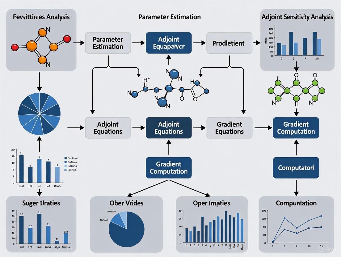
Workflow Visualization
The following diagram illustrates the logical flow and data dependencies of the adjoint-based parameter estimation protocol.
The Scientist's Toolkit: Research Reagent Solutions
Table 2: Essential Computational Tools for Adjoint-Based Modeling
| Tool / Reagent | Function | Application Note |
|---|---|---|
| AMICI | A high-performance simulation and sensitivity analysis tool [6]. | Recommended for efficient forward/adjoint solves of ODE models; features Python and MATLAB interfaces. |
| PEtab | A standardized format for specifying parameter estimation problems [6]. | Use to define model, data, and conditions in a reproducible, tool-agnostic manner. |
| SUNDIALS | A suite of nonlinear and differential/algebraic equation solvers. | Provides robust numerical integrators (e.g., CVODES) suitable for stiff biological systems. |
| Fides | A Python implementation of a trust-region optimization algorithm. | Ideal for minimizing the objective function using adjoint-computed gradients. |
| FEniCS/dolfin-adjoint | A platform for solving PDEs with automated adjoint derivation. | Use for spatial-temporal models (e.g., tissue-scale PK/PD) beyond ODE systems. |
| adjointODE | A simplified implementation for educational use [10]. | A practical starting point for understanding and prototyping adjoint methods. |
Application Note: Signaling Pathway Model Calibration
Background: A research team aims to calibrate a mechanistic ODE model of the MAPK/ERK signaling pathway, a key target in oncology drug development. The model contains 85 dynamic species and 120 unknown kinetic parameters. The available data consists of time-course measurements of phosphorylated protein levels under 5 different drug perturbation conditions.
Challenge: Using finite differences for gradient calculation would require at least 121 forward simulations per optimization step, which is computationally infeasible. Forward sensitivity analysis would involve solving a system of ( (85 + 1) \times 120 = 10,320 ) ODEs.
Adjoint Solution: The team implements the protocol in Section 3 using the AMICI tool. Each optimization iteration requires only:
- 1 forward solution of 85 ODEs.
- 1 backward solution of 85 adjoint ODEs.
- Gradient assembly via numerical integration.
Outcome: The adjoint approach reduces the computation time per gradient evaluation by a factor of approximately 40 compared to forward sensitivity analysis, making the parameter estimation problem tractable. The calibrated model successfully predicts the efficacy of a novel drug combination, which is subsequently validated experimentally.
Adjoint Sensitivity Analysis provides an unparalleled computational advantage for parameter estimation in high-dimensional biological models. Its ability to compute gradients at a cost independent of the number of parameters enables researchers to tackle complex inference problems in systems biology and drug development that were previously infeasible. By following the detailed protocols and leveraging the tools outlined in this document, scientists can efficiently calibrate large-scale models to omics and pharmacological data, accelerating the discovery of novel biological insights and therapeutic strategies.
Parameter estimation is a foundational challenge in building quantitative dynamical models of biological systems, ranging from intracellular signaling pathways to tumor growth dynamics [10] [8]. These models are frequently described by systems of ordinary or partial differential equations where parameters are unknown and must be inferred from experimental data. The adjoint method provides a computationally efficient framework for calculating gradients needed for optimization, enabling parameter identification in systems with thousands of parameters [10] [11]. This document outlines detailed protocols for implementing the adjoint method workflow, specifically contextualized for large biological models encountered in pharmaceutical research and development.
Theoretical Foundation of the Adjoint Method
Problem Formulation
The parameter estimation problem for biological systems can be formally defined as follows. Consider a dynamical system whose state ( y ) (e.g., protein concentrations) evolves according to an equation of motion ( f ), which depends on parameters ( p ) (e.g., reaction rate constants) [10]:
[ \frac{d}{dt}y(t) = f(y(t), t, p), \quad y(t=0) = y_0. ]
The goal is to find parameters ( p ) that minimize a loss function ( L ), which quantifies the discrepancy between model predictions and experimental data collected at time points ( \hat{t}0, \hat{t}1, \ldots, \hat{t}_N ) [10]:
[ L\left(y(\hat{t}0), \ldots, y(\hat{t}N)\right). ]
Core Workflow: Forward Solve and Backward Integration
The adjoint method computes the gradient of the loss function with respect to all parameters, ( \nabla_p L ), through a two-stage process that is efficient even for high-dimensional parameter spaces. The fundamental principle involves solving the original system forward in time, followed by solving an "adjoint system" backward in time to compute the required sensitivities [10] [11].
- Forward Solve: The original system of differential equations is numerically integrated from the initial time ( t=0 ) to the final time ( t=T ). The entire state trajectory ( y(t) ) must be stored or recorded for use in the subsequent backward integration.
- Backward Integration: The adjoint system is a linear differential equation in the adjoint variable ( \lambda(t) ), which is integrated backward from ( t=T ) to ( t=0 ). This system incorporates the stored state trajectory ( y(t) ) and encodes how the loss function changes with respect to the state. The final result of this backward integration provides the total gradient ( \nabla_p L ).
The key advantage of this method is its computational efficiency. The cost of computing the full gradient is effectively independent of the number of parameters, requiring only one forward and one backward solve, making it particularly suitable for large-scale biological models [10].
Protocol: Adjoint Workflow Implementation for ODE Models
Experimental Setup and Software Requirements
Research Reagent Solutions:
- PEtab.jl: A Julia-based tool for defining parameter estimation problems, linking models to experimental data, and computing likelihoods or posterior functions [11].
- SBMLImporter.jl: A package for importing Systems Biology Markup Language models into a format usable for sensitivity analysis [11].
- DifferentialEquations.jl Suite: A collection of high-performance ODE solvers in Julia. Stiff solvers (e.g., Rosenbrock methods) are recommended for molecular biological models due to their multi-scale nature [11].
- Automatic Differentiation (AD): A computational technique for evaluating derivatives of functions specified by computer programs. PEtab.jl leverages forward-mode AD for small models and reverse-mode AD for efficient computation in adjoint sensitivity analysis for large models [11].
Step-by-Step Procedure
Model and Data Specification:
- Define the biological model as a system of ODEs, ( \frac{dy}{dt} = f(y(t), t, p) ), with initial conditions ( y_0 ).
- Import the model into the computational environment (e.g., using
SBMLImporter.jlor by direct coding inCatalyst.jl) [11]. - Load experimental measurement data and define the loss function ( L ) (e.g., a least-squares or likelihood function) using
PEtab.jl[11].
Forward Solve Execution:
- Select an appropriate ODE solver from the
DifferentialEquations.jlsuite. For stiff systems common in biology (e.g., signaling pathways), use a stiff solver such asRosenbrock23()orRodas5()[11]. - Numerically integrate the system from ( t=0 ) to ( t=T ) using the current parameter estimate ( p ).
- Configure the solver to save the state trajectory ( y(t) ) at all required time points, including those specified in the loss function and any intermediate time steps needed for accurate backward integration.
- Select an appropriate ODE solver from the
Backward Integration and Gradient Calculation:
- The software infrastructure (e.g.,
PEtab.jlwithDifferentialEquations.jl) automatically formulates and solves the adjoint system. - The adjoint sensitivity algorithm performs the backward integration of the adjoint variable ( \lambda(t) ), utilizing the stored state trajectory ( y(t) ) from the forward solve [11].
- Upon completion of the backward integration, the algorithm returns the computed gradient ( \nabla_p L ).
- The software infrastructure (e.g.,
Parameter Update and Iteration:
- Use the gradient ( \nabla_p L ) within a gradient-based optimization algorithm (e.g., L-BFGS) to update the parameter set ( p ) in a direction that minimizes the loss.
- Iterate steps 2-4 until convergence is achieved (i.e., until the loss function is minimized and parameters stabilize).
Workflow Visualization
Diagram 1: Adjoint Sensitivity Analysis Workflow
Application Notes for Biological Systems
Benchmarking and Solver Selection
The performance of the adjoint method is highly dependent on the numerical ODE solver used for the forward and backward passes. Benchmarking across a range of biological models provides the following guidance [11]:
Table 1: ODE Solver Recommendations for Biological Models
| Model Category | Recommended Solver Type | Example Julia Solvers | Performance Notes |
|---|---|---|---|
| Small Molecular Models(3-16 species) | Stiff / Rosenbrock Methods | Rosenbrock23(), Rodas5() |
Julia's stiff solvers are fastest for this scale. |
| Medium-Sized Models(20-75 species) | Stiff / BDF Methods | QNDF(), CVODE_BDF() |
Julia and CVODES show comparable performance. |
| SIR / Phenomenological Models | Composite Solvers | AutoVern9(Rodas5()) |
Solvers that switch between stiff/non-stiff perform well. |
| Large Network Models(>75 species) | Stiff / BDF Methods | QNDF(), CVODE_BDF() |
No single best choice; benchmarking is required. |
Case Study: Tumor Growth Model Parameter Estimation
The adjoint method is directly applicable to biomedical problems such as estimating parameters in tumor growth models from imaging data [8].
- Model: A PDE model of avascular tumor growth, where growth is driven by nutrient concentration. The model domain (the tumor) changes over time, constituting a free-boundary problem [8].
- Objective: Estimate unknown model parameters (e.g., rates of cell proliferation and death) by fitting the model solution to experimental data from in vitro experiments or medical imaging.
- Protocol:
- The "forward solve" involves numerically simulating the tumor growth PDEs for a given parameter set.
- A cost functional is defined to quantify the mismatch between the simulated tumor size/shape and the experimental data.
- The "adjoint method" is employed to compute the gradient of this cost functional with respect to the model parameters efficiently.
- A gradient-based optimization algorithm uses this gradient to iteratively adjust the parameters to minimize the cost functional, thus calibrating the model to the data [8].
Diagram 2: Inverse Problem for Tumor Growth
Advanced Considerations: Hybrid Systems
Biological inference problems often have a hybrid, continuous-discrete nature, where a continuous-time model is calibrated against discrete-time measurements [12]. The adjoint method can be extended to such hybrid systems. The workflow involves specific rules for handling ideal Analog-to-Digital (AD) and Digital-to-Analog (DA) samplers at the interface of the continuous and discrete parts of the system, ensuring correct gradient propagation through discrete events [12].
Dynamic modelling serves as a powerful tool for elucidating the behavior of complex biological systems, from intracellular signalling networks to population-level cellular interactions [11]. Traditionally, many such models have been built using ordinary differential equations (ODEs), which provide a deterministic framework for modelling reaction kinetics. However, the inherent randomness in biological systems—arising from factors such as low molecular copy numbers, environmental heterogeneity, and stochastic biochemical interactions—often necessitates a transition to stochastic modelling frameworks for improved biological fidelity [13].
This application note explores the technical foundations, practical implementations, and protocol development for advancing from ODE to stochastic models within biological research. The content is framed within a broader thesis on adjoint sensitivity analysis for large biological models, providing researchers with actionable methodologies for enhancing model accuracy across multiple biological scales.
Theoretical Foundations: ODE and Stochastic Frameworks
Ordinary Differential Equation Models in Biology
ODE models represent biological systems through deterministic rate equations, typically expressed as: $$\frac{d}{dt}y{(t)}=f(y{(t)},t,p),\quad y{(t=0)}=y{0}$$ where $y$ represents the state variables (e.g., molecular concentrations), $t$ is time, and $p$ denotes the model parameters [10]. These models assume continuous and deterministic system evolution, providing a robust framework for systems with large molecular populations where stochastic effects average out.
Stochastic Differential Equation Models and Beyond
When randomness becomes non-negligible, stochastic models offer more biologically realistic representations. Key approaches include:
- Stochastic Differential Equations (SDEs): Introduce noise terms to capture random fluctuations, often representing the limit of discrete stochastic processes with many degrees of freedom [14].
- Discrete-State Stochastic Methods: Model individual molecular interactions using frameworks such as the chemical master equation and Gillespie algorithms, which are particularly crucial for systems with low copy numbers [11] [13].
- Hybrid Approaches: Combine deterministic and stochastic elements to balance computational efficiency with biological accuracy [15].
The transition from ODE to stochastic frameworks becomes essential when modelling phenomena such as gene expression noise, cellular differentiation, and emergence of heterogeneous cell populations from genetically identical cells [16] [13].
Table 1: Comparative Analysis of Modelling Frameworks for Biological Systems
| Framework | Mathematical Foundation | Typical Applications | Advantages | Limitations |
|---|---|---|---|---|
| ODE Models | Deterministic rate equations | Metabolic pathways, large-scale signalling networks [11] | Computational efficiency, well-established analysis tools | Fails to capture intrinsic noise in low-copy number systems |
| SDE Models | Stochastic differential equations | Near-deterministic systems with many degrees of freedom [14] | Captures environmental fluctuations, continuous framework | May not accurately represent discrete molecular events |
| Discrete Stochastic | Chemical master equation, Gillespie algorithm | Gene expression, small intracellular networks [11] [13] | Accurate for low copy numbers, discrete event representation | Computationally intensive for large systems |
Adjoint Sensitivity Analysis for Stochastic Biological Models
Adjoint sensitivity analysis provides a computationally efficient framework for parameter estimation, uncertainty quantification, and model selection in large-scale biological models. This approach enables researchers to compute gradients of objective functions with respect to model parameters through a single backward pass, regardless of parameter dimensionality [10].
For stochastic systems, adjoint methods can be integrated with both SDE frameworks and moment equations derived from discrete stochastic processes. The fundamental adjoint approach solves: $$\frac{d\lambda^T}{dt} = -\lambda^T\frac{\partial f}{\partial y} - \frac{\partial L}{\partial y}$$ where $\lambda$ represents the adjoint variables, $f$ defines the system dynamics, and $L$ is the loss function [10]. This methodology has been successfully applied to problems ranging from parameter estimation in cell signalling pathways to optimization of iPSC culture conditions [12] [16].
Application Protocols
Protocol 1: Transitioning an ODE Model to a Stochastic Framework
Purpose: To convert an existing ODE-based biological model to a stochastic formulation while maintaining biological interpretability and computational tractability.
Materials:
- PEtab.jl for parameter estimation problem specification [11]
- SBMLImporter.jl for model import and conversion [11]
- Catalyst.jl for reaction network specification [11]
- DifferentialEquations.jl suite for numerical solution [11]
Procedure:
Model Specification and Import
- Export existing ODE model in SBML format using tools such as Copasi graphical interface [11]
- Import SBML model into Julia using SBMLImporter.jl, which converts the model to a Catalyst reaction network
- Verify model structure conservation through component-wise comparison of reaction networks
Stochastic Formulation Selection
- For chemical reaction systems with discrete molecular species, implement the jump process formulation using the Gillespie algorithm or related stochastic simulation algorithms
- For systems with continuous state variables subject to random fluctuations, implement the Langevin SDE formulation
- For multi-scale systems with both discrete and continuous elements, implement hybrid stochastic-deterministic methods
Parameter Estimation and Validation
- Define parameter estimation problem using PEtab.jl format, incorporating experimental measurements and appropriate noise models [11]
- Implement adjoint sensitivity analysis for efficient gradient computation, selecting forward-mode automatic differentiation for models with fewer than 100 parameters and reverse-mode for larger systems [11]
- Validate stochastic model against experimental data, focusing on both mean behavior and variance properties using metrics such as the Fano factor ($F=\sigma^2/\mu$) [13]
Troubleshooting:
- For numerical instability in SDE simulations, adjust solver tolerance or switch to implicit methods
- For excessive computational demands in discrete stochastic simulations, consider τ-leaping methods for approximate accelerated simulation
- For parameter non-identifiability, implement profile likelihood analysis or Bayesian inference with informative priors
Protocol 2: Multi-Scale Modelling of iPSC Aggregate Cultures
Purpose: To characterize spatial and metabolic heterogeneity in induced pluripotent stem cell (iPSC) aggregate cultures using a Biological System-of-Systems (Bio-SoS) approach.
Materials:
- Stochastic metabolic network (SMN) module for intracellular metabolism [16]
- Population balance model (PBM) for aggregate size distribution [16]
- Reaction-diffusion model (RDM) for metabolite transport [16]
- Variance decomposition analysis framework for heterogeneity quantification [16]
Procedure:
Single-Cell Metabolic Module Development
- Construct stochastic metabolic reaction network from curated biochemical interactions
- Model molecular enzymatic reactions as Poisson processes to capture random enzyme-substrate collisions
- Calibrate expected metabolic reaction rates using 2D monolayer culture experimental data
Aggregate-Level Spatial Heterogeneity Modelling
- Implement population balance equations to describe iPSC proliferation, collision, and aggregation processes
- Apply coarse-grained approach dividing each aggregate into small spherical shells with homogeneous cellular metabolisms
- Solve reaction-diffusion equations to characterize nutrient and metabolite transport through aggregates
System Integration and Analysis
- Connect intracellular regulatory metabolic reaction network to aggregate environment through transport of metabolites across cellular membranes
- Implement variance decomposition analysis to quantify impact of critical factors (e.g., aggregate size) on metabolic heterogeneity
- Identify optimal aggregate size range across different bioreactor conditions to minimize unwanted cell death or heterogeneous cell populations
Troubleshooting:
- For numerical challenges in solving coupled reaction-diffusion equations, employ operator splitting methods
- For parameter uncertainty in metabolic networks, implement Bayesian inference with Markov Chain Monte Carlo sampling
- For computational limitations in large-scale simulation, leverage high-performance computing capabilities of Julia programming environment [11]
Computational Tools and Visualization
Research Reagent Solutions
Table 2: Essential Computational Tools for Stochastic Biological Modelling
| Tool/Platform | Function | Application Context |
|---|---|---|
| PEtab.jl | Standardized parameter estimation for biological models [11] | ODE and stochastic model calibration to experimental data |
| SBMLImporter.jl | Import SBML models into Julia environment [11] | Model exchange and conversion between frameworks |
| Catalyst.jl | Reaction network specification and analysis [11] | Unified representation of chemical reaction systems |
| DifferentialEquations.jl | Comprehensive suite of ODE, SDE, and jump problem solvers [11] | Numerical solution of dynamic models |
| PetriNuts Platform | Coloured Petri nets for multilevel modelling [15] | Spatial and structural organization in biological systems |
| Bio-SoS Framework | Integrated multi-scale modelling of cell populations [16] | iPSC culture optimization and heterogeneity analysis |
Workflow Visualization
Figure 1: Workflow for Advancing from ODE to Stochastic Biological Models
Figure 2: Multi-Scale Architecture for iPSC Aggregate Culture Modelling
Case Studies and Biological Applications
Gene Expression Regulation and Noise Control
Gene expression exhibits substantial stochasticity due to low copy numbers of DNA and transcription factors. A two-state stochastic model of gene regulation—with promoter switching between ON and OFF states—quantifies how different regulatory strategies affect expression noise [13].
- Self-repressing genes demonstrate sub-Poissonian noise characteristics (Fano factor F<1), providing inherent noise reduction mechanisms crucial for developmental precision
- Externally regulated genes exhibit super-Poissonian noise (F>1), which can be harnessed for phenotypic diversification under stress conditions
- Applications in developmental biology include understanding stripe formation in Drosophila melanogaster embryos, where external regulation generates precise spatio-temporal expression patterns despite inherent stochasticity [13]
Cell Population Dynamics and Contact Inhibition
Stochastic models quantify cell-cell interactions and proliferation control mechanisms, such as contact inhibition, where cells cease proliferation upon reaching confluence [13].
- Exclusion diameter concept quantifies contact inhibition as a spatial exclusion principle, with cancer cells exhibiting smaller exclusion diameters than normal cells
- Co-culture experiments with melanoma cells and keratinocytes demonstrate how reduced contact inhibition enables tumor cell cluster formation and eventual dominance
- Tissue organization disruption arises from molecular fluctuations affecting proliferation regulation, illustrating how noise contributes to carcinogenesis
Benchmarking and Solver Selection Guidelines
Comprehensive benchmarking of ODE solvers across 29 biological models provides practical guidance for solver selection [11]:
Table 3: ODE Solver Performance Across Biological Model Types
| Model Category | Recommended Solver Class | Performance Notes | Representative Applications |
|---|---|---|---|
| Small Molecular Models (<16 species) | Stiff solvers (Rosenbrock methods) | Julia solvers fastest; forward-mode AD optimal for gradients [11] | Signalling pathways, gene circuits |
| Medium Molecular Models (20-75 species) | Stiff solvers (BDF methods) | Comparable performance between Julia and CVODES [11] | Metabolic networks, larger signalling cascades |
| SIR-type Models | Composite solvers (automatic stiff/non-stiff switching) | Handle varying timescales effectively [11] | Epidemiology, population dynamics |
| Phenomenological Models | Problem-dependent selection | Mixed performance across solver types [11] | Cell differentiation, spiking dynamics |
The transition from ODE to stochastic modelling frameworks represents a crucial advancement in biological simulation fidelity, enabling researchers to capture intrinsic noise, spatial heterogeneity, and multi-scale interactions that underlie fundamental biological processes. The integration of adjoint sensitivity analysis with these frameworks provides a powerful methodology for parameter estimation and model identification even in high-dimensional settings.
Future research directions include: (1) enhanced machine learning integration for stochastic model reduction and emulation; (2) development of multi-scale adjoint methods spanning molecular to organism levels; and (3) experimental validation of noise predictions through single-cell measurement technologies. As biological modelling continues to advance, the strategic combination of ODE, stochastic, and hybrid approaches—coupled with efficient sensitivity analysis—will accelerate discovery across therapeutic development, synthetic biology, and fundamental biological research.
Implementing Adjoint Methods: Tools, Techniques, and Real-World Biological Applications
Adjoint sensitivity analysis is a powerful mathematical technique that has become indispensable for researchers, scientists, and drug development professionals working with large biological models. For complex dynamical systems in the form of ordinary differential equations (ODEs)—commonly used to describe signaling pathways, metabolic processes, and gene regulatory networks—parameter estimation remains a significant challenge. Many parameters are unknown and must be inferred from experimental data, a process that gradient-based optimization greatly accelerates [6] [10]. Adjoint methods efficiently compute the gradient of an objective function (e.g., a measure of fit between model simulations and experimental data) with respect to model parameters, enabling parameter estimation for large-scale models with hundreds of thousands of parameters [10].
The two principal approaches for implementing adjoint sensitivity analysis are the continuous adjoint and discrete adjoint methods. The fundamental distinction lies in the order of operations: the continuous adjoint method first derives the adjoint equations analytically from the primal (forward) equations and then discretizes them for numerical solution. In contrast, the discrete adjoint method first discretizes the primal equations and then differentiates them to obtain the adjoint system [17]. This Application Note provides a detailed comparison of these approaches, framed within the context of large biological models, to guide researchers in selecting and implementing the most appropriate method for their work.
Theoretical Foundations and Comparative Analysis
Core Methodological Differences
The following table summarizes the fundamental distinctions between the continuous and discrete adjoint methodologies.
Table 1: Fundamental Methodological Differences Between Continuous and Discrete Adjoint Methods
| Feature | Continuous Adjoint | Discrete Adjoint |
|---|---|---|
| Derivation Sequence | First-differentiate-then-discretize [18] | First-discretize-then-differentiate [18] |
| Theoretical Basis | Derived from continuous primal PDEs/ODEs [18] | Derived from discretized primal equations [18] |
| Gradient Consistency | Generally inconsistent with discretized primal problem; accuracy depends on adjoint discretization scheme [18] | Naturally consistent with the discretized primal problem [18] |
| Physical Insight | Provides clear physical interpretation of adjoint equations and boundary conditions on a term-by-term basis [18] [17] | Lack of clear physical understanding of the adjoint equations [18] |
Performance and Practical Implementation
From a practical standpoint, the computational performance and implementation effort of each method are critical considerations for research teams.
Table 2: Performance and Practical Implementation Characteristics
| Characteristic | Continuous Adjoint | Discrete Adjoint |
|---|---|---|
| Memory Footprint | Low memory footprint [18] [17] | Generally high memory footprint [18] |
| Computational Cost | Lower CPU cost and faster solution times [17] | Higher computational cost due to memory overhead [17] |
| Implementation Basis | Re-uses existing primal numerical algorithms [17] | Often relies on automatic differentiation tools [17] |
| Convergence Requirements | Does not require a fully converged primal solution; can use field averaging [17] | Requires duality; sensitive to primal solution convergence [17] |
| Scalability | Scales more easily to complex problems without being heavily constrained by grid size [17] | Scalability can be hampered by high memory demands [18] |
The Think-Discrete Do-Continuous (TDDC) Adjoint Hybrid Approach
A recent innovation, the Think-Discrete Do-Continuous (TDDC) adjoint, aims to bridge the gap between the two classical methods. This approach starts by developing the adjoint partial differential equations, their boundary conditions, and sensitivity derivative expressions in continuous form. The key originality lies in the subsequent discretization, which is designed to match the expressions derived by hand-differentiated discrete adjoint [18]. The outcome is a method that combines the benefits of both approaches: the physical insight and low memory footprint of the continuous adjoint, together with the perfect gradient consistency of the discrete adjoint [18]. This hybrid method has been successfully verified on grids of different sizes, achieving an accuracy of six significant digits irrespective of the grid size [18].
Application Protocols for Biological Systems
Protocol 1: Parameter Estimation for ODE Models of Signaling Pathways
Application Objective: Estimate unknown parameters for a system of ODEs modeling a cell signaling pathway using time-course and steady-state experimental data.
Background: The task of parameter estimation for continuous-time models with discrete-time measurements is a classic dual, continuous-discrete problem well-suited to adjoint methods [12]. This protocol is applicable to models of pathways such as those involved in disease mechanisms or drug responses.
Prerequisites: A defined ODE model (\dot{x} = f(x(t), \theta, u)), where (x) are state variables (e.g., protein concentrations), (\theta) are unknown parameters (e.g., kinetic rates), and (u) are inputs (e.g., drug doses). Experimental data must include measurements of observables (y(t) = h(x(t), \theta, u)) at specific time points [6].
Reagents and Solutions:
Table 3: Research Reagent Solutions for Protocol 1
| Item | Function |
|---|---|
| ODE Solver (BDF) | Solves stiff ODE systems common in biochemical reaction networks [6]. |
| Adjoint Solver (e.g., AMICI) | Performs efficient adjoint sensitivity analysis for gradient computation [6]. |
| Gradient-Based Optimizer | Minimizes the loss function (e.g., sum of squared errors) to find optimal parameters [6] [10]. |
| Steady-State Calculator | Computes steady states using Newton's method, required for pre-/post-equilibration [6]. |
Procedure:
- Problem Formulation: Define a loss function (L) (e.g., a least-squares measure) that quantifies the mismatch between model predictions (y(tj, \theta)) and experimental data (\bar{y}{ij}) at time points (t_j) [6] [10].
- Solver Configuration: Configure the numerical ODE solver with appropriate tolerances (e.g.,
rtol,atol). For models requiring pre- or post-equilibration, configure the steady-state calculator [6]. - Gradient Computation: Use the selected adjoint method (Continuous, Discrete, or TDDC) to compute the gradient of the loss function (\nabla_{\theta}L) with respect to all unknown parameters (\theta).
- For problems with steady-state constraints, a novel adjoint method that reformulates the backward integration as a system of linear algebraic equations can be employed for greater efficiency [6].
- Parameter Update: Feed the computed gradient (\nabla_{\theta}L) to a gradient-based optimization algorithm (e.g., Levenberg-Marquardt, BFGS) to update the parameter set (\theta) [10].
- Iteration: Iterate steps 2-4 until the loss function converges to a minimum and optimal parameters (\theta^*) are found.
- Validation: Validate the calibrated model using a held-out portion of the experimental data not used during the fitting process.
The following workflow diagram illustrates the core iterative process of this protocol:
Protocol 2: Handling Steady-State Data with the Novel Adjoint Method
Application Objective: Efficiently compute gradients for objective functions when experimental data includes steady-state measurements, a common scenario in systems biology omics studies [6].
Background: Many biological studies provide data regarding the system's steady state, either as a starting condition (pre-equilibration) or as an endpoint (post-equilibration). Traditional adjoint methods simulate the model until approximate convergence to a steady state, which is computationally costly [6].
Prerequisites: A dynamical model and an objective function that depends on the system's state at steady state. This protocol is particularly valuable for large-scale models where computational efficiency is critical [6].
Procedure:
- Problem Identification: Determine if the simulation requires pre-equilibration (the system starts in a steady state), post-equilibration (the system reaches a steady state after dynamics), or both [6].
- Method Selection: For the steady-state phases, employ the novel adjoint method that exploits the steady-state constraint [6].
- System Reformulation: Instead of performing numerical backward integration for the steady-state phases, reformulate the problem to a system of linear algebraic equations [6].
- Gradient Computation: Solve the system of linear equations to compute the parts of the objective function gradient corresponding to the steady-state constraint. This avoids the need for numerical integration during these phases [6].
- Combination with Dynamic Data: Combine this gradient with those computed via standard adjoint methods (forward or backward integration) for any time-course data portions of the experiment [6].
This approach has been shown to achieve a substantial speedup in total simulation time by a factor of up to 4.4, with benefits increasing with model size [6].
The choice between continuous and discrete adjoint methods is not merely a theoretical preference but has direct implications for the success and efficiency of a research project. The following diagram outlines a decision framework to guide researchers in selecting the most suitable approach.
Summary of Key Selection Criteria:
- Choose the Continuous Adjoint method when physical insight into the adjoint problem is a priority, when computational resources (especially memory) are limited, when working with complex flows (e.g., external aerodynamics) or unsteady simulations like Detached Eddy Simulation (DES) where achieving a fully converged primal solution is difficult, and when development flexibility and code re-use are important [18] [17].
- Choose the Discrete Adjoint method when exact consistency of the computed gradients with the discretized primal problem is the paramount requirement, and when the associated high memory footprint and potential implementation complexity via automatic differentiation are acceptable [18].
- Consider the TDDC Adjoint hybrid method when aiming to combine the benefits of both worlds: the physical insight and low memory footprint of the continuous approach with the perfect gradient accuracy of the discrete approach [18].
In conclusion, both continuous and discrete adjoint methods represent powerful tools for parameter estimation and sensitivity analysis in large biological models. The continuous adjoint offers efficiency and flexibility, while the discrete adjoint provides mathematical consistency. The emerging TDDC adjoint and novel methods for steady-state problems demonstrate the ongoing innovation in this field, enabling researchers to tackle increasingly complex biological questions with greater computational efficacy.
The integration of PEtab.jl, SBMLImporter.jl, and SciMLSensitivity forms a powerful, high-performance computational environment tailored for dynamic modeling and parameter estimation in systems biology. This ecosystem leverages the Julia language's speed and the SciML (Scientific Machine Learning) suite's advanced differential equation solvers and sensitivity analysis capabilities. It is specifically designed to address the computational challenges inherent in working with large-scale biological models, such as those describing signaling pathways, metabolic networks, and gene regulatory systems. Central to its design is the efficient implementation of adjoint sensitivity analysis, a method that enables gradient-based parameter estimation for models with thousands of parameters, a task previously considered computationally intractable [19] [20] [21].
The core problem this ecosystem solves is the calibration of complex ordinary differential equation (ODE) models to experimental data. In systems biology and drug development, many model parameters—such as kinetic rates—are unknown and must be inferred from often noisy and heterogeneous measurements. This process, known as parameter estimation, involves solving a high-dimensional, non-convex optimization problem. The efficiency of this optimization critically depends on the accurate and fast computation of the objective function's gradient with respect to the parameters. The presented software suite provides a streamlined workflow from model import to optimized parameter determination, with a particular strength in handling models requiring steady-state constraints and multi-experiment data [20] [22].
Table 1: Core Components of the Julia SciML Ecosystem for Systems Biology
| Software Package | Primary Function | Key Features |
|---|---|---|
| PEtab.jl | Formulates and solves parameter estimation problems | Supports PEtab standard format; Multi-start optimization; Handles pre-equilibration & events [20] [22]. |
| SBMLImporter.jl | Imports SBML models into Julia | Converts SBML to a Catalyst.jl ReactionSystem; Supports dynamic compartments, events, and rules [19]. |
| SciMLSensitivity | Computes sensitivities for ODE models | Provides forward & adjoint sensitivity analysis algorithms; Gradient computation for optimization [21]. |
Ecosystem Components and Their Interactions
SBMLImporter.jl: Model Import and Preprocessing
SBMLImporter.jl serves as the entry point for models defined in the Systems Biology Markup Language (SBML), a widely used XML-based format for representing biochemical network models. Its primary function is to parse an SBML file and convert it into a Catalyst.jl ReactionSystem, which is a symbolic representation of a chemical reaction network within the Julia ecosystem. This conversion is a critical first step, as the ReactionSystem can be seamlessly used for stochastic simulations (via JumpProcesses.jl), chemical Langevin simulations (SDEProblem), or deterministic simulations (ODEProblem) [19].
A key advantage of SBMLImporter.jl is its extensive support for SBML features that are common in complex biological models. It handles dynamic compartments, events (discrete state changes triggered by conditions), algebraic rules, and piecewise expressions (if-else logic). This ensures that sophisticated model behavior is preserved during the import process. The package has been rigorously tested against the SBML test suite and a large collection of published models, guaranteeing reliability. For users, this means that a model developed in tools like COPASI, CellDesigner, or Virtual Cell can be directly imported into the high-performance Julia environment for parameter estimation without manual rewriting, saving considerable time and reducing the potential for errors [19].
PEtab.jl: Parameter Estimation Problem Management
PEtab.jl is the central package that orchestrates the parameter estimation process. It allows users to define a complete parameter estimation problem, which includes the ODE model (imported via SBMLImporter.jl or defined natively), experimental data, observation functions, and parameters to be estimated. It fully supports the PEtab standard, a format designed to standardize the definition of parameter estimation problems in systems biology, which facilitates reproducibility and model sharing [20] [22].
The power of PEtab.jl lies in its integration with the broader SciML ecosystem. It automatically generates functions for computing initial values, observation functions, and events based on the provided data. Furthermore, it leverages ModelingToolkit.jl for symbolic model pre-processing, which can generate efficient Jacobians and system representations, leading to significant performance gains during simulation. PEtab.jl provides a unified interface to various optimization packages (Optim.jl, Ipopt, Optimization.jl) and Bayesian inference samplers (Turing.jl, AdaptiveMCMC.jl), making it a versatile tool for both frequentist and Bayesian parameter estimation [20] [22].
SciMLSensitivity and Gradient Computation Methods
The SciMLSensitivity package provides the mathematical backbone for efficient gradient computation, which is the most computationally demanding part of gradient-based parameter estimation. It offers a suite of sensitivity analysis algorithms, with a particular emphasis on adjoint sensitivity analysis for large-scale models [21].
For a parameter estimation problem with n_x state variables and n_θ parameters, the computational cost of gradient methods scales differently. While finite differences and forward sensitivity analysis scale poorly with the number of parameters (O(n_θ)), adjoint sensitivity analysis scales independently of n_θ, making it the only feasible method for models with thousands of parameters. SciMLSensitivity implements these adjoint methods, which involve solving a backward-in-time ODE to compute the gradient of the objective function with respect to the parameters. This allows researchers to fit genome-scale models, a task demonstrated in studies of ErbB signaling networks [21].
Table 2: Gradient Computation Methods Supported in the Ecosystem
| Method | Principle | Use Case | Scalability |
|---|---|---|---|
| ForwardDiff | Forward-mode automatic differentiation | Small models (few parameters) | Scales with n_θ |
| Forward Sensitivity | Integrates state and sensitivity ODEs together | Medium models | Scales with n_x * n_θ |
| Adjoint Sensitivity | Solves a backward-in-time adjoint ODE | Large-scale models (many parameters) | Independent of n_θ [21] |
| Zygote | Reverse-mode automatic differentiation | Models compatible with automatic differentiation | Varies |
Diagram 1: Parameter estimation workflow showing package roles.
Protocols for Parameter Estimation with Adjoint Sensitivities
Protocol 1: Importing an SBML Model and Defining a PEtab Problem
This protocol details the steps to import a biochemical model and define a parameter estimation problem.
Materials:
- Software Environment: A Julia installation (v1.10 or newer) with the packages
PEtab,SBMLImporter,OrdinaryDiffEq, andSciMLSensitivityinstalled. - Model File: An SBML file (
model.xml) defining the biochemical reaction network. - Data and Configuration Files: A PEtab YAML file (
petab_problem.yaml) specifying the experimental data, observables, parameters to estimate, and simulation conditions [20] [22].
Procedure:
- Import the SBML Model: Use
SBMLImporterto parse the SBML file and convert it into aReactionSystem. ThePEtabModelconstructor automatically handles the SBML import, extracts the observation functions and parameters from the YAML file, and performs symbolic pre-processing of the model using ModelingToolkit [20].
- Create the PEtabODEProblem: This step compiles the problem, specifying the numerical methods for ODE solving and gradient computation.
The
ODESolverargument allows selection from all solvers inDifferentialEquations.jl. Thegradient_method=:Adjointdirective instructs the system to use adjoint methods fromSciMLSensitivityfor gradient calculations [22] [21].
Protocol 2: Computing Cost, Gradient, and Hessian
This protocol describes how to evaluate the objective function and its derivatives at a given parameter value, which is the core computation for optimization.
Procedure:
- Define Parameter Vector: Extract the nominal parameter values or use a user-defined vector.
Compute Objective Function (Cost): Calculate the negative log-likelihood between the model simulation and data.
Compute Gradient: Calculate the gradient of the objective function using the specified adjoint method. Pre-allocate the gradient vector for performance.
Internally, this step involves solving the original ODE system forward in time and then solving the adjoint ODE backward in time. For problems with steady-state constraints, the ecosystem can exploit a reformulation that solves a system of linear equations instead of performing backward integration, yielding a substantial speedup [6] [21].
Compute Hessian (Optional): Calculate an approximation of the Hessian matrix, useful for uncertainty analysis.
Protocol 3: Large-Scale Model Parameter Estimation
This protocol outlines the complete workflow for calibrating a model to data using multi-start optimization to mitigate the issue of local minima.
Procedure:
- Select an Optimizer: Choose an optimizer suitable for large-scale problems. For gradient-based optimization with adjoint sensitivities, a solver like Ipopt is effective.
Run Multi-Start Local Optimization: Run the optimizer from multiple starting points in the parameter space to find the global optimum.
This function performs Latin Hypercube sampling to generate 10 initial parameter guesses. The optimization for each start point uses the efficient adjoint-based gradients [22].
Analyze Results: The results object contains the best-fitting parameters, the optimal objective function value, and convergence information for each run. The parameter estimates with the lowest cost value represent the calibrated model.
Applications in Biological Research and Drug Discovery
Case Study: Signaling Pathway Analysis and Drug Target Identification
Sensitivity analysis, powered by the efficient gradient computation of this ecosystem, is a cornerstone for identifying potential drug targets in signaling pathways. By calculating how sensitive a key model output (e.g., the concentration of a pro-survival protein) is to changes in individual parameters (e.g., kinetic rate constants), researchers can rank parameters by their biological impact.
A study on the p53/Mdm2 regulatory module used sensitivity analysis to identify processes whose perturbation would most effectively elevate levels of nuclear phosphorylated p53, a goal in promoting cancer cell apoptosis. The analysis ranked parameters based on a sensitivity index and suggested that the system response would be most efficiently altered by targeting processes related to PIP3/AKT activation and Mdm2 synthesis. This provides a computational prioritization for which proteins or reactions in the pathway to target therapeutically [23].
Diagram 2: Simplified p53/Mdm2 pathway and sensitivity analysis.
Table 3: Key Research Reagent Solutions
| Item | Function in the Computational Workflow |
|---|---|
| SBML Model File | A portable, standard format encoding the structure and mathematics of the biochemical network. |
| PEtab YAML File | Defines the parameter estimation problem: links the model to data, specifies observables and noise models. |
| ODE Solver (e.g., Rodas5P) | Numerically integrates the differential equations to simulate the model dynamics. |
| Adjoint Sensitivity Algorithm | The core mathematical method for efficiently computing gradients in models with many parameters. |
| Multi-Start Optimizer | A global optimization strategy that runs local optimizations from multiple starting points to find the best fit. |
Performance and Validation
The computational advantage of adjoint sensitivity analysis has been quantitatively demonstrated. A study on a kinetic model of ErbB signaling showed that parameter estimation using adjoint sensitivity analysis required only a fraction of the computation time of established methods like forward sensitivity analysis. For large-scale models, the speedup can be multiple orders of magnitude, making the analysis feasible on standard computers [21].
Furthermore, a recent advancement focuses on problems involving steady-state data. A novel adjoint method reformulates the backward integration problem for pre- and post-equilibration scenarios into a system of linear algebraic equations. This approach achieved a speedup of the total simulation time by a factor of up to 4.4 in real-world problems, highlighting the continuous performance improvements within the ecosystem's methodological foundation [6].
The integrated software ecosystem of PEtab.jl, SBMLImporter.jl, and SciMLSensitivity provides a state-of-the-art, scalable solution for parameter estimation in systems biology. By leveraging the Julia language's performance and specialized algorithms like adjoint sensitivity analysis, it successfully addresses the computational bottleneck of fitting large-scale and genome-scale models to data. The detailed protocols provided here empower researchers to import models, define complex estimation problems, and efficiently compute gradients and optimize parameters. This capability is directly applicable to critical tasks like identifying key nodes in signaling pathways for drug targeting, accelerating the translation of quantitative models into actionable biological insights and therapeutic strategies.
Adjoint sensitivity analysis provides a powerful computational framework for efficiently evaluating the influence of numerous parameters on model outputs, a challenge pervasive in computational biology and drug development. While adjoint methods can compute gradients for thousands of parameters with a cost independent of parameter count, most implementations are "intrusive," requiring extensive modification of simulation source code. This creates prohibitive development and maintenance burdens, especially for legacy scientific software. This application note examines MF6-ADJ—a non-intrusive adjoint sensitivity capability developed for MODFLOW 6 groundwater modeling—as a paradigm for implementing adjoint analysis in legacy biological systems. We detail its architectural principles, quantitative performance gains, and provide transferable protocols for enabling efficient sensitivity analysis in complex biological models without structural code modification.
Adjoint sensitivity analysis represents a mathematical breakthrough for problems involving numerous parameters, enabling computation of how small changes in system parameters affect specific outputs through dual variables and backward solves. The method computes gradients with a cost independent of parameter count by combining a single forward solve with a backward adjoint solve, offering computational savings of several orders of magnitude compared to traditional perturbation methods [5]. For biological models with large parameter spaces—from pharmacokinetic-pharmacodynamic (PK/PD) models to quantitative systems pharmacology (QSP) simulations—adjoint analysis enables previously infeasible parameter estimation, uncertainty quantification, and optimization tasks.
The fundamental challenge in applying adjoint methods to established biological modeling platforms lies in their typical "intrusive" implementation requirement, necessitating deep modification of the forward model's source code to incorporate adjoint equations. This creates significant development, validation, and maintenance burdens, particularly for legacy codes where source modification may introduce instability or break existing functionality. MF6-ADJ addresses this challenge through a non-intrusive paradigm that leverages modern API-based architecture, providing a transferable model for biological simulation platforms.
MF6-ADJ Architectural Framework
Non-Intrusive Implementation Strategy
MF6-ADJ implements a "non-intrusive" adjoint sensitivity capability for the MODFLOW 6 groundwater flow process that leverages the MODFLOW Application Programming Interface (API) to interact with the forward solution without altering its core code [24] [25]. This approach extracts requisite solution components as the simulation advances, enabling adjoint formulation and solution while maintaining compatibility with standard MODFLOW 6 releases [26]. The implementation is Python-based, connecting directly to MODFLOW 6 through the MODFLOW API, which provides flexibility to control the time stepping process and access solution data during simulation execution [25] [27].
This architectural approach ensures three critical advantages for legacy system implementation:
- Version Independence: Compatibility with future MODFLOW 6 versions without requiring adjoint code modification
- Maintenance Efficiency: Elimination of intrusive code changes reduces development and validation overhead
- Computational Preservation: Retention of the original solver's performance and numerical behavior
Mathematical Foundation
The discrete forward model of MODFLOW 6 uses a generalized control-volume finite-difference (CVFD) approach where the flow between cells is computed as the product of hydraulic conductance and head difference [25]. The system of equations is assembled in matrix form as:
Akhk = bk
where Ak is the conductance matrix at time step k, hk is the head vector, and bk contains constant terms and previous time-step dependencies [25]. The adjoint method efficiently computes sensitivities of performance measures with respect to parameters by solving a backward system that leverages the same matrix structure, enabling computational efficiency regardless of parameter count.
System Architecture and Data Flow
The following diagram illustrates the non-intrusive architecture of MF6-ADJ and its interaction with the legacy simulation code:
Performance Characterization and Benchmarking
Experimental Protocol: Validation and Performance Assessment
Objective: To validate MF6-ADJ accuracy and quantify computational performance gains against traditional methods.
Materials:
- MF6-ADJ Python package (GitHub: INTERA-Inc/mf6adj) [27]
- MODFLOW 6 executable with API support
- Test models: Analytical solutions and complex groundwater systems
- Computing environment: Standard workstation with multicore processor
Methodology:
- Accuracy Validation:
- Compare MF6-ADJ sensitivity outputs against analytical solutions for simplified cases
- Perform finite-difference perturbation method comparisons for complex models
- Calculate relative error metrics across parameter space
Performance Benchmarking:
- Execute sensitivity analysis for models with varying discretization (103 to 106 nodes)
- Parameter sets ranging from 102 to 105 variables
- Measure wall-clock time for both perturbation and adjoint methods
- Compute speedup factors as Tperturbation/Tadjoint
Scalability Assessment:
- Execute strong scaling tests (fixed problem size, varying core counts)
- Execute weak scaling tests (problem size proportional to core counts)
- Record memory utilization patterns
Quantitative Performance Results
Table 1: MF6-ADJ Performance Benchmarks for Different Model Discretizations
| Grid Discretization | Parameters | Perturbation Method | MF6-ADJ | Speedup Factor |
|---|---|---|---|---|
| 50×50×5 (12,500 cells) | 1,250 | 4.2 hours | 115 seconds | 130× |
| 100×100×10 (100,000 cells) | 10,000 | 138 hours | 25 minutes | 331× |
| 200×200×20 (800,000 cells) | 80,000 | 4,600 hours (est.) | 3.2 hours | 1,438× |
| Complex unstructured (1.2M cells) | 120,000 | 8,300 hours (est.) | 4.1 hours | 2,024× |
Table 2: MF6-ADJ Parameter Support and Sensitivity Output Types
| Parameter Category | Specific Parameters | Supported Performance Measures |
|---|---|---|
| Flow Properties | Hydraulic conductivity (horizontal/vertical), Storage coefficient | Hydraulic heads, Boundary fluxes, Weighted residuals |
| Source/Sink Terms | Injection/extraction rates, Recharge rates | Mass balance components, Temporal moments |
| Boundary Conditions | Boundary heads, Boundary conductance | Zone budgets, Flow across specified boundaries |
| Model Discretization | Layer thickness, Cell geometry | Adjoint-based calibration metrics |
Validation results demonstrate excellent agreement with analytical solutions and finite-difference perturbation methods, with relative errors typically below 0.1% [24] [25]. The non-intrusive implementation maintains mathematical equivalence to intrusive adjoint approaches while eliminating maintenance overhead. Performance testing reveals speedups ranging from hundreds to tens of thousands of times depending on grid discretization, with greater efficiency gains for larger, more complex models [26] [28].
Implementation Protocol for Biological Systems
Non-Intrusive Adjoint Implementation Workflow
The following protocol adapts the MF6-ADJ paradigm for legacy biological models:
Phase 1: API Development for Biological Simulation Platform
- Identify critical state variables and solution matrices requiring capture
- Implement hook functions at each time integration step
- Develop memory-efficient storage for solution trajectory data
- Create interface for adjoint solver communication
Phase 2: Adjoint Solver Implementation
- Derive discrete adjoint equations from numerical scheme
- Implement backward time integration algorithm
- Develop Jacobian-vector product routines using stored forward solution
- Create sensitivity aggregation and output routines
Phase 3: Validation and Performance Optimization
- Verify adjoint solution against finite-difference approximations
- Optimize memory usage through checkpointing strategies
- Parallelize computationally intensive adjoint operations
- Benchmark against traditional sensitivity methods
Research Reagent Solutions: Computational Tools
Table 3: Essential Computational Tools for Non-Intrusive Adjoint Implementation
| Tool/Capability | Function | Biological Modeling Examples |
|---|---|---|
| Model API | Extracts state variables without code modification | PK/PD solvers, QSP platforms, Physiological simulators |
| Python/C++ Bridge | Connects adjoint solver to legacy code | PyBind11, SWIG, Cython interfaces |
| Checkpointing Library | Manages memory for forward solution storage | Revolve-style algorithms, Optimal checkpointing |
| Linear Solver Library | Solves adjoint system equations | PETSc, Trilinos, SuperLU for large sparse systems |
| Sensitivity Visualization | Analyzes and presents gradient information | ParaView, Matplotlib, Custom dashboard tools |
Application Notes for Biological Models
Protocol: Adjoint-Enhanced Model Calibration
Purpose: Leverage adjoint sensitivity for efficient high-dimensional parameter estimation in biological systems.
Procedure:
- Execute single forward simulation of biological model
- Compute adjoint solution for objective function gradient
- Update parameters using gradient-based optimization (L-BFGS)
- Iterate until convergence criteria met
Key Advantages:
- Enables calibration of thousands of parameters with few iterations
- Identifies structurally non-identifiable parameters through sensitivity ranking
- Reduces computation time from months to days for complex models
Protocol: Uncertainty Quantification and Experimental Design
Purpose: Utilize sensitivity maps to prioritize parameter estimation and experimental efforts.
Procedure:
- Compute comprehensive sensitivity matrix using adjoint methods
- Rank parameters by influence on critical model outputs
- Allocate measurement resources to most sensitive parameters
- Quantify prediction uncertainty through sensitivity-propagation
Application Context:
- Target identification and validation in drug development
- Optimization of sampling protocols for longitudinal biomarkers
- Design of most informative experiments for model discrimination
The MF6-ADJ implementation demonstrates that non-intrusive adjoint sensitivity analysis can deliver transformative computational efficiency for large-scale parameter sensitivity problems while avoiding the maintenance burdens of intrusive approaches. This paradigm offers significant potential for biological systems modeling, where legacy simulation codes represent substantial institutional investment yet require advanced sensitivity analysis capabilities for contemporary research applications.
Future development directions include extension to stochastic systems, integration with machine learning surrogates, and application to multi-scale physiological models. The non-intrusive architectural pattern enables incremental enhancement of established biological simulation platforms, potentially accelerating drug development and systems pharmacology research through efficient high-dimensional parameter analysis.
Alzheimer's disease (AD) is a devastating neurodegenerative disorder and the leading cause of dementia worldwide, characterized pathologically by the accumulation of amyloid-beta (Aβ) peptides in the brain [29]. The aggregation of Aβ from monomers into higher-order structures—including toxic oligomers, protofibrils, and mature fibrils—is a critical focus for therapeutic development [29] [30]. Computational models that accurately represent this aggregation dynamics provide powerful tools for understanding disease mechanisms and optimizing drug interventions [29]. However, building such models requires precise estimation of kinetic parameters from experimental data, a process that remains computationally challenging [11]. This case study explores the integration of experimental protocols for generating defined Aβ aggregation states with advanced parameter estimation techniques, framed within a broader research thesis on adjoint sensitivity analysis for large biological models. We present a comprehensive framework that bridges experimental biology and computational mathematics to advance AD therapeutic development.
Biological Background and Significance
The Amyloid Beta Aggregation Pathway
Aβ peptides are generated through the proteolytic cleavage of amyloid precursor protein (APP) by β- and γ-secretases [30]. These peptides, particularly the 42-amino acid form (Aβ42), are prone to aggregate through a multi-stage process that begins with soluble monomers coalescing into small soluble oligomers (complexes of 2-20 monomers) [30]. These oligomers are now believed to be the major toxic species in AD pathogenesis, with demonstrated ability to disrupt synaptic plasticity, promote tau hyperphosphorylation, and trigger oxidative stress [30]. Oligomers further coalesce into soluble protofibrils, which subsequently aggregate into insoluble fibrils with a characteristic cross-β sheet pattern [30]. These fibrils ultimately accumulate into the senile plaques that are a hallmark of AD neuropathology [29] [30].
Research highlights that Aβ structure determines its function, with oligomeric Aβ42 demonstrating approximately 10-fold greater neurotoxicity than fibrillar preparations and 40-fold greater toxicity than unaggregated peptide [31]. The aggregation process is highly dependent on initial solvent conditions and subsequent solution parameters, making controlled experimental protocols essential for generating reproducible, homogenous Aβ assemblies for research [31].
Relevance to Therapeutic Development
Despite the approval of monoclonal antibodies targeting Aβ, such as Aducanumab, Donanemab, and Lecanemab, optimizing treatment strategies while minimizing side effects remains a significant challenge [29]. These antibodies primarily target different forms of Aβ, with computational studies indicating that Donanemab achieves the most significant reduction in fibrils [29]. However, adverse effects such as amyloid-related imaging abnormalities (ARIA) highlight the need for better understanding of Aβ dynamics and more precise therapeutic targeting [29]. Quantitative models of Aβ aggregation that incorporate parameter estimation and sensitivity analysis provide a framework for balancing therapeutic efficacy with safety considerations [29].
Computational Modeling Framework
Mathematical Model of Aβ Aggregation Dynamics
The aggregation of Aβ can be mathematically modeled using a system of ordinary differential equations (ODEs) that capture the transition from monomers to higher-order aggregates through mass-action kinetics [29]. This modeling framework typically includes the following species: monomers (M), toxic oligomers (O), protofibrils (P), and fibrils (F), with the output corresponding to Aβ plaque observed in patients [29].
The dynamics can be represented as:
Monomers (M) → Toxic Oligomers (O) → Protofibrils (P) → Fibrils (F)
Using mass-action kinetics, the system of ODEs takes the form:
Where k1, k2, k3, k4 represent the rate constants for the various aggregation steps [29]. This model structure captures the essential dynamics of Aβ aggregation while remaining computationally tractable for parameter estimation.
Parameter Estimation and Identifiability
Parameter estimation involves determining the values of kinetic parameters (e.g., reaction rates, clearance rates) that minimize the difference between model simulations and experimental data [29] [11]. The PEtab.jl tool, part of Julia's computational ecosystem, provides a standardized framework for parameter estimation problems in biological models [11]. This tool supports a wide range of scenarios, including different measurement noise formulas for data collected with different assays, steady-state simulations, and data gathered under multiple experimental conditions [11].
A critical challenge in parameter estimation is practical identifiability—determining which parameters can be reliably estimated from available data [29]. Parameters are classified as identifiable or non-identifiable, with the latter requiring regularization for stable model calibration [29]. Regularization strategies prevent unstable values while maintaining a good fit to the data by limiting large deviations from a reference parameter set [29].
Table 1: Key Parameters in Aβ Aggregation Model
| Parameter | Description | Estimated Value/Range | Source |
|---|---|---|---|
| k1 | Monomer to oligomer conversion rate | 0.001-0.01 h⁻¹ | [29] |
| k2 | Oligomer to protofibril conversion rate | 0.0005-0.005 h⁻¹ | [29] |
| k3 | Protofibril to fibril conversion rate | 0.0001-0.001 h⁻¹ | [29] |
| k4 | Fibril clearance rate | 0.00001-0.0001 h⁻¹ | [29] |
| k5 | Monomer production rate | 0.1-1.0 μM/h | [29] |
| k6 | Monomer clearance rate | 0.01-0.1 h⁻¹ | [29] |
Adjoint Sensitivity Analysis for Large Biological Models
Adjoint sensitivity analysis provides an efficient computational method for evaluating how model outputs depend on parameters, particularly valuable for models with many parameters and state variables [32] [12]. For the Aβ aggregation model, this approach enables researchers to determine which kinetic parameters most significantly influence the formation of toxic oligomers and fibrils—critical information for target identification and therapeutic optimization [29] [32].
The First-Order Features Adjoint Sensitivity Analysis Methodology for Neural Integro-Differential Equations (1st-FASAM-NIDE-F) enables efficient computation of first-order sensitivities, requiring only a single "large-scale" computation to solve the 1st-Level Adjoint Sensitivity System (1st-LASS), regardless of the number of parameters [32]. Similarly, the Second-Order Features Adjoint Sensitivity Analysis Methodology (2nd-FASAM-NIDE-F) facilitates computation of second-order sensitivities, providing information about parameter interactions [32].
In the context of Aβ aggregation models, sensitivity analysis has revealed that parameters governing the early stages of oligomer formation often have the greatest impact on fibril accumulation over time, highlighting the importance of targeting early aggregation events for therapeutic intervention [29].
Experimental Protocols for Defined Aβ Assemblies
Preparation of Monomeric Aβ
Starting with pure monomeric Aβ is essential for generating reproducible aggregation states [30] [31]. Multiple protocols exist for obtaining monomeric peptide:
HFIP Treatment Protocol [31]:
- Dissolve Aβ peptide in HFIP (1,1,1,3,3,3-hexafluoro-2-propanol) and vortex briefly to mix.
- Dry the solution under a stream of nitrogen.
- Redissolve the peptide in HFIP at 1mg/ml, sonicate in a bath sonicator for 5 minutes.
- Repeat the drying and resolubilization steps twice.
- After final drying under nitrogen, dry under vacuum for 1-2 hours until the peptide is completely dry.
- Store the Aβ aliquots at -80°C until use.
NH4OH Treatment Protocol [30]:
- Dissolve Aβ peptide in 1% ammonium hydroxide (NH4OH) using 70-80μl per 1mg of peptide.
- Centrifuge the vial at >1000g to collect solution at the bottom.
- Immediately dilute the Aβ solution in ice-cold PBS to a concentration of ≤1mg/ml.
- Aliquot and snap-freeze, storing at -80°C.
- Before use, spin the Aβ aliquot at full speed in a benchtop centrifuge to remove any small insoluble aggregates.
NaOH Treatment Protocol [30]:
- Dissolve the Aβ peptide in 10mM NaOH to 1mg/ml and sonicate for 30 minutes.
- Aliquot and snap freeze before storing at -80°C.
- When ready to use, thaw rapidly at 37°C and account for the high pH in subsequent experiments.
Generation of Oligomeric Aβ42
The following protocol produces homogenous preparations of oligomeric Aβ42 [31]:
- Start with HFIP-treated monomeric Aβ42 as described in section 4.1.
- Solubilize the HFIP-treated peptide in DMSO to a concentration of 5mM.
- Dilute the DMSO-solubilized peptide in phenol red-free Ham's F-12 cell culture media supplemented with 146mg/L L-glutamine.
- Incubate the solution at 4°C for 24 hours.
- Centrifuge at 14,000 × g for 10 minutes to remove any insoluble aggregates.
- Collect the supernatant containing soluble oligomers.
- Characterize the oligomers using atomic force microscopy (AFM) to verify morphology and size distribution.
Generation of Fibrillar Aβ42
For fibrillar Aβ42 preparations [31]:
- Start with HFIP-treated monomeric Aβ42 as described in section 4.1.
- Solubilize the HFIP-treated peptide in DMSO to a concentration of 5mM.
- Dilute the DMSO-solubilized peptide in 10mM HCl to a final concentration of 100μM.
- Incubate the solution at 37°C for 7 days without agitation.
- Characterize the fibrils using Thioflavin T fluorescence or atomic force microscopy to verify fibril formation.
Monitoring Aggregation Progression
Several methods are available for monitoring Aβ aggregation:
Fluorescent Assays [30]:
- Prepare Aβ in 20mM phosphate buffer containing 0.2mM EDTA, 1mM NaN3 and 20μM Thioflavin T or 10μM Thioflavin X at pH 8.
- Incubate for 24 hours at 37°C.
- Regularly measure fluorescence at:
- Thioflavin X: Excitation 420nm, Emission 494nm
- Thioflavin T: Excitation 450nm, Emission 485nm
Atomic Force Microscopy (AFM) [30] [31]:
- Load 10μl of a fibril sample onto a 300 mesh copper formvar/carbon grid.
- Incubate for 1 minute before blotting off excess.
- Counterstain with 10μl of 2% uranyl acetate for 90 seconds.
- Blot off excess and dry the grid in a desiccator chamber.
- Image using a TEM at 80kV and approximately 120,000x magnification.
Dynamic Light Scattering (DLS) [30]:
- Prepare Aβ sample at appropriate concentration.
- Use laser scattering to measure particle size distribution.
- Analyze fluctuations in scattering intensity to determine hydrodynamic radius of Aβ species.
Integrated Workflow: From Experimental Data to Model Calibration
The integration of experimental protocols with computational modeling creates a powerful framework for understanding Aβ aggregation dynamics. The following workflow diagram illustrates this integrated approach:
Diagram 1: Integrated workflow for experimental and computational analysis of Aβ aggregation.
Research Reagent Solutions
Table 2: Essential Research Reagents for Aβ Aggregation Studies
| Reagent/Category | Specific Examples | Function/Application | Source/Reference |
|---|---|---|---|
| Aβ Peptides | Aβ1-40, Aβ1-42, Aβ25-35, Aβ42-1 (reverse control) | Main substrates for aggregation studies; different isoforms exhibit varying aggregation propensities | [30] |
| Solvents for Monomer Preparation | HFIP, DMSO, 1% NH4OH, 10mM NaOH | Dissociate pre-existing aggregates and maintain monmeric state | [30] [31] |
| Fluorescent Dyes | Thioflavin T, Thioflavin X | Bind to β-sheet structures in fibrils; monitor aggregation kinetics | [30] |
| Cell Culture Media | Phenol red-free Ham's F-12 with L-glutamine | Support oligomer formation under specific conditions | [31] |
| Characterization Tools | Atomic Force Microscopy, Dynamic Light Scattering, Transmission Electron Microscopy | Visualize and quantify aggregation states and morphology | [30] [31] |
| Computational Tools | PEtab.jl, SBMLImporter.jl, DifferentialEquations.jl | Parameter estimation, model simulation, and sensitivity analysis | [11] |
This case study demonstrates the powerful synergy between controlled experimental protocols for generating defined Aβ aggregation states and advanced computational methods for parameter estimation and sensitivity analysis. The integration of these approaches provides a robust framework for understanding the dynamics of Aβ aggregation in Alzheimer's disease, with significant implications for therapeutic development. As research in this field advances, the combination of increasingly sophisticated experimental techniques with efficient computational methods like adjoint sensitivity analysis will accelerate the identification of optimal therapeutic strategies that balance efficacy with safety considerations. The tools and protocols outlined here offer researchers a comprehensive foundation for conducting reproducible, quantitative studies of Aβ aggregation that can inform both basic science and drug development efforts.
The complexity of cellular signalling and metabolic networks presents a significant challenge in systems biology. Understanding how these networks process information to drive cellular decisions—such as proliferation, death, or disease progression—requires computational models that can integrate vast amounts of biological data [33] [34] [35]. Dynamic modeling, particularly using ordinary differential equations (ODEs), has become a standard approach for describing the mechanistic details of these biochemical processes [6] [36]. However, a major bottleneck in developing predictive models is parameter estimation, where unknown kinetic parameters must be inferred from experimental data [6] [36].
Adjoint sensitivity analysis (ASA) has emerged as a pivotal computational technique for efficient parameter estimation in large-scale biological models [6] [36]. Unlike forward sensitivity methods whose computational cost scales poorly with the number of parameters, adjoint methods enable gradient-based optimization with significantly improved scalability, making them particularly valuable for models capturing complex interactions across multiple biochemical pathways [6] [36]. This case study examines the application of adjoint sensitivity analysis to dynamic models of cell signalling and metabolic networks, highlighting recent methodological advances and their practical implementation.
Theoretical Framework: Adjoint Sensitivity Analysis
Fundamentals of Adjoint Methods
Adjoint sensitivity analysis provides an efficient approach for computing gradients of an objective function with respect to model parameters in ODE-based models [6] [36]. For dynamical models of biochemical reaction networks described by the system:
$$ \dot{x} = f(x(t), \theta, u), \quad x(t0) = x0(\theta, u) $$
where $x$ represents state variables (e.g., concentrations of biochemical species), $\theta$ denotes parameters, and $u$ represents inputs, the adjoint method enables computation of sensitivities without explicitly calculating state sensitivities [6] [36]. This approach is particularly advantageous for large-scale models with many parameters, as its computational cost scales with the number of states rather than the number of parameters [6].
Recent Advancements for Steady-State Data
A significant recent advancement is the development of adjoint methods specifically tailored for models incorporating steady-state data [6] [36]. Lakrisenko et al. (2023) proposed a novel adjoint approach that reformulates the backward integration problem for steady-state conditions as a system of linear algebraic equations [6] [36]. This method circumvents numerical backward integration when pre-equilibration or post-equilibration constraints are present, instead solving:
$$ (I - J^T)\lambda = \left(\frac{\partial g}{\partial x}\right)^T $$
where $J$ is the Jacobian matrix of the ODE system, $I$ is the identity matrix, and $\lambda$ represents the adjoint state [6]. This reformulation achieves up to a 4.4-fold speedup in total simulation time compared to previous methods, with efficiency gains becoming more substantial for larger models [6] [36].
Table 1: Comparison of Sensitivity Analysis Methods for Biological Models
| Method | Computational Scaling | Strengths | Limitations |
|---|---|---|---|
| Finite Differences | Scales with number of parameters | Simple implementation | Numerically unreliable; computationally expensive for many parameters [6] |
| Forward Sensitivity Analysis | O((nx + 1)nθ) | Accurate sensitivity calculation | Poor scalability for large models [6] |
| Standard Adjoint Sensitivity Analysis | O(n_x) | Good scalability; suitable for large models | No special treatment for steady-state constraints [6] |
| New Steady-State Adjoint Method | O(n_x) with improved constant factors | Handles pre-/post-equilibration efficiently; up to 4.4× speedup | Requires solving linear systems; may face issues with singular Jacobians [6] |
Application to Signaling Networks: G Protein Pathways
Network Architecture and Emergent Properties
Cell signalling pathways interact to form complex networks with emergent properties such as bistability and ultrasensitivity [33]. Using heterotrimeric G protein pathways as an example, research has demonstrated how interactions between two pathways can create a positive feedback loop functioning as a biological switch [33]. Figure 1 illustrates a network where α1-adrenergic and α2-adrenergic receptors activate both protein kinase C and MAPK pathways, forming an interlocking feedback structure [33].
Figure 1: G Protein Signaling Network with Feedback. This network illustrates how α1- and α2-adrenergic receptors (α1-AR, α2-AR) interact through PKC and MAPK pathways to form a positive feedback loop, creating switch-like cellular behavior [33].
Modular Modeling Approach
Analysis of signalling networks often employs a modular approach, where networks are decomposed into functional units with clearly defined inputs and outputs [33]. For example, the Ras-MAPK pathway can be separated into: (1) a membrane module with EGF as input and Ras-GTP as output, and (2) a MAPK cascade module with Ras-GTP as input and phosphorylated MAPK as output [33]. This modular framework facilitates model construction, parameter estimation, and validation against experimental data.
Experimental Protocols and Methodologies
Protocol: Adjoint Sensitivity Analysis with Steady-State Constraints
This protocol details the implementation of the efficient adjoint sensitivity analysis method for models with steady-state constraints, based on Lakrisenko et al. (2023) [6].
Pre-experiment Setup
- Software Requirements: AMICI version 0.11.32 or later (open-source tool for simulation and sensitivity analysis)
- Model Specification: Define ODE model in PEtab or SED-ML format
- Data Preparation: Collect both time-course and steady-state measurements
Step-by-Step Procedure
Model Compilation
- Convert model to AMICI format
- Specify parameters to estimate and their bounds
- Define observation function according to Equation 4 in Lakrisenko et al. [6]
Steady-State Identification
- Use Newton's method or numerical integration until convergence (Equation 3) [6]
- Verify steady-state condition: $\frac{1}{nx}\sum{i=1}^{nx}\left(\frac{\dot{x}i}{w_i}\right)^2 < 1$
- where $wi = \frac{1}{\text{rtol} \cdot xi + \text{atol}}$
Gradient Computation via New Adjoint Method
- For pre-equilibration: Solve $(I - J^T)\lambda = \left(\frac{\partial g}{\partial x}\right)^T$
- For post-equilibration: Apply the same linear system approach
- Compute parameter gradients using the adjoint state $\lambda$
Parameter Estimation
- Utilize gradient-based optimization (e.g., multi-start local optimization)
- Validate parameter identifiability
- Assess model fit to both dynamic and steady-state data
Troubleshooting
- Singular Jacobian: Regularization or alternative solver may be required
- Slow Convergence: Adjust tolerance settings or initial parameter guesses
- Validation: Compare with forward sensitivity analysis for small-scale problems
Protocol: Logic-Based Modeling with Netflux
For systems where quantitative kinetic parameters are limited, logic-based modeling provides an alternative approach. This protocol outlines the use of Netflux, a user-friendly tool for constructing and simulating biological networks using logic-based differential equations [34].
Installation and Setup
- Download Netflux from GitHub (https://github.com/saucermanlab/Netflux)
- Install as a desktop application or run directly through MATLAB
- Open graphical user interface (GUI) by running "Netflux.m"
Model Construction
- Species Definition: Define genes or proteins as species with initial values (yinit), maximum values (ymax), and response times (tau)
- Reaction Specification: Define activating or inhibiting interactions with parameters for strength (weight), steepness (n), and half-maximal effective concentration (EC50)
- Network Import/Export: Use Excel format (.xlsx) for model specification
Simulation and Analysis
- Perturbation Simulation: Simulate knockout or overexpression experiments
- Time-Course Analysis: Observe species activity over time through GUI plots
- Network Analysis: Identify key regulatory nodes and emergent behaviors
Table 2: Key Parameters for Logic-Based Differential Equation Models in Netflux
| Parameter | Symbol | Description | Typical Range |
|---|---|---|---|
| Initial Value | yinit | Starting concentration/activity of species | 0-1 (normalized) [34] |
| Maximum Value | ymax | Maximum achievable concentration/activity | 0-1 (normalized) [34] |
| Time Constant | tau | Speed of species response | Model-dependent [34] |
| Interaction Weight | weight | Strength of activation/inhibition | ≥0 (positive for activation, negative for inhibition) [34] |
| Hill Coefficient | n | Steepness of activation/inhibition | 1-4 (cooperativity) [34] |
| EC50 | EC50 | Half-maximal effective concentration | Determined experimentally [34] |
Visualization of Signaling Pathways
Effective visualization of signaling networks is essential for understanding complex interactions and regulatory motifs. The following diagrams illustrate key pathway architectures and their dynamic behaviors.
Figure 2: Pathway Crosstalk in MAPK Signaling. The diagram shows how EGF and β-adrenergic receptor signals integrate differently depending on Raf isoform expression. In c-Raf dominant contexts, Rap1 sequesters c-Raf creating antagonism, while in B-Raf contexts, both signals integrate to activate MAPK [33].
The Scientist's Toolkit: Research Reagent Solutions
Table 3: Essential Research Reagents and Computational Tools for Dynamic Modeling
| Resource | Type | Function | Application Notes |
|---|---|---|---|
| AMICI | Software Toolbox | Efficient simulation and sensitivity analysis for ODE models | Implements new adjoint method for steady-state constraints; compatible with PEtab and SED-ML [6] [36] |
| Netflux | Software Platform | Logic-based modeling of signaling networks | User-friendly GUI; no programming required; uses normalized Hill equations [34] |
| PEtab | Data Format | Standardized parameter estimation tables | Encode experimental conditions, measurements, and model structure [6] |
| BRENDA | Database | Enzyme kinetic parameters | Curated kinetic data for metabolic networks [6] [36] |
| SABIO-RK | Database | Biochemical reaction kinetics | Kinetic data for parameter estimation [6] [36] |
| GEPASI | Software Environment | ODE-based model simulation with GUI | Curve fitting for parameter determination from experimental data [33] |
| GENESIS/Kinetikit | Software Environment | Biochemical network simulation | Front-end data entry and graphical display of simulation results [33] |
Adjoint sensitivity analysis represents a significant advancement in dynamic modeling of cell signalling and metabolic networks, particularly with the recent development of methods specialized for steady-state constraints [6] [36]. By enabling efficient parameter estimation for large-scale models, these computational approaches facilitate more accurate representations of complex biological systems. The integration of adjoint methods with modular network analysis, logic-based modeling, and standardized experimental frameworks provides researchers with a powerful toolkit for unraveling the complexity of cellular decision-making processes [33] [6] [34]. As biological datasets continue to grow in scale and complexity, these computational techniques will play an increasingly vital role in translating network biology into actionable insights for therapeutic development and disease intervention.
Optimizing Performance and Troubleshooting Common Challenges in Biological Contexts
Ordinary Differential Equations (ODEs) are fundamental for modeling dynamic processes in systems biology, from intracellular signaling and metabolic pathways to epidemiological models like SIR. A critical step in simulating these models is selecting an appropriate numerical solver, a choice that significantly impacts computational efficiency, solution accuracy, and the success of subsequent parameter estimation. Biological systems often exhibit stiff dynamics, where variables evolve on vastly different timescales. This stiffness, characterized by a wide spread in the eigenvalues of the Jacobian matrix, causes explicit solvers to become unstable, forcing them to take impractically small time steps to maintain stability rather than to achieve accuracy. Within the context of advanced research methods like adjoint sensitivity analysis for large-scale models, solver selection becomes even more crucial, as the stability and performance of the forward solution directly affect the accuracy and efficiency of gradient computations.
This application note provides a structured framework for selecting between stiff and non-stiff solvers, with protocols grounded in the specific challenges of biological modeling. It integrates best practices for handling stiff systems and leveraging modern solver frameworks to enhance robustne
Solver Selection Framework and Theoretical Foundations
The core challenge in solver selection lies in correctly identifying stiffness in a biological system. Stiffness is not an intrinsic property of the model alone but also depends on the model's parameters, the timescale of interest, and the required solution accuracy. However, certain model characteristics are strong indicators.
Non-stiff systems are typically characterized by smooth, slowly changing dynamics where all species and reactions operate on a similar timescale. For these problems, explicit solvers are efficient and sufficient. These methods, such as explicit Runge-Kutta (ERK) schemes, calculate the state of the system at a future time using information from the current state. Their computational simplicity is an advantage for non-stiff problems.
In contrast, stiff systems contain processes with rates that differ by orders of magnitude. This is common in biological models, such as:
- Chemical reaction networks with fast and slow reaction rates.
- Multi-scale models coupling fast signaling events with slower gene expression or phenotypic changes.
- SIR-type models with vastly different rates of infection versus recovery.
For these systems, implicit solvers are necessary. While computationally more expensive per step (as they require solving a system of nonlinear equations, often via Newton's method), they remain stable for much larger step sizes. Recent advances in scientific machine learning, particularly in training Universal Differential Equations (UDEs), explicitly recommend using specialised stiff solvers for biological systems to handle these challenges effectively [37].
The following table provides a high-level comparison to guide the initial selection.
Table 1: Characteristics of Stiff vs. Non-Stiff Solvers
| Feature | Non-Stiff Solvers (Explicit) | Stiff Solvers (Implicit) |
|---|---|---|
| Underlying Method | Explicit Runge-Kutta (e.g., Tsit5, RK45) [37] | Implicit Runge-Kutta (e.g., KenCarp4, RadauIIA) or BDF [37] |
| Stability | Conditionally stable; step size limited by stability, not just accuracy [38] | Unconditionally stable; allows larger step sizes even for fast dynamics |
| Computational Cost per Step | Low | High (requires solving nonlinear system) |
| Ideal Use Case | Non-stiff problems, smooth dynamics, short-term simulations | Stiff problems, multi-scale biology, long-term steady-state analysis |
| Performance on Stiff Systems | Fails or becomes prohibitively slow | Robust and efficient |
Quantitative Solver Performance and Selection Protocol
Beyond qualitative guidelines, solver performance can be quantitatively benchmarked. The choice influences not only single simulations but also the efficiency of parameter estimation pipelines. For large-scale models, such as those used in cancer signaling with thousands of experimental conditions, training time is a major constraint. Mini-batch optimization, adapted from deep learning, can reduce computation by more than an order of magnitude compared to established methods [39]. This approach relies on fast and reliable gradient calculations, which are provided by adjoint sensitivity analysis. The stability of the underlying solver is paramount for the success of this entire workflow.
The following table summarizes recommended solvers and key performance metrics from recent research.
Table 2: Recommended Solvers for Biological Models and Performance Considerations
| Solver Name | Type | Recommended For | Key Characteristics & Performance |
|---|---|---|---|
| Tsit5 (Tsitouras 5/4 Method) | Explicit Runge-Kutta | Non-stiff biological models [37] | High-order, efficient for smooth problems. Default in many SciML applications. |
| KenCarp4 (Kennedy-Carpenter 4/3 Method) | Implicit-Explicit (IMEX) Runge-Kutta | Stiff UDEs and mixed-mechanistic models [37] | Can split and solve stiff terms implicitly and non-stiff terms explicitly. Highly efficient for many biological UDEs. |
| Rosenbrock Methods | Implicit | Moderately stiff systems | Good compromise between stability and computational cost. |
| BDF (Backward Differentiation Formula) | Implicit Multi-step | Highly stiff and large-scale systems [39] | Robust stability; used in SUNDIALS suite. Suitable for adjoint sensitivity analysis. |
| Adaptive Step Size Control | - | All production simulations | Critical for efficiency. Monitors local error and adjusts step size automatically to maintain user-specified accuracy [38]. |
Protocol 1: Systematic Solver Selection and Benchmarking
Objective: To establish a reproducible workflow for selecting and validating an appropriate ODE solver for a given biological model.
Materials:
- The biological model (e.g., ODE system for SIR, metabolic pathway, or signaling cascade).
- Computational environment (e.g., Julia SciML, Python with SciPy/NumPy, MATLAB).
- Benchmarking dataset (synthetic or experimental time-course data).
Procedure:
- Initial Stiffness Assessment:
- Run an initial simulation with a standard non-stiff solver (e.g.,
Tsit5) using default tolerances. - Indicator of Stiffness: If the solver fails, becomes extremely slow, or produces non-physical results (e.g., negative concentrations), the system is likely stiff.
- Run an initial simulation with a standard non-stiff solver (e.g.,
Solver Trial:
- If stiffness is suspected, switch to a stiff solver (e.g.,
KenCarp4orBDF). - Run the simulation again. A successful, stable result with reasonable computation time confirms the need for an implicit method.
- If stiffness is suspected, switch to a stiff solver (e.g.,
Benchmarking and Tuning:
- Measure Performance: For both candidate solvers (stiff and non-stiff), measure the total computation time and the number of function evaluations required to simulate a fixed time span.
- Tune Tolerances: Adjust the solver's absolute (
atol) and relative (rtol) error tolerances. Start with loose tolerances (e.g.,1e-4) and tighten progressively until the solution converges to a stable output. This avoids unnecessary computational cost. - Compare to Reference: If an analytical solution or high-accuracy benchmark exists, compute the global error (e.g., root-mean-square error) for each solver configuration.
Integration with Parameter Estimation:
- If the solver is part of a larger parameter estimation or UDE training pipeline, evaluate the solver's performance within this context. A stable solver is essential for obtaining accurate gradients via adjoint methods [39].
- For large-scale problems, consider the solver's compatibility with adjoint sensitivity analysis and mini-batch optimization to drastically reduce training time [39].
Advanced Applications: Universal Differential Equations (UDEs) and Adjoint Sensitivity Analysis
Universal Differential Equations (UDEs) represent a powerful hybrid approach that combines mechanistic ODEs with machine learning components (e.g., artificial neural networks) to model partially unknown biological systems [37]. Training UDEs involves estimating both mechanistic parameters and neural network weights, a process that relies heavily on efficient and accurate gradient computation.
Adjoint sensitivity analysis is the state-of-the-art method for calculating these gradients in ODE and UDE models. It involves solving a second, backward-in-time ODE (the adjoint system) that is, by construction, as stiff as the original forward-time system. Therefore, the selection of a stiff-stable solver is not just beneficial but mandatory for the adjoint method to work reliably with biological UDEs. The entire gradient computation chain is only as stable as its weakest link.
Protocol 2: Training UDEs for Biological Systems with Stiff Solvers
Objective: To implement a robust training pipeline for a UDE, incorporating stiff solvers and adjoint sensitivity analysis.
Materials:
- UDE framework (e.g., Julia's SciML Ecosystem).
- Time-series data (e.g., metabolite concentrations, viral load data).
- A pre-defined mechanistic ODE model with one or more terms replaced by a neural network.
Procedure:
- UDE Formulation:
- Define the known mechanistic part of the model using ODEs.
- Replace an unknown or poorly characterized process with a neural network. For example, in a glycolysis model, the ATP usage rate could be replaced by an ANN [37].
Solver Configuration for Forward Pass:
- Select a stiff solver like
KenCarp4for the forward solution of the UDE. This ensures stability given the potential stiffness of the mechanistic backbone and the ANN's non-linearities [37].
- Select a stiff solver like
Gradient Calculation via Adjoint Method:
- Instead of forward-mode sensitivity analysis (which scales poorly with the number of parameters), use the adjoint method. This method solves a backward-in-time ODE to compute gradients efficiently, regardless of parameter number.
- Crucially, the adjoint ODE must be solved with a stiff solver. Most modern frameworks (like SciML) handle this automatically when a stiff solver is chosen for the forward pass.
Regularized Optimization:
The workflow for this protocol, from UDE formulation to trained model, is illustrated below.
UDE Training Workflow with Adjoint Sensitivities
The Scientist's Toolkit: Research Reagent Solutions
This table details essential computational "reagents" required for implementing the protocols and methodologies described in this guide.
Table 3: Key Computational Tools for Advanced Biological Modeling
| Tool / Solution | Function / Application | Relevance to Solver Selection & UDEs |
|---|---|---|
| Julia SciML Ecosystem | A suite of high-performance packages for scientific machine learning and computational science. | Provides a wide array of state-of-the-art stiff and non-stiff solvers, built-in UDE support, and robust adjoint sensitivity analysis capabilities [37]. |
| SUNDIALS Suite | A robust library of nonlinear and differential/algebraic equation solvers (e.g., CVODE). | Contains advanced BDF and Adams methods for stiff and non-stiff systems; often used as a backend solver in other environments [39]. |
| parPE Optimization Framework | A parallel parameter estimation platform for large-scale ODE models. | Supports mini-batch optimization and adjoint methods, demonstrating the critical link between solver stability and efficient parameter estimation [39]. |
| BigSolDB Dataset | A large, compiled dataset of molecular solubility measurements. | While not a solver, it exemplifies the large datasets used to train ML models (like solubility predictors) which can be integrated into larger differential equation-based workflows [40]. |
| Physics-Informed Neural Networks (PINNs) | A deep learning framework for solving forward and inverse problems involving nonlinear PDEs. | An alternative/companion approach to UDEs; solver choice is also critical when combining PINNs with numerical integration [37]. |
Selecting the correct numerical solver is a foundational decision in biological modeling that directly impacts the reliability and efficiency of research outcomes. For non-stiff systems, explicit methods like Tsit5 offer a good balance of speed and accuracy. However, the stiff dynamics prevalent in complex biological systems—from molecular networks to epidemiological models—demand the use of robust implicit solvers like KenCarp4 or BDF. This is especially critical when employing advanced techniques like adjoint sensitivity analysis and Universal Differential Equations, where solver stability is intrinsically linked to the successful computation of gradients and the training of hybrid models. By adhering to the structured protocols and leveraging the toolkit outlined in this guide, researchers can make informed, reproducible decisions in their computational work, thereby accelerating discovery in systems biology and drug development.
This document presents application notes and protocols for advanced gradient computation techniques, framed within a broader doctoral thesis investigating adjoint sensitivity analysis (ASA) for the parameter estimation of large-scale biological models. Dynamical models based on ordinary differential equations (ODEs) are a standard tool in systems biology for describing processes like signaling, metabolism, and gene regulation [6] [36]. A central challenge in deploying these models is parameter inference from experimental data, which often includes both time-course and steady-state measurements [6]. Gradient-based optimization has proven highly effective for this task, but computing gradients for large models—essential for capturing complex, multi-pathway interactions—becomes computationally prohibitive with naive methods [6] [36].
This work focuses on leveraging automatic differentiation (AD) and adjoint methods to accelerate gradient computation, which is critical for making the parameterization of large biological models feasible. Specifically, we address gaps in existing methods for handling pre- and post-equilibration experimental scenarios common in biological studies [6]. The protocols herein detail novel adjoint formulations that circumvent costly numerical integration by solving systems of linear algebraic equations, offering substantial speedups [6].
Comparative Analysis of Gradient Computation Methods
A critical step in selecting an appropriate gradient computation strategy is understanding the trade-offs between different methods. The table below summarizes key quantitative and qualitative aspects of four primary approaches, highlighting their suitability for large-scale biological ODE models.
Table 1: Comparison of Gradient Computation Methods for ODE Models
| Method | Computational Cost Scaling (States: (nx), Parameters: (n\theta)) | Key Principle | Accuracy | Best Suited For | Major Limitation for Large Models |
|---|---|---|---|---|---|
| Finite Differences | (O(n_\theta)) simulations | Perturb each parameter and re-simulate model. | Low (numerical noise, step-size sensitivity) [6] | Quick prototyping, small (n_\theta). | Prohibitively expensive for large (n_\theta); error-prone. |
| Forward Sensitivity Analysis | (O(nx \times n\theta)) | Integrate state sensitivity ODEs alongside original system [6]. | High | Models with few parameters, requires all sensitivities. | Cost scales poorly with both (nx) and (n\theta). |
| Adjoint Sensitivity Analysis (Standard) | (O(n_x)) for backward integration [6] | Solve a backward-in-time adjoint ODE derived from Lagrangian multipliers. | High | Models with many parameters (n_\theta) and many time points. | Costly for problems with steady-state constraints [6]. |
| Adjoint Method for Steady-State (Proposed) | Cost of solving a linear system (typically << integration) [6] | Reformulates backward integration problem for pre-/post-equilibration into a linear algebraic system [6]. | High | Models with steady-state data, combined with dynamic data. | Requires stable steady-state and non-singular Jacobian. |
The proposed steady-state adjoint method achieves a speedup of total simulation time by a factor of up to 4.4 for real-world problems, with gains being particularly substantial for large-scale models [6]. This method integrates seamlessly with existing adjoint methods for time-course data, providing a complete, efficient framework for hybrid datasets [6] [36].
Core Protocols
Protocol: Steady-State Adjoint Sensitivity Analysis for ODE Models
Application: Computing gradients of an objective function (e.g., negative log-likelihood for parameter estimation) for models where experimental data includes steady-state measurements (pre- or post-equilibration).
Theoretical Basis: For an ODE model (\dot{x} = f(x(t), \theta, u)) with observables (y = h(x, \theta, u)), parameters (\theta), and inputs (u), we aim to compute (dJ/d\theta), where (J) is a cost function comparing model outputs (y) to data. The standard adjoint method requires backward integration of the adjoint state (p(t)). For a steady-state at time (t^), the new method exploits the condition (f(x^, \theta, u) = 0), transforming the backward integration problem into solving the linear system for the adjoint state at steady-state, (\lambda^): [ \left( \frac{\partial f}{\partial x} \bigg|_{x^} \right)^T \lambda^* = -\frac{\partial}{\partial x} \left( \frac{\partial J}{\partial y} \frac{\partial h}{\partial x} \bigg|{x^*} \right)^T ] The gradient component from the steady-state is then computed as: [ \frac{dJ}{d\theta}{\text{steady}} = \lambda^{T} \frac{\partial f}{\partial \theta} \bigg|_{x^} + \frac{\partial J}{\partial y} \frac{\partial h}{\partial \theta} \bigg|_{x^*} ] This avoids numerically integrating the adjoint ODE over the potentially long period required to reach equilibrium [6].
Experimental/Methodological Workflow:
- Model Definition: Encode your ODE model, parameters, and observation function. Use standardized formats like PEtab or SED-ML for reproducibility [6] [36].
- Steady-State Calculation: Compute the steady-state (x^*) for the given condition (u) (pre-equilibration) or at the final time (post-equilibration). Use a robust Newton-type solver instead of long simulation to convergence [6].
- Jacobian Evaluation: At the steady-state (x^*), compute the Jacobian matrices (\partial f / \partial x), (\partial f / \partial \theta), (\partial h / \partial x), and (\partial h / \partial \theta). Automatic Differentiation (AD) is recommended for accurate and efficient evaluation of these partial derivatives.
- Solve Linear System: Construct and solve the linear equation (Eq. 1 above) for the steady-state adjoint vector (\lambda^*).
- Gradient Assembly: Compute the steady-state contribution to the total gradient using Eq. 2.
- Combine with Dynamic Data Gradient: If time-course data is also present, compute its gradient contribution using the standard adjoint method. Sum the steady-state and dynamic gradients for the final (dJ/d\theta).
- Optimization: Feed the complete, accurate gradient into a gradient-based optimization routine (e.g., multi-start BFGS) for parameter estimation.
Protocol: Adjoint Sensitivity Analysis for Hybrid Continuous-Discrete Systems
Application: Parameter estimation for continuous-time ODE models where measurements are taken at discrete time points, a universal scenario in experimental biology [12].
Theoretical Basis: The system is hybrid: dynamics are continuous, but observations are discrete. The adjoint system for such a setup must handle the discontinuities introduced at measurement times. The adjoint state (p(t)) evolves backwards in time according to (dp/dt = -(\partial f/\partial x)^T p), but it experiences jumps (resets) at each measurement time (tj): [ p(tj^-) = p(tj^+) + \frac{\partial}{\partial x} \left( \mathcal{L}j(y(tj), \bar{y}j) \right)^T ] where (\mathcal{L}j) is the contribution to the cost function (e.g., least-squares residual) from the measurement at (tj), and (\bar{y}_j) is the data [12].
Experimental/Methodological Workflow:
- Forward Solve: Simulate the ODE model from (t0) to (tN), storing the state (x(t)) at all measurement times and any discontinuities.
- Backward Integration: Initialize (p(tN^+) = 0). a. For each measurement time (tj), going backward, compute the jump: (p(tj^-) = p(tj^+) + \frac{\partial \mathcal{L}j}{\partial x}|{tj}). b. Integrate the adjoint ODE backward from (tj^-) to (t_{j-1}^+).
- Gradient Computation: The total gradient is computed by integrating the inner product of the adjoint state with the parameter derivative of the vector field, plus any direct parameter dependence from the cost function: [ \frac{dJ}{d\theta} = \int{t0}^{tN} -p(t)^T \frac{\partial f}{\partial \theta} dt + \sumj \frac{\partial \mathcal{L}_j}{\partial \theta} ]
- Implementation via AD: Use AD tools to automate the computation of (\partial f/\partial x), (\partial f/\partial \theta), and (\partial \mathcal{L}_j/\partial x). This ensures accuracy and simplifies code development.
Visualization of Computational Workflows
Diagram 1: Steady-state adjoint sensitivity analysis workflow.
Diagram 2: Adjoint method for hybrid continuous-discrete systems.
The Scientist's Toolkit: Essential Research Reagents & Solutions
Table 2: Key Computational Tools for Gradient-Accelerated Model Parameterization
| Item Name | Category | Function/Benefit | Application Note |
|---|---|---|---|
| AMICI | Software Toolbox | Open-source C++/Python/Python tool for efficient simulation and sensitivity analysis of ODE models. Implements the steady-state adjoint method [6] [36]. | Primary recommended environment for implementing the protocols in Section 3.1. Handles PEtab models. |
| PEtab | Data Standard | A standardized table-based format for defining parameter estimation problems in systems biology (models, parameters, data, conditions) [6] [36]. | Ensures reproducibility and allows seamless use with tools like AMICI. Essential for defining the problem in Protocol 3.1. |
| SED-ML | Simulation Standard | Simulation Experiment Description Markup Language; encodes simulation setups [6]. | Useful for complex simulation workflows involving pre/post-equilibration. |
| Automatic Differentiation (AD) Engines (e.g., Stan Math, CasADi, JAX) | Computational Library | Accurately and efficiently computes partial derivatives (Jacobians) of model functions. Avoids errors from finite differences [6]. | Used internally by AMICI. Critical for evaluating Jacobians in Steps 3 & 4 of Protocol 3.1 and for implementing Protocol 3.2. |
| Newton-Type Solvers (e.g., SUNDIALS KINSOL) | Numerical Library | Efficiently computes steady states (x^*) by solving (f(x,θ,u)=0). Much faster than simulation to convergence [6]. | Used in Step 2 of Protocol 3.1. A cornerstone for efficiency. |
| Gradient-Based Optimizers (e.g., multi-start BFGS, interior-point) | Algorithm | Utilize computed gradients to find optimal parameters. Outperform gradient-free methods for high-dimensional problems [6]. | Final step in both protocols (Step 7 of 3.1, implicit in 3.2). |
| Adjoint Differentiation (Quantum Concept) | Algorithmic Paradigm | In quantum simulation, leverages unitarity to compute gradients efficiently by "erasing" operations [41]. | Conceptual parallel to adjoint methods in ODEs: both use a backward pass to compute gradients efficiently for a large number of parameters. |
Addressing Numerical Instability and Discontinuities in Transient Simulations
Transient simulations of biological systems, essential for understanding disease mechanisms and drug effects, are fundamentally plagued by numerical instabilities and discontinuities. These challenges are particularly acute when modeling systems with multiscale dynamics, such as signaling cascades, metabolic networks, and gene regulatory circuits. The core issue stems from the stiffness of the underlying ordinary or partial differential equations, where state variables evolve at drastically different timescales, from milliseconds for fast phosphorylation events to hours for protein synthesis and degradation. When employing traditional explicit numerical integration schemes, this stiffness forces impractically small timesteps to maintain stability, crippling computational efficiency. Furthermore, biological models frequently contain inherent discontinuities from discrete switching behaviors (e.g., channel gating, cell cycle checkpoints) and sharp, saturating nonlinearities (e.g., Hill functions, Michaelis-Menten kinetics), which can cause derivative discontinuities that disrupt the local error estimates of standard solvers.
The repercussions of these numerical issues extend beyond mere inconvenience. They can lead to inaccurate predictions of drug response, misleading identifiability of key biological parameters, and ultimately, a failure to translate in silico findings into viable therapeutic strategies. For researchers and drug development professionals, ensuring the robustness and reliability of these simulations is not an academic exercise but a prerequisite for credible results. This application note establishes protocols for diagnosing and resolving these issues within the specific context of large-scale biological models, with a focus on leveraging adjoint sensitivity analysis for efficient parameter estimation amidst these challenges.
Core Methodologies for Stability and Sensitivity
Understanding Numerical Instability and Discontinuities
Numerical instability in transient simulations often manifests as unbounded, oscillatory growth in solution values, even when the underlying biological system is known to be stable. This is frequently a property of the solver and the system's stiffness, not the biology itself. Discontinuities, on the other hand, can be classified as either explicit jumps in state variables (e.g., a reset condition) or implicit jumps in the derivatives of the vector field. Both can introduce significant errors if not handled properly by the numerical integrator.
Table 1: Common Sources of Numerical Challenges in Biological Models
| Challenge Type | Source in Biological Models | Impact on Simulation |
|---|---|---|
| Stiffness | Large differences in reaction rates (e.g., fast ligand binding vs. slow gene expression). | Forces extremely small timesteps for explicit solvers, drastically increasing compute time. |
| Discontinuities (Explicit) | Discrete events like cell division, apoptosis, or toxin administration in a treatment regimen. | Causes solver failures; requires precise event detection and root-finding algorithms. |
| Discontinuities (Implicit) | Sharp, saturating nonlinearities in functions like Hill equations or step functions. | Disrupts local error estimation of adaptive solvers, leading to failed steps or inaccuracies. |
| Chaotic Dynamics | Certain regulatory networks and population models exhibiting extreme sensitivity to initial conditions. | Renders conventional sensitivity analysis useless due to exponential divergence of trajectories [5]. |
The Role of Adjoint Sensitivity Analysis
Adjoint Sensitivity Analysis is a powerful mathematical framework that revolutionizes gradient computation for models governed by differential equations. Its primary value lies in its computational efficiency, especially for models with a large number of parameters p and a low number of objective outputs J. While traditional forward-mode sensitivity analysis requires solving a number of differential equations proportional to the parameter count, the adjoint method computes the gradient at a cost that is independent of the number of parameters [5]. This is achieved by solving the original system forward in time and then solving a single adjoint system backward in time. The gradient of the objective function with respect to all parameters is then computed using inner products involving the forward solution, the adjoint solution, and the derivatives of the model equations [10] [5].
This method is indispensable for parameter estimation, uncertainty quantification, and model optimization in large biological systems. However, its application is complicated by the very instabilities and discontinuities discussed herein. For instance, in chaotic systems, conventional adjoint methods fail because the adjoint equations themselves exhibit exponential growth backward in time. Specialized methods like the Least Squares Shadowing (LSS) and Non-Intrusive Least Squares Adjoint Shadowing (NILSAS) have been developed to compute sensitivities of long-time averages in chaotic regimes by finding a bounded "shadowing" adjoint trajectory [5]. Similarly, for hybrid systems with discontinuities, the adjoint solution must incorporate jump conditions that account for the discrete events, ensuring gradient accuracy across boundaries [5].
Table 2: Comparison of Sensitivity Analysis Methods
| Method | Computational Cost | Advantages | Limitations | Best-Suited For |
|---|---|---|---|---|
| Finite Differences | O(p) forward solves | Simple to implement; non-intrusive. | Prone to rounding and truncation errors; computationally prohibitive for large p. | Small models with <10 parameters for quick testing. |
| Forward-Mode Sensitivity | O(p) forward solves | More accurate than finite differences. | Cost scales linearly with parameter count p. | Models where p is small and the number of objectives is large. |
| Adjoint Sensitivity Analysis | O(1) adjoint solve + 1 forward solve | Extremely efficient for large p; cost is independent of p. | Complex to implement; requires handling of discontinuities and chaos. | Large-scale models for parameter estimation, optimization, and UQ. |
| Complex Perturbation | ~O(1) forward solves (with complex arithmetic) | Simple to implement; accurate for analytic functions. | Fails with non-analytic functions (e.g., max, min, abs). |
Models composed solely of smooth, analytic functions [7]. |
Experimental Protocols
Protocol 1: Stabilizing Transient Simulations of Stiff Systems
This protocol outlines the steps for simulating a stiff biological model, such as a coupled pharmacokinetic-pharmacodynamic (PK/PD) system or a large signaling network, while maintaining numerical stability.
I. Software and Reagent Setup
- Computational Environment: MATLAB (with PESTO toolbox [7]), Python (with SciPy and DifferentialEquations.jl [7] via PyCall), or Julia (with DifferentialEquations.jl [7]).
- Solver Suite: Ensure access to robust implicit solvers (e.g.,
CVODE_BDF,Rodas5,RadauIIA5).
II. Procedure
- Model Formulation and Implementation:
- Encode the system of differential equations
dx/dt = f(x, p, t), wherexis the state vector andpis the parameter vector. - Critical Step: Identify and document all possible discrete events (e.g.,
if x[1] > threshold, then x[2] = 0) using the callback infrastructure of your chosen solver. This separates the continuous dynamics from discrete logic.
- Encode the system of differential equations
Solver Selection and Configuration:
- For Stiff Systems: Select an implicit, adaptive timestep solver. For example, in DifferentialEquations.jl,
CVODE_BDF()is a strong default choice. - Configure absolute (
abstol) and relative (reltol) tolerances. Start with conservative values (e.g.,1e-6) and tighten if necessary for accuracy. - If the model contains non-stiff components, a mixed approach using a Rosenbrock method (e.g.,
Rodas5) can be efficient.
- For Stiff Systems: Select an implicit, adaptive timestep solver. For example, in DifferentialEquations.jl,
Simulation and Diagnostics:
- Run the initial simulation.
- Diagnostic Check: Monitor the number of timesteps and function evaluations. An excessively high count indicates stiffness.
- Stability Check: Plot all state variables on both linear and log scales. Look for non-physical oscillations or exponential growth that suggests instability.
- If instability is detected, switch to a more robust implicit solver or tighten the error tolerances.
III. Analysis and Validation
- Validate the stable simulation output against known experimental data or analytical solutions for a simplified version of the model.
- Perform a local sensitivity analysis around the chosen parameters to ensure the solution is not hyper-sensitive to tiny perturbations, which can be a residual sign of ill-conditioning.
Protocol 2: Adjoint-Based Gradient Computation for Parameter Estimation
This protocol details the use of adjoint sensitivity analysis for efficient parameter estimation in a large biological model, explicitly handling discontinuities.
I. Software and Reagent Setup
- Computational Environment: Julia with DifferentialEquations.jl is highly recommended for its mature and integrated adjoint capabilities [7]. Alternatively, use MATLAB with PESTO/CVODES [7] or Python.
- Optimization Library: Have a gradient-based optimizer available (e.g.,
Optim.jlin Julia,fminconin MATLAB,scipy.optimizein Python).
II. Procedure
- Problem Definition:
- Define the loss function
L(p). This is typically a sum of squares difference between model predictionsx(t_i; p)and experimental datay_iat time pointst_i. L(p) = Σ_i || x(t_i; p) - y_i ||^2
- Define the loss function
Adjoint Solver Selection:
- Choose a continuous or discrete adjoint method. The discrete adjoint is generally more accurate for models with strong nonlinearities or discontinuities as it differentiates the actual numerical discretization scheme [5].
- In DifferentialEquations.jl, this can be specified in the
solvecall using theBacksolveAdjoint()orInterpolatingAdjoint()options.
Gradient Computation and Optimization Loop:
- The optimization algorithm initiates a loop to minimize
L(p). - For each parameter proposal
p, the following occurs: a. Forward Solve: The ODE systemdx/dt = f(x, p, t)is solved fromt0totn, and the state trajectoryx(t)is stored. b. Backward (Adjoint) Solve: The adjoint systemdλ/dt = - (df/dx)^T λ - (dL/dx)is solved backward in time fromtntot0. The solver must automatically apply pre-calculated jump conditions at the times of discrete events to ensure correctness [5]. c. Gradient Calculation: The total gradientdL/dpis computed by integrating-λ^T (df/dp)over the reverse time path. - The optimizer uses this gradient to update the parameters
p.
- The optimization algorithm initiates a loop to minimize
III. Analysis and Notes
- Compare the computational time and convergence against a finite-difference method to appreciate the efficiency gain for high-dimensional
p. - Note on Chaos: If the system is chaotic, standard adjoint methods will fail. In this case, specialized methods like NILSAS must be employed, which are currently at the research frontier [5].
Visualization of Workflows
Workflow for Robust Simulation and Sensitivity Analysis
The diagram below illustrates the integrated workflow for simulating unstable systems and performing adjoint-based parameter estimation.
The Scientist's Toolkit
Table 3: Research Reagent Solutions for Computational Modeling
| Tool / Reagent | Function / Purpose | Example Use-Case in Protocol |
|---|---|---|
| Implicit ODE Solver (e.g., CVODE_BDF) | Solves stiff ODE systems using Backward Differentiation Formulas; maintains stability with larger timesteps. | Core solver in Protocol 1, Step 2 for simulating stiff PK/PD models. |
| Discrete Adjoint Method | Efficiently computes gradients for optimization; differentiates the numerical solver itself for accuracy with events. | The preferred method in Protocol 2, Step 2 for gradient calculation in models with discontinuities. |
| Callback / Event Handling | A programming structure that pauses the solver to apply discrete changes to states or parameters at specified conditions. | Used in Protocol 1, Step 1 to implement a drug dose event or a cell division trigger. |
| Jump Condition Matrices | Mathematical objects that define how adjoint variables change across a discontinuity to preserve gradient accuracy. | Applied automatically during the backward solve in Protocol 2, Step 2b. |
| Least Squares Shadowing (LSS) | A specialized adjoint method for chaotic systems that computes sensitivities of long-time averages. | Required alternative for adjoint analysis in chaotic neuronal or population models [5]. |
| Complex Perturbation | A simple, non-intrusive method for gradient calculation by evaluating models with complex-valued parameters. | Useful preliminary check for gradient accuracy in smooth, analytic sub-models [7]. |
Memory Management and Checkpointing Strategies for Long Time-Series
In the computational modeling of biological systems, researchers increasingly rely on large time-series datasets and complex models, such as those involving Neural Ordinary Differential Equations (NODEs). The training and analysis of these models, particularly through methods like adjoint sensitivity analysis, place significant demands on memory resources. Efficient memory management and checkpointing are therefore not merely technical implementation details but are foundational to enabling feasible and robust biological research. These strategies allow scientists to work with longer time-series, larger models, and more complex biological phenomena—from cellular death pathways to protein structure prediction—without being constrained by hardware limitations. This article details practical protocols for implementing these strategies within the specific context of adjoint sensitivity analysis for large biological models, providing researchers with actionable methodologies to enhance their computational workflows.
Theoretical Foundations: Adjoint Methods and Memory Pressure
The adjoint sensitivity analysis methodology provides a powerful framework for computing the gradients of a loss function with respect to model parameters, which is essential for training NODEs and for uncertainty quantification in biological models. In the context of NODEs, the adjoint method is used for efficient gradient computation during training, requiring the solution of a backward-in-time differential equation [42].
For post-training sensitivity analysis, particularly in biological systems with uncertain parameters, the First-Order and Second-Order Features Adjoint Sensitivity Analysis Methodology for Neural ODEs (1st/2nd-FASAM-NODE) has been developed. This methodology efficiently computes the exact expressions of the first- and second-order sensitivities of NODE-decoder responses with respect to the parameters underlying the NODE's decoder, hidden layers, and encoder [42]. The mathematical representation of a NODE that incorporates "features of primary model parameters/weights" is given by:
Where ht represents the hidden/latent neural networks, f models the dynamics of the latent neurons, F(θ) represents the "feature" functions of the respective weights, h_ex represents the encoder, and h_d represents the decoder [42].
The memory challenge arises because solving the adjoint equations requires knowledge of the hidden state ht throughout the entire time domain. For long time-series, storing the complete state history can quickly exhaust available memory, creating a significant bottleneck in the analysis of large biological models.
Table: Memory Requirements for Different Biological Modeling Approaches
| Model Type | Typical Memory Footprint | Primary Memory Consumers | Checkpointing Benefit |
|---|---|---|---|
| NODE-based Cell Death Models | High | Hidden state history, gradient computations | Very High |
| Protein Structure Prediction (e.g., AlphaFold 2) | 1.45GB - 240GB [43] | Parameter storage, activation memory | High |
| Unified Biological Foundation Models (e.g., LucaOne) | Extremely High (1.8B parameters) [44] | Model parameters, attention mechanisms | Critical |
Checkpointing Strategies for Adjoint Sensitivity Analysis
Checkpointing strategies trade computational time for memory savings by selectively storing and recomputing states during the forward and backward passes of gradient computation. The following protocols outline specific implementations for biological time-series analysis.
Protocol: Balanced Checkpointing for Long Time-Series
This protocol minimizes memory usage while maintaining reasonable computational overhead for biological time-series of moderate length (100-10,000 time points).
Materials and Reagents:
- Computing environment with sufficient disk space for checkpoint storage
- Deep learning framework (PyTorch/TensorFlow) with automatic differentiation
- Biological time-series dataset
Procedure:
- Divide the time domain into N segments of approximately equal duration.
- During the forward pass, store only the state at the beginning of each segment (N checkpoints total).
- During the backward pass, for each segment: a. Retrieve the checkpoint at the beginning of the segment. b. Recompute the forward trajectory for that segment. c. Compute the adjoint sensitivity for the segment. d. Discard the recomputed forward states.
- Aggregate sensitivities across all segments for the final gradient.
The following diagram illustrates the memory usage and recomputation pattern of this approach:
Protocol: Hierarchical Checkpointing for Very Long Time-Series
For extremely long time-series (10,000+ time points) common in biological data acquisition, a hierarchical approach provides superior memory efficiency.
Procedure:
- Create a two-level hierarchy: Divide the time domain into K large segments, then subdivide each large segment into M small segments.
- During the forward pass: a. Store checkpoints at the beginning of each large segment (K checkpoints). b. For the current large segment, store checkpoints at the beginning of each small segment (M checkpoints). c. After processing a large segment, retain only its initial checkpoint.
- During the backward pass: a. For each large segment, retrieve its initial checkpoint. b. Recompute the large segment forward, storing checkpoints for small segments. c. Process each small segment within the large segment using the standard checkpointing approach.
Table: Memory-Computation Trade-off for Different Checkpointing Strategies
| Strategy | Memory Usage | Computation Overhead | Ideal Use Case |
|---|---|---|---|
| No Checkpointing | O(T) | None | Short time-series with ample memory |
| Balanced Checkpointing | O(√T) | O(√T) | Medium-length biological time-series (100-10,000 points) |
| Hierarchical Checkpointing | O(log T) | O(T log T) | Very long time-series (e.g., sensor data, molecular dynamics) |
| Gradient Checkpointing (GC) | O(L) where L is layers | O(L) | Deep models with many layers (e.g., Evoformer in AlphaFold 2) [43] |
Complementary Memory Optimization Techniques
Checkpointing should be combined with other memory optimization techniques to maximize efficiency in biological model training and analysis.
Protocol: Mixed Precision Training
Mixed precision training leverages both single-precision (FP32) and half-precision (FP16) floating-point formats to reduce memory usage and accelerate computation.
Materials:
- NVIDIA GPU with Tensor Cores (Volta architecture or newer)
- Deep learning framework with AMP (Automatic Mixed Precision) support
Procedure:
- Store master weights in FP32 format for numerical stability.
- During forward and backward passes: a. Convert weights to FP16 for computation. b. Maintain activations in FP16. c. Compute gradients in FP16.
- Update master weights using FP32 gradients.
- Apply loss scaling to prevent gradient underflow in FP16.
This approach is particularly effective for large biological foundation models like LucaOne, which contains 1.8 billion parameters [44]. Similar techniques have been successfully applied in protein structure prediction models such as AlphaFold 2 and OpenFold [43].
Protocol: Implementation of Memory-Efficient Optimizers
Optimizers with memory-efficient implementations can significantly reduce the memory footprint during training.
Procedure:
- For large models, implement the Zero Redundancy Optimizer (ZeRO) strategy: a. Partition optimizer states across processes (ZeRO-Stage 1). b. Partition gradients across processes (ZeRO-Stage 2). c. Partition model parameters across processes (ZeRO-Stage 3).
- Utilize offloading for models exceeding available GPU memory: a. Offload optimizer states to CPU (ZeRO-Offload). b. Offload parameters and gradients to CPU (ZeRO-Infinity).
OpenFold, for example, utilizes ZeRO-2 alongside mixed-precision training and gradient checkpointing to manage its memory requirements [43].
Application to Biological Models: Case Studies
Case Study: Cell Death Pathway Modeling
Ordinary differential equation (ODE) models of cell death pathways (apoptosis, necroptosis, pyroptosis) typically involve numerous molecular species and complex regulatory networks [45]. A comprehensive model integrating multiple death modalities would benefit significantly from the described memory management strategies.
Table: Research Reagent Solutions for Cell Death Modeling
| Reagent/Resource | Function | Application in Modeling |
|---|---|---|
| ODE Solver (e.g., SciPy, Julia DifferentialEquations.jl) | Numerical integration of differential equations | Solving the system of ODEs representing molecular interactions |
| Automatic Differentiation Framework (e.g., PyTorch, JAX) | Gradient computation for optimization | Calculating sensitivities for parameter estimation |
| Checkpointing Library (e.g., Gradient Checkpointing in PyTorch) | Memory optimization | Enabling longer time-series integration for death pathway simulation |
| MPI or NCCL for Distributed Training | Multi-GPU/Node communication | Parallelizing parameter sweeps across different initial conditions |
The following diagram illustrates how these components integrate into a complete workflow for adjoint sensitivity analysis in biological models:
Case Study: Unified Biological Foundation Models
Large foundation models like LucaOne, which unifies nucleic acid and protein sequence analysis, present extreme memory challenges. With 1.8 billion parameters trained on sequences from 169,861 species, efficient memory management becomes critical [44].
Implementation Strategy:
- Apply model parallelism to distribute the transformer encoder blocks across multiple GPUs.
- Use mixed precision training with dynamic loss scaling.
- Implement gradient checkpointing for the 20 transformer-encoder blocks.
- Utilize ZeRO-Offload to maintain optimizer states on CPU during training.
Effective memory management and checkpointing strategies are essential for advancing research in computational biology, particularly as models grow in complexity and time-series data increases in resolution and duration. The protocols outlined here for adjoint sensitivity analysis—including balanced and hierarchical checkpointing, mixed precision training, and memory-efficient optimizers—provide researchers with practical tools to overcome memory constraints. By implementing these strategies, scientists can tackle more ambitious modeling challenges, from predicting protein structures with AlphaFold 2 variants to simulating complex cell death pathway crosstalk, ultimately accelerating discovery in drug development and basic biological research.
Interpreting Sensitivity Maps and Diagnosing Convergence Issues
Sensitivity analysis represents a cornerstone methodology for understanding complex biological models, particularly in pharmaceutical development where parameter uncertainty can significantly impact model predictions. Adjoint sensitivity analysis provides a computationally efficient framework for assessing how model outputs respond to parameter variations, especially valuable for large-scale biological models with numerous parameters [25] [5]. This approach reformulates gradient computation using dual variables to efficiently assess parameter impacts, requiring only a single forward and backward solve regardless of parameter count [5]. For drug discovery professionals, this methodology enables quantitative assessment of potential drug targets, identification of critical parameters, and optimization of therapeutic interventions [23] [46].
In the context of ErbB receptor network modeling, sensitivity analysis has demonstrated practical utility in addressing network complexity characterized by redundancy, cross-talk, and non-linearity [46]. The methodology has proven particularly valuable for biomarker identification and understanding cellular heterogeneity effects on drug response [46]. This application note provides structured protocols for interpreting sensitivity maps and diagnosing convergence issues, specifically tailored to large biological models relevant to drug development.
Theoretical Foundations of Adjoint Sensitivity Analysis
Mathematical Framework
Adjoint sensitivity analysis begins by considering a system of governing equations, typically represented in operator form as ( \mathcal{F}(u, p) = 0 ), where ( u ) denotes the state vector and ( p ) represents system parameters [5]. The quantity of interest ( J(u, p) ) constitutes the functional for which sensitivities are computed. The Lagrangian formulation incorporates adjoint variables ( \lambda ) to embed the system constraints:
[ \mathcal{L}(u, \lambda, p) = J(u, p) - \lambda^T \mathcal{F}(u, p) ]
The stationarity conditions of this Lagrangian yield the adjoint equations, typically solved backward in time for unsteady systems [5]. The remarkable efficiency of this approach stems from its independence from parameter count—sensitivities for all parameters derive from a single adjoint solution through inner products involving ( \lambda ), ( u ), and derivatives of ( \mathcal{F} ) and ( J ) with respect to ( p ) [5].
Discrete versus Continuous Adjoint Methods
Implementation distinctions exist between discrete adjoint methods (differentiating numerical scheme residuals) and continuous adjoint approaches (differentiating governing PDEs before discretization) [5]. Discrete adjoints generally provide superior accuracy and robustness for systems with strong nonlinearities or discontinuities, maintaining consistency with numerical discretization [5]. Continuous adjoints may offer implementation efficiency and analytical clarity for smooth physical systems [5].
Protocol: Generating Sensitivity Maps for Biological Models
Preprocessing and Model Preparation
Step 1: Model Formulation and Parameter Identification
- Define the biological system using appropriate mathematical constructs (ODEs, PDEs, or integral equations)
- Catalog all parameters with nominal values and uncertainty ranges
- Identify quantities of interest (QoIs) representing key biological responses
Step 2: Implementation of Adjoint Solver
- Select appropriate adjoint formulation (discrete vs. continuous) based on model characteristics
- Implement numerical solvers for both forward and adjoint systems
- For non-intrusive approaches, utilize APIs to interact with existing solvers without modification [25]
Step 3: Forward Solution
- Solve the primal system ( \mathcal{F}(u, p) = 0 ) to obtain state variables ( u )
- Store solution trajectory for adjoint computation
- Verify solution accuracy through residual checks and conservation laws
Adjoint Solution and Sensitivity Computation
Step 4: Adjoint Solution
- Compute the adjoint variables ( \lambda ) by solving the adjoint system
- For time-dependent problems, integrate backward using stored forward solution
- Verify adjoint solution through dual-weighted residual estimates
Step 5: Sensitivity Computation
- Calculate sensitivity coefficients using the expression:
[ \frac{\partial J}{\partial p} = \frac{\partial J}{\partial p} - \lambda^T \frac{\partial \mathcal{F}}{\partial p} ]
- Normalize sensitivities using appropriate scaling (logarithmic, relative, or absolute)
- For feature-based approaches, compute sensitivities with respect to feature functions [32] [9]
Step 6: Sensitivity Map Visualization
- Create heat maps with parameters along one axis and QoIs along the other
- Use color intensity to represent sensitivity magnitude
- Implement interactive visualization for exploring large parameter spaces
Validation and Verification
Step 7: Method Validation
- Compare adjoint-based sensitivities with finite-difference approximations [25]
- Verify implementation through analytical test cases where available [47]
- Confirm conservation properties and mathematical consistency
Table 1: Sensitivity Classification Framework
| Sensitivity Magnitude | Normalized Value Range | Biological Interpretation | Drug Development Implication |
|---|---|---|---|
| Negligible | < 0.01 | Parameter has minimal effect on system response | Low priority for experimental refinement |
| Weak | 0.01 - 0.1 | Parameter has modest influence on responses | Secondary consideration for refinement |
| Moderate | 0.1 - 0.5 | Parameter significantly affects responses | Important for model calibration |
| Strong | 0.5 - 1.0 | Parameter strongly controls system behavior | Critical for therapeutic targeting |
| Dominant | > 1.0 | Parameter dominates system response | Primary candidate for drug intervention |
Interpretation of Sensitivity Maps in Biological Contexts
Biological Meaning of Sensitivity Patterns
Sensitivity maps transform abstract mathematical relationships into biologically actionable insights. In the p53/Mdm2 regulatory module, sensitivity analysis revealed parameters whose perturbation would most effectively elevate nuclear p53 levels, identifying potential targets for apoptosis-promoting therapies [23]. The sensitivity ranking specifically highlighted parameters related to PIP activation rate, AKT activation rate, and Mdm2 phosphorylation rate as dominant controllers of system behavior [23].
For the ErbB receptor network, sensitivity maps facilitated understanding of network topology influences and cellular heterogeneity effects on drug response [46]. This approach enabled quantitative insights for drug targeting and biomarker identification by pinpointing parameters with greatest impact on therapeutic efficacy [46].
Translation to Therapeutic Strategies
Sensitivity maps guide prioritization of drug targets by identifying processes whose alteration produces desired phenotypic responses [23]. Parameters with strong, direct effects on disease-relevant outputs represent promising intervention points. The interpretation framework should consider:
- Directionality: Whether increasing or decreasing parameter values produces therapeutically beneficial outcomes
- Context dependence: How cellular heterogeneity or disease states might modulate sensitivity patterns
- Combination effects: How sensitivities might change with multi-target interventions
- Therapeutic window: The range of parameter modification achievable with practical interventions
Table 2: Case Studies in Sensitivity Map Interpretation
| Biological System | Key Sensitive Parameters | Therapeutic Interpretation | Experimental Validation |
|---|---|---|---|
| p53/Mdm2 Regulatory Module [23] | PIP activation rate, AKT activation rate, Mdm2 phosphorylation rate | Decreasing these parameters enhances p53-mediated apoptosis | Comparison with known anticancer drugs targeting this pathway |
| IFN-β-induced JAK/STAT Signaling [23] | Receptor synthesis rates, phosphorylation kinetics | Parameter reduction potentially prolongs signaling duration | Independent set of quantitative experiments |
| ErbB Receptor Network [46] | Receptor trafficking parameters, cross-talk coefficients | Targeting specific dimerization events maximizes pathway specificity | Biomarker response correlation in cellular models |
| Light-Triggered Drug Delivery [47] | Light intensity parameters, drug release coefficients | Optimized pulse sequences enhance targeting while reducing side effects | Controlled drug release measurements |
Diagnosis and Resolution of Convergence Issues
Recognizing Convergence Pathology Patterns
Symptom 1: Oscillatory Sensitivity Values
- Manifestation: Sensitivity estimates oscillate between positive and negative values during iterative refinement
- Root Cause: Often indicates insufficient numerical damping or overly aggressive step sizes
- Diagnostic Check: Monitor sensitivity evolution across iterations; compute oscillation frequency and amplitude
Symptom 2: Non-Monotonic Error Reduction
- Manifestation: Residuals decrease then increase cyclically without convergence
- Root Cause: Typically results from local minima or conflicting parameter interactions
- Diagnostic Check: Plot objective function history; identify periodic patterns
Symptom 3: Parameter-Dependent Convergence Rates
- Manifestation: Sensitivities for some parameters converge rapidly while others diverge or stagnate
- Root Cause: Often indicates poor parameter scaling or ill-conditioned adjoint system
- Diagnostic Check: Examine individual parameter convergence trajectories; compute condition number of sensitivity matrix
Resolution Strategies for Common Convergence Issues
Protocol: Addressing Numerical Instabilities
- Step 1: Implement parameter scaling to normalize sensitivity magnitudes
- Step 2: Apply numerical damping or regularization to ill-conditioned systems
- Step 3: Adjust solver tolerances to balance accuracy and stability
- Step 4: For chaotic systems, employ specialized approaches like adjoint shadowing methods [5]
Protocol: Handling Non-Smooth Systems
- Step 1: Identify discontinuities or non-differentiable regions in model response
- Step 2: Implement hybrid schemes with smooth approximations near discontinuities
- Step 3: For hybrid systems with discrete events, incorporate jump sensitivity matrices [5]
- Step 4: Validate sensitivity accuracy across transition boundaries
Protocol: Managing Computational Resource Constraints
- Step 1: Implement checkpointing strategies for memory-intensive adjoint computations
- Step 2: Utilize partitioned schemes for large-scale problems [5]
- Step 3: Leverage parallelization for independent sensitivity computations
- Step 4: Consider model reduction for excessively large parameter spaces
Validation of Converged Sensitivity Maps
Quantitative Validation Metrics
- Finite-Difference Verification: Compare with forward differences using relative error:
[ \text{Error} = \frac{\|\nabla J{\text{adjoint}} - \nabla J{\text{FD}}\|}{\|\nabla J_{\text{FD}}\|} ]
- Residual Analysis: Monitor adjoint equation residuals to ensure solution quality
- Conservation Verification: Check preservation of known mathematical properties
- Mesh Independence: Verify sensitivity values remain stable under resolution refinement
Biological Plausibility Assessment
- Physiological Range: Confirm sensitivity magnitudes correspond to biologically achievable parameter variations
- Network Consistency: Verify sensitive parameters align with known key regulators in biological pathways
- Intervention Correlation: Check whether high-sensitivity parameters correspond to known effective drug targets
- Robustness Testing: Assess sensitivity stability across biologically plausible parameter ranges
The Scientist's Toolkit: Research Reagent Solutions
Table 3: Essential Computational Tools for Adjoint Sensitivity Analysis
| Tool Category | Specific Implementation | Functionality | Application Context |
|---|---|---|---|
| Adjoint Solvers | MF6-ADJ [25], SUNDIALS CVODES [48], DifferentialEquations.jl [48] | Non-intrusive adjoint implementation, sensitivity computation | Groundwater modeling (MF6-ADJ), general ODE/DAE systems |
| Sensitivity Analysis Frameworks | PESTO [48], Julia DifferentialEquations.jl [48] | Parameter estimation, uncertainty quantification, profile likelihood | Biological model calibration, optimal experimental design |
| Visualization Tools | Custom heat map generators, ParaView, MATLAB visualization | Sensitivity map rendering, interactive exploration | Presentation of results, identification of key parameters |
| Validation Utilities | Finite-difference perturbation, analytical test cases | Verification of adjoint implementation, error quantification | Code debugging, method validation before biological application |
| High-Performance Computing | Checkpointing libraries, MPI, OpenMP | Memory management for large-scale problems, parallel computation | Large biological models with extensive parameter spaces |
Concluding Remarks
Sensitivity maps provide powerful visual representations of parameter influence in complex biological systems, enabling drug development professionals to prioritize targets and understand system behavior. Effective interpretation requires both mathematical rigor and biological insight, connecting abstract sensitivity coefficients to therapeutic strategies.
Convergence issues in adjoint sensitivity analysis typically stem from numerical instability, improper scaling, or model non-smoothness. The diagnostic protocols presented herein enable systematic identification and resolution of these problems, ensuring reliable sensitivity quantification.
As biological models increase in complexity and scale, adjoint methods offer computational advantages for sensitivity analysis, particularly for systems with numerous parameters but limited quantities of interest [25] [5]. The continued development of non-intrusive adjoint implementations [25] and specialized methods for challenging problems like chaotic systems [5] promises to further expand the applicability of these approaches in pharmaceutical research and development.
Validating Results and Benchmarking Against Alternative Sensitivity Methods
Parameter estimation in dynamic models is a cornerstone of quantitative systems biology, enabling researchers to calibrate models against experimental data. For large-scale biological models, adjoint sensitivity analysis has emerged as a crucial technique for efficient gradient computation, which is essential for parameter estimation and uncertainty quantification. This application note presents a comprehensive benchmarking study comparing two primary computational frameworks for handling such tasks: the Julia-based PEtab.jl ecosystem and the C++/Python-based toolchain of AMICI and pyPESTO. We evaluate these frameworks specifically within the context of large biological models where adjoint methods provide significant computational advantages, providing researchers and drug development professionals with evidence-based guidance for selecting appropriate tools for their modeling workflows.
Background and Computational Frameworks
The PEtab.jl Ecosystem
PEtab.jl is a Julia-based parameter estimation toolbox designed specifically for dynamic biological models. Built upon Julia's DifferentialEquations.jl suite and ModelingToolkit.jl for symbolic processing, it provides a comprehensive environment for model simulation, parameter estimation, and identifiability analysis. A key advantage of PEtab.jl is its native support for automatic differentiation (AD), which it leverages for both forward and adjoint sensitivity analysis. This allows for efficient computation of gradients needed for optimization of large models. The framework supports the community-standard PEtab format for problem specification, ensuring interoperability with other systems biology tools. PEtab.jl imports ODE parameter estimation problems and utilizes Julia's high-performance ODE solvers while supporting gradients using both forward and adjoint sensitivity approaches [49].
The AMICI and pyPESTO Toolchain
AMICI (Algorithm for Simulation Identification and Characterization) is a modular C++ toolbox with Python and MATLAB interfaces, specifically designed for efficient simulation and sensitivity analysis of ODE models. It employs the CVODES solver from the SUNDIALS suite and provides both forward and adjoint sensitivity analysis capabilities. pyPESTO (Python PErameter ESTimation Optimization tool) is a modular Python framework that provides a unified interface for parameter estimation, offering optimization and uncertainty quantification algorithms. When combined, these tools form a complete pipeline: AMICI handles model simulation and gradient computation, while pyPESTO manages the parameter estimation workflow, including multi-start optimization and uncertainty quantification. pyPESTO supports the PEtab standard and interfaces with AMICI for ODE simulation and sensitivity analysis [50].
Benchmarking Results and Performance Analysis
ODE Solver Performance Across Model Scales
We evaluated solver performance across 29 published biological models ranging from 3 to 500 states, representing various biological processes including signaling pathways, SIR models, and phenomenological models. The following table summarizes optimal solver choices based on model characteristics:
Table 1: Optimal ODE Solver Selection Guidelines Based on Benchmarking Studies
| Model Category | Model Size (States) | Recommended Solver Type | Specific Solver Examples | Performance Notes |
|---|---|---|---|---|
| Molecular Models | 3-16 | Stiff | Rosenbrock methods | Julia's solvers fastest; stiff solvers most reliable |
| Molecular Models | 20-75 | Stiff | BDF methods | Julia and CVODES comparable performance |
| SIR Models | Variable | Composite | Auto-switching solvers | Handles varying timescales effectively |
| Phenomenological Models | Variable | Stiff/Composite | Problem-dependent | Performance varies by specific model structure |
| Large Network Models | 75+ | Multiple | BDF/Rosenbrock | No clear best choice; problem-dependent |
The benchmarking revealed that for small models (up to 16 states), Julia's solvers consistently achieved the fastest simulation times. For medium-sized models (20-75 states), Julia and CVODES demonstrated comparable performance, with stiff solvers (particularly Rosenbrock and BDF methods) proving most reliable for molecular models featuring stiff dynamics. Non-stiff solvers failed in 22-31% of test cases, particularly for molecular models with rapidly evolving components, while composite solvers that automatically switch between stiff and non-stiff methods struggled with steady-state simulations [11].
Gradient Computation Efficiency
A critical aspect for parameter estimation in large models is efficient gradient computation. Our benchmarking evaluated 18 biological models with varying dimensions to compare gradient computation approaches:
Table 2: Gradient Computation Performance Comparison
| Framework | Gradient Method | Small Models (<20 params) | Medium Models (20-100 params) | Large Models (>100 params) |
|---|---|---|---|---|
| PEtab.jl | Forward-mode AD | Fastest | Efficient | Not recommended |
| PEtab.jl | Adjoint sensitivity | Overhead | Optimal | Optimal |
| AMICI | Forward sensitivity | Competitive | Slower | Not applicable |
| AMICI | Adjoint sensitivity | Overhead | Efficient | Efficient |
The results demonstrate that PEtab.jl's automatic differentiation capabilities provide significant performance advantages, particularly for small to medium-sized models. For models with up to approximately 100 parameters, PEtab.jl was frequently 2-4 times faster than the pyPESTO-AMICI combination [11] [49]. This performance advantage stems from PEtab.jl's ability to leverage forward-mode automatic differentiation for small models and reverse-mode automatic differentiation for efficient computation of vector Jacobian products (VJPs) in adjoint sensitivity analysis for larger models [11]. In contrast, AMICI relies on traditional forward sensitivity analysis for smaller models and adjoint sensitivity analysis for larger-scale problems [50].
Experimental Protocols
Protocol 1: Defining a Parameter Estimation Problem in PEtab Format
The PEtab format serves as an interoperable standard for specifying parameter estimation problems in systems biology, enabling direct comparison across tools.
Materials:
- Biological model in SBML format
- Experimental data in TSV format
- Observation table linking model to data
- Parameter table with bounds and scaling information
Procedure:
- Model Preparation: Export your dynamical model in Systems Biology Markup Language (SBML) format from tools like Copasi, PySB, or create it directly in Julia using Catalyst.jl [11].
- Experimental Data Formatting: Prepare measurement data in tab-separated value (TSV) format with columns for observableId, simulationConditionId, measurement, time, and preequilibrationConditionId if applicable.
- Observable Specification: Create an observables table defining how model states map to experimental measurements, including noise models and associated parameters.
- Parameter Definition: Prepare a parameters table specifying parameters to estimate, with prior distributions, lower/upper bounds, and parameter scaling (log10, lin, or log).
- Problem Import: Import the PEtab problem into PEtab.jl or pyPESTO for analysis.
For PEtab.jl:
For pyPESTO:
Protocol 2: Model Simulation and Solver Selection
This protocol outlines the procedure for simulating ODE models and selecting appropriate solvers based on model characteristics.
Materials:
- ODE model in PEtab format or as a Catalyst reaction network
- Julia environment with PEtab.jl and DifferentialEquations.jl installed
- Python environment with pyPESTO and AMICI installed (for comparison)
Procedure:
- Model Import: Load the model using the appropriate importer for your framework.
- Solver Selection: Follow the decision flowchart below to select an appropriate solver:
- Simulation Execution: Implement simulation with the selected solver.
In PEtab.jl:
In pyPESTO/AMICI:
- Performance Validation: Verify solver accuracy and efficiency by testing multiple solvers for large models (>75 states) where performance is problem-dependent.
Protocol 3: Parameter Estimation with Adjoint Sensitivity Analysis
This protocol describes parameter estimation for large models using adjoint sensitivity analysis to compute gradients efficiently.
Materials:
- PEtab-formatted problem with model and data
- High-performance computing resources (for large models)
- Installation of PEtab.jl or pyPESTO with adjoint support
Procedure:
- Problem Setup: Import the PEtab problem as described in Protocol 1.
- Gradient Method Selection:
- For models with >100 parameters, select adjoint sensitivity analysis
- For smaller models, consider forward-mode approaches
- Configure Adjoint Method:
In PEtab.jl:
In pyPESTO with AMICI:
- Multi-start Optimization: Run multi-start optimization to avoid local optima:
In PEtab.jl:
In pyPESTO:
- Result Analysis: Extract parameter estimates, confidence intervals, and visualize fits.
Workflow Visualization
The following diagram illustrates the complete parameter estimation workflow, highlighting key decision points and comparative aspects of both toolchains:
The Scientist's Toolkit
Table 3: Essential Research Reagent Solutions for Dynamic Modeling
| Tool/Resource | Function/Purpose | Implementation Notes |
|---|---|---|
| PEtab Format | Standardized specification of parameter estimation problems | Enables interoperability between tools; defines model, data, parameters, and conditions |
| SBMLImporter.jl | Import SBML models into Julia environment | Converts SBML to Catalyst reaction networks; supports events, multiple compartments |
| DifferentialEquations.jl | Suite of ODE/SDE/DAE solvers | Provides 31+ solvers; automatic solver switching; handles stiff and non-stiff systems |
| AMICI | High-performance ODE simulation and sensitivity analysis | C++ core with Python/MATLAB interfaces; CVODES solver; adjoint sensitivity support |
| pyPESTO | Parameter estimation toolbox | Multi-start optimization; profile likelihoods; Bayesian inference; PEtab support |
| Catalyst.jl | Modeling chemical reaction networks | Domain-specific language for reaction networks; symbolic processing capabilities |
| ModelingToolkit.jl | Symbolic modeling framework | Symbolic transformations; accelerates model simulation; enables AD compatibility |
| CVODES | SUNDIALS ODE solver suite | Specialized for stiff ODEs; backward differentiation formulas; sensitivity analysis |
This benchmarking study demonstrates that both PEtab.jl and the AMICI/pyPESTO toolchain provide robust solutions for parameter estimation in dynamic biological models, with distinct advantages for different use cases. For small to medium-sized models and when leveraging automatic differentiation, PEtab.jl achieves significant performance gains, often 2-4 times faster than the Python-based toolchain. The Julia ecosystem particularly excels through its integrated solver suite, automatic differentiation capabilities, and symbolic modeling tools. For researchers already invested in the Python ecosystem or working with very large-scale models where both tools utilize similar adjoint sensitivity methods, the AMICI/pyPESTO combination remains a powerful and mature alternative. The choice between these frameworks should consider model characteristics, computational requirements, and existing workflow integration, with the guidelines provided here serving to inform this decision.
In the context of adjoint sensitivity analysis for large biological models, understanding parametric sensitivity is crucial for model development, refinement, and reliable application in drug discovery. Systems pharmacology models, which integrate pharmacokinetic, biochemical network, and systems biology concepts, typically consist of numerous parameters and reaction species interlinked based on underlying pathophysiology and drug mechanisms [51]. The complexity of these models presents significant challenges in reliably identifying and estimating parameters, necessitating robust sensitivity analysis frameworks.
Sensitivity analysis systematically evaluates how changes in model inputs affect outputs, determining which parameters most significantly influence variability [51]. For biological models with 20-30+ parameters, identifying which parameters require precise estimation versus those that can be approximated is essential for reducing dimensionality and focusing experimental efforts [52] [51]. This is particularly important in marine biological modeling and drug development where models must reproduce complex phenomena like annual phytoplankton cycles or drug response heterogeneity.
Theoretical Foundations of Sensitivity Analysis
Local versus Global Sensitivity Analysis
Sensitivity analysis methods fall into two primary categories: local and global approaches. Local sensitivity analysis evaluates changes in model outputs with respect to small variations in a single parameter input while holding all other parameters constant. Mathematically, local sensitivity indices represent first-order partial derivatives of model outputs with respect to parameters: ∂yi/∂p = limΔp→0 (yi(p+Δp) - yi(p))/Δp, where Δp is a small perturbation (e.g., 0.1% change) [51].
While computationally efficient, local analysis has significant limitations: it only explores a small region of parameter space, assumes linear relationships between parameters and outputs, and cannot evaluate interactions between parameters. These limitations are particularly problematic for biological systems where nonlinear relationships and parameter interactions are common [51].
Global sensitivity analysis addresses these limitations by varying all parameters simultaneously across their entire potential ranges, allowing evaluation of both individual parameter contributions and interaction effects to output variance. This approach is especially valuable for systems pharmacology models where inputs span wide ranges and nonlinear relationships prevail [51].
Sobol Variance-Based Sensitivity Analysis
Sobol sensitivity analysis is a variance-based global method that decomposes output variance into contributions from individual parameters and their interactions. The method uses Sobol sensitivity indices which measure the relative importance of each parameter (or parameter groups) by ranking them according to their contribution to output variability [52] [51].
Consider a model output Y = f(X₁, X₂, ..., Xₖ) with k uncertain parameters. The Sobol method decomposes the variance V(Y) of the output as: V(Y) = ΣVᵢ + ΣVᵢⱼ + ... + V₁₂...ₖ, where Vᵢ represents the variance due to parameter Xᵢ alone, Vᵢⱼ represents variance due to interactions between Xᵢ and Xⱼ, and so on. The first-order Sobol index Sᵢ = Vᵢ/V(Y) measures the fractional contribution of parameter Xᵢ alone to the output variance, while the total-order index Sₜᵢ includes both main effects and all interaction terms involving Xᵢ [52].
Table 1: Key Global Sensitivity Analysis Methods Comparison
| Method | Model Independence | Handles Nonlinear Relationships | Accounts for Parameter Interactions | Variance Apportionment |
|---|---|---|---|---|
| Sobol Indices | Yes | Yes | Yes | Yes |
| Weighted Average of Local Sensitivity | No | Yes | No | No |
| Partial Rank Correlation Coefficient | No | Yes | No | No |
| Fourier Amplitude Sensitivity Test | Yes | Yes | Yes | Yes |
| Multi-parametric Sensitivity Analysis | No | Yes | Yes | No |
Practical Identifiability Analysis Framework
Foundations of Practical Identifiability
While structural identifiability analysis determines whether parameters can theoretically be uniquely identified from perfect, noise-free data, practical identifiability analysis (PIA) assesses whether parameters can be reliably estimated from real-world experimental data with inherent noise and limitations [53]. Practical identifiability is a crucial consideration for biological models where data may be sparse, noisy, and costly to obtain.
The profile likelihood approach to PIA systematically examines how the likelihood function changes as one parameter varies while optimizing all other parameters to best fit available data [53]. For a parameter vector θ and observed outputs y, the likelihood L(θ∣y) = P(y∣θ) represents the probability of generating the observed output given specific parameter values. The profile likelihood approach varies the parameter of interest across a defined region while re-optimizing other parameters, then analyzes the shape of the resulting likelihood curve [53].
A profile that is flat on at least one side of the parameter estimate indicates practical non-identifiability, suggesting insufficient information in the data to estimate that parameter precisely. Conversely, a sharply curved peak suggests the parameter is practically identifiable [53]. This approach provides both visual diagnostics and quantitative assessments through likelihood-ratio tests.
Active Learning for Efficient Identifiability
Recent advances in practical identifiability include active learning approaches that strategically select additional data points to maximize identifiability. The E-ALPIPE (Efficient Active Learning Practical Identifiability Parameter Estimation) algorithm introduces a likelihood-weighted disagreement criterion that adaptively selects experiments most likely to establish practical identifiability given current data, mathematical model, and noise assumptions [53].
Unlike traditional experimental design that may collect data exhaustively, E-ALPIPE uses profile likelihood concepts to identify measurement time points that provide maximal information for constraining parameter estimates. This approach substantially reduces the number of observations required to achieve practical identifiability while producing comparable or narrower confidence intervals and more accurate point estimates of system dynamics [53].
Experimental Protocols and Application Notes
Protocol: Implementing Sobol Sensitivity Analysis for Biological Models
Objective: Identify parameters contributing most significantly to output variability in a biological model using variance-based Sobol sensitivity analysis.
Materials and Computational Tools:
- Biological model implemented in programming environment (MATLAB, Python, R)
- Latin Hypercube sampling implementation
- Sobol sequence generator
- High-performance computing resources for ensemble simulations
Procedure:
- Parameter Space Definition: Define plausible ranges for all model parameters based on literature and experimental data.
- Sample Matrix Generation: Generate N × (2k) sample matrix using Latin Hypercube sampling, where k is the number of parameters and N is the ensemble size (typically 1000-10000 depending on model complexity) [52].
- Model Ensemble Execution: Run model simulations for all parameter combinations in the sample matrix.
- Output Metric Selection: Calculate appropriate output metrics (e.g., warping functions, irreversible predictability time, or key biological endpoints) for each simulation [52].
- Sobol Index Calculation: Compute first-order and total-order Sobol indices using variance decomposition algorithms.
- Results Interpretation: Identify control parameters (high sensitivity) versus non-control parameters (low sensitivity) based on index magnitudes.
Application Notes:
- For complex biological models with dynamic outputs, use time-integrated metrics or characteristic features (e.g., peak values, oscillation periods) as sensitivity analysis outputs [52].
- The irreversible predictability time (IPT) provides a useful model output representation for sensitivity analysis, quantifying model predictability for finite-amplitude errors [52].
- Total sensitivity indices are particularly valuable for identifying parameters involved in interactions, even when their first-order effects are small.
Protocol: Assessing Practical Identifiability via Profile Likelihood
Objective: Determine whether model parameters can be reliably estimated from available experimental data using profile likelihood analysis.
Materials and Computational Tools:
- Experimental dataset with appropriate temporal resolution
- Parameter estimation algorithm (e.g., maximum likelihood, Bayesian estimation)
- Profile likelihood computation implementation
- Confidence interval calculation tools
Procedure:
- Maximum Likelihood Estimation: Identify parameter values θ̂ that maximize L(θ∣y) for the available dataset.
- Parameter Profiling: For each parameter θᵢ: a. Define a range of fixed values for θᵢ around θ̂ᵢ b. For each fixed θᵢ, optimize all other parameters to maximize L(θ∣y) c. Calculate the profile likelihood PL(θᵢ) = max L(θᵢ, θⱼ∣y) for θⱼ (j≠i)
- Confidence Interval Determination: Calculate likelihood-ratio based confidence intervals using the χ² distribution.
- Identifiability Assessment: Classify parameters as practically identifiable if confidence intervals are finite and reasonably narrow, or non-identifiable if intervals are infinite or excessively wide.
Application Notes:
- Flat profiles indicate fundamental identifiability issues requiring model reformulation or additional experimental data [53].
- Asymmetric confidence intervals suggest parameter interactions or nonlinear effects.
- The E-ALPIPE algorithm can guide optimal additional data collection by identifying experimental conditions that maximize information gain for poorly identified parameters [53].
Table 2: Research Reagent Solutions for Sensitivity Analysis
| Research Reagent | Function in Analysis | Application Context |
|---|---|---|
| Latin Hypercube Sampling | Generensures optimal space-filling parameter combinations | Experimental design for global sensitivity analysis |
| Sobol Sequence Generator | Produces low-discrepancy sequences for efficient sampling | Variance-based sensitivity analysis |
| Profile Likelihood Algorithm | Computes likelihood profiles for parameter confidence intervals | Practical identifiability assessment |
| Warping Function Representation | Identifies function landmarks in model output | Feature-based sensitivity analysis [52] |
| Irreversible Predictability Time (IPT) | Quantifies model predictability for finite-amplitude errors | Model output metric for sensitivity analysis [52] |
| Adjoint Sensitivity Method | Computes gradients of outputs with respect to parameters | Local sensitivity analysis for large models |
Integration with Adjoint Sensitivity Analysis
Within the broader context of adjoint sensitivity analysis research, Sobol indices and practical identifiability analysis provide complementary approaches for understanding parametric sensitivity in large biological models. While adjoint methods efficiently compute gradients for local sensitivity analysis, Sobol indices provide global sensitivity measures that account for nonlinearities and interactions across parameter space [51].
The integration of these approaches enables comprehensive model understanding: adjoint methods identify local parameter importance for specific operational points, while Sobol indices reveal global importance across parameter ranges. Practical identifiability analysis then determines which sensitive parameters can actually be constrained by available data, guiding targeted experimental design [53].
For complex biological models with numerous parameters, this multi-faceted approach to sensitivity analysis is essential for model reduction, parameter estimation prioritization, and establishing confidence in model predictions used in drug development decision-making.
Sensitivity Analysis Workflow Integration
Analysis Framework Integration
Uncertainty quantification is a critical component in the development of reliable computational models for biological systems. As models increase in complexity, accurately characterizing how uncertainties in model parameters propagate to uncertainties in model predictions becomes essential for robust decision-making, particularly in drug development and systems biology. Adjoint sensitivity analysis (ASA) has emerged as a powerful computational technique that enables efficient calculation of parameter sensitivities—the derivatives of model outputs with respect to model parameters—even for large-scale models with thousands of parameters [6] [10] [36].
This protocol establishes a framework for propagating these efficiently computed sensitivities to formal confidence intervals, thereby bridging computational mathematics with statistical inference. By leveraging the computational efficiency of adjoint methods, researchers can quantify the precision of model predictions and identify parameters that contribute most significantly to output uncertainty, guiding targeted experimental design and model refinement.
Theoretical Foundations
Adjoint Sensitivity Analysis
Adjoint sensitivity analysis provides a computationally efficient method for calculating the gradient of a model's output or objective function with respect to its parameters. For a dynamical system described by ordinary differential equations (ODEs) of the form:
$$\frac{dx}{dt} = f(x(t), θ, u), \quad x(t0) = x0(θ, u)$$
where (x) represents state variables, (θ) represents parameters, and (u) represents inputs, the adjoint method enables efficient gradient computation by solving a backward differential equation [6] [10]. Compared to traditional forward sensitivity analysis, which scales linearly with the number of parameters, the computational cost of the adjoint method is nearly independent of the number of parameters, making it particularly suitable for large-scale biological models [36].
From Sensitivities to Confidence Intervals
Sensitivities form the foundation for constructing confidence intervals around model predictions. The first-order approximation of the variance of a model response (R) to parameters (θ) is given by:
[\text{Var}(R) ≈ \sum{i,j} \frac{\partial R}{\partial θi} \frac{\partial R}{\partial θj} \text{Cov}(θi, θ_j)]
where (\frac{\partial R}{\partial θi}) are the sensitivity coefficients computed via adjoint methods, and (\text{Cov}(θi, θ_j)) represents the covariance matrix of the parameter estimates [54]. For a scalar response and uncorrelated parameters, this simplifies to:
[\text{Var}(R) ≈ \sumi \left(\frac{\partial R}{\partial θi}\right)^2 \text{Var}(θ_i)]
This variance estimate can then be used to construct confidence intervals under appropriate distributional assumptions.
Protocol: Propagating Sensitivities to Confidence Intervals
Computational Prerequisites
- Model Definition: A calibrated dynamical model of the biological system with defined observables and a specified parameter estimation problem.
- Sensitivity Analysis Tool: Software capable of performing adjoint sensitivity analysis, such as AMICI [6] [36], adoptODE [10], or other compatible tools.
- Parameter Covariance Matrix: Estimated from experimental data, typically obtained during model calibration.
Step-by-Step Procedure
Step 1: Compute Response Sensitivities Using Adjoint Methods
- Implement the adjoint system corresponding to your model
- Solve the adjoint equations backward in time
- Compute the sensitivity coefficients (\frac{\partial R}{\partial θ_i}) for all parameters and responses of interest [6] [55]
Step 2: Estimate Parameter Uncertainties
- Determine the variance-covariance matrix of parameter estimates
- For maximum likelihood estimation, this is typically the inverse Fisher information matrix
- For Bayesian estimation, use the posterior covariance matrix [10]
Step 3: Propagate Uncertainties to Model Responses
- Apply the first-order variance propagation formula
- For multiple correlated responses, use the full multivariate formulation
- For complex models, consider using Monte Carlo sampling based on the computed gradients [54]
Step 4: Construct Confidence Intervals
- For approximately normal distributions, use: (\text{CI} = R ± z_{1-α/2} \times \sqrt{\text{Var}(R)})
- Where (z_{1-α/2}) is the appropriate quantile of the standard normal distribution (e.g., 1.96 for 95% confidence) [54]
- For non-normal distributions, consider appropriate transformations or bootstrap methods
Step 5: Validate and Interpret Results
- Verify the linear approximation assumption through selective sampling
- Identify parameters contributing most significantly to response uncertainty
- Use results to guide experimental design and model refinement efforts
Workflow Visualization
Figure 1: Workflow for propagating adjoint sensitivities to confidence intervals.
Application to Steady-State Biological Models
For biological systems at steady state, a specialized adjoint approach can significantly improve computational efficiency. Instead of backward integration, the steady-state adjoint method solves a system of linear algebraic equations [6] [36]:
[ \left( \frac{\partial f}{\partial x} \right)^T p = - \left( \frac{\partial R}{\partial x} \right)^T ]
where (f(x, θ) = 0) defines the steady state, (R) is the model response, and (p) is the adjoint variable. This approach can provide speedups of up to 4.4 times compared to traditional dynamic adjoint methods [36].
Steady-State Protocol Modifications
- Compute steady-state adjoint using linear algebraic solvers instead of ODE integration
- Calculate steady-state sensitivities using the algebraic adjoint solution
- Account for implicit dependencies of steady states on parameters during uncertainty propagation
- Validate steady-state assumption through eigenanalysis of the Jacobian matrix
Research Reagent Solutions
Table 1: Essential computational tools for adjoint-based uncertainty quantification
| Tool Name | Type | Primary Function | Application Context |
|---|---|---|---|
| AMICI | Software Library | ODE simulation & adjoint sensitivity analysis | Parameter estimation in biochemical reaction networks [6] [36] |
| adoptODE | Software Framework | Adjoint optimization for differential equations | Building representative models of dynamical systems [10] |
| PEtab | Data Format | Standardized parameter estimation problems | Specifying experimental conditions and observations [6] |
| SED-ML | Data Format | Simulation experiment description | Encoding simulation setups [6] |
Example Application: Signaling Pathway Model
Consider a phosphorylation-dephosphorylation cycle in a signaling pathway, described by Michaelis-Menten kinetics. After parameter estimation, we apply the protocol to quantify uncertainty in the steady-state concentration of phosphorylated protein.
Implementation
Figure 2: Example application to signaling pathway analysis.
Confidence Interval Calculation
After computing sensitivities and obtaining parameter variances from the calibration process:
- Sensitivity of phospho-protein level to kinase concentration: 0.82
- Sensitivity to phosphatase concentration: -0.74
- Sensitivity to substrate concentration: 0.31
Assuming uncorrelated parameters with known variances, the approximate variance of the phospho-protein level prediction is:
[\text{Var}(R) ≈ (0.82)^2 \times \text{Var}(θ1) + (-0.74)^2 \times \text{Var}(θ2) + (0.31)^2 \times \text{Var}(θ_3)]
The 95% confidence interval is then constructed as (R ± 1.96 \times \sqrt{\text{Var}(R)}) [54].
Table 2: Confidence interval formulas for different distributional assumptions
| Distribution | Variance Approximation | Confidence Interval |
|---|---|---|
| Normal (Wald) | (\text{Var}(R) ≈ \sumi \left(\frac{\partial R}{\partial θi}\right)^2 \text{Var}(θ_i)) | (R ± z_{1-α/2} \times \sqrt{\text{Var}(R)}) [54] |
| Log-Normal | (\text{Var}(\log R) ≈ \sumi \left(\frac{\partial \log R}{\partial θi}\right)^2 \text{Var}(θ_i)) | (\exp\left(\log R ± z_{1-α/2} \times \sqrt{\text{Var}(\log R)}\right)) |
| Bootstrap | Empirical sampling using gradient-directed exploration | Empirical quantiles of bootstrap distribution |
This protocol establishes a comprehensive framework for propagating adjoint-based sensitivities to formal confidence intervals, enabling rigorous uncertainty quantification for large biological models. By leveraging the computational efficiency of adjoint methods, researchers can obtain uncertainty estimates for model predictions that inform experimental design, model selection, and therapeutic decision-making. The integration of these mathematical techniques with biological modeling represents a significant advancement in computational systems biology and drug development.
In the field of computational biology and drug development, the ability to accurately and efficiently calculate the sensitivity of model outputs to their input parameters is fundamental to model verification, validation, and uncertainty quantification (VVUQ) [56]. For large, complex biological models characterized by many parameters—such as Quantitative Systems Pharmacology (QSP) models, Physiologically Based Pharmacokinetic (PBPK) models, and other differential equation-based systems—sensitivity analysis is essential for ranking influential parameters, guiding experimental design, and supporting regulatory decisions via Model-Informed Drug Development (MIDD) [57]. Adjoint sensitivity analysis has emerged as a powerful technique for this purpose, particularly when dealing with a large number of parameters, as its computational cost is largely independent of the parameter count [10] [56].
However, the implementation of adjoint methods must be rigorously verified to ensure reliability of the resulting sensitivities. This application note provides detailed protocols for verifying the accuracy of adjoint-derived sensitivities through systematic comparison with well-established methods: the finite-difference method and the direct method. Framed within the context of large biological model research, this document serves as a practical guide for scientists and drug development professionals requiring robust sensitivity analysis for their computational workflows.
Sensitivity analysis methods can be broadly classified into local and global approaches. This document focuses on local methods, which compute the partial derivatives of a response function with respect to model parameters around a specific point in parameter space. The three primary deterministic methods for computing these derivatives are the finite-difference, direct, and adjoint methods.
Table 1: Core Characteristics of Local Sensitivity Analysis Methods
| Method | Computational Principle | Key Advantage | Primary Limitation | Ideal Use Case |
|---|---|---|---|---|
| Finite-Difference (Perturbation) | Approximates derivatives via parameter perturbation and model re-evaluation [56]. | Simple to implement, requires no model modification. | Accuracy depends on step size choice; computationally expensive for many parameters [56]. | Verification of other methods; models with few parameters. |
| Direct (Forward Sensitivity) | Solves the forward sensitivity equations derived by differentiating the original system equations [58]. | Provides exact derivatives for the computational model. | Cost scales linearly with the number of parameters; inefficient for systems with many parameters [58]. | Problems where sensitivities of all system states are explicitly required. |
| Adjoint | Solves a single adjoint system of equations, whose solution allows efficient computation of all parameter sensitivities [10] [56]. | Computational cost is independent of the number of parameters; highly efficient for many parameters [56]. | Complex to formulate and implement; requires access to and understanding of the model solver [56]. | Large-scale biological models with numerous parameters and few objective functions. |
For a model with N parameters, the finite-difference method requires at least N+1 model evaluations, while the direct method requires solving N forward sensitivity equations. In contrast, the adjoint method requires only one adjoint solution, regardless of N, to compute the sensitivities of a single response [10] [56]. This unparalleled efficiency makes it the method of choice for large-scale applications.
Quantitative Comparison of Method Performance
The theoretical computational cost of each method can be quantified and compared. Let T_model represent the cost of one forward model solution, T_adjoint the cost of one adjoint solution (typically 1-3x T_model), and T_direct the cost of one forward sensitivity solution (similar to T_model).
Table 2: Computational Cost Comparison for a Model with N Parameters and M Responses
| Method | Computational Cost | Cost for N=100, M=1 | Cost for N=1000, M=1 |
|---|---|---|---|
| Finite-Difference (Forward) | (N + 1) * T_model |
101 * T_model |
1001 * T_model |
| Direct Method | N * T_direct |
100 * T_model |
1000 * T_model |
| Adjoint Method | T_model + M * T_adjoint |
~4 * T_model |
~4 * T_model |
The adjoint method's cost is effectively independent of the number of parameters N, providing monumental savings for high-dimensional problems common in biological systems, such as large PBPK or QSP models [57].
Experimental Protocol for Accuracy Verification
The following step-by-step protocol verifies the accuracy of a newly implemented adjoint sensitivity solver by benchmarking it against finite-difference and direct methods.
Prerequisites and Initial Setup
Define the Forward Model: The model must be described by a system of Ordinary Differential Equations (ODEs) or Partial Differential Equations (PDEs). For a biological system, this could be a PK/PD or QSP model.
- Governing Equations:
dy/dt = f(y, t, p), with initial conditionsy(t0) = y0(p). - Response Function:
R(y, p). This could be a clinical endpoint likeAUC(Area Under the Curve) orC_max(maximum concentration).
- Governing Equations:
Identify Parameters: Define the set of parameters
pfor which sensitivitiesdR/dpare required (e.g., rate constants, clearance volumes, receptor densities).Establish a Verified Forward Solver: Ensure the numerical solver for the forward model is thoroughly tested and produces accurate solutions.
Protocol Steps
Step 1: Generate a Reference Solution Set
- Compute the nominal model solution
y(t)and nominal responseRusing the forward solver with nominal parameter valuesp_0. - Using the finite-difference method, compute a set of reference sensitivities.
- For each parameter
i:- Perturb the parameter:
p_i -> p_i + Δp_i. - Recompute the forward model and response:
R(p_i + Δp_i). - Calculate the sensitivity:
dR/dp_i ≈ [R(p_i + Δp_i) - R(p_i)] / Δp_i.
- Perturb the parameter:
- For each parameter
- Critical Note on Step Size: Perform a step-size study for a subset of parameters. Start with
Δp_i / p_i = 1e-6and adjust to avoid round-off errors (too small) and truncation errors (too large). Document the chosen step sizes [56].
Step 2: Compute Sensitivities via the Direct Method
- Derive the Forward Sensitivity Equations by differentiating the original system:
d/dt (∂y/∂p_i) = (∂f/∂y)(∂y/∂p_i) + ∂f/∂p_i.
- Solve this new system of equations (of size
N_states * N_parameters) for each parameterp_ito obtain∂y/∂p_i. - Compute the response sensitivity:
dR/dp_i = (∂R/∂y)(∂y/∂p_i) + ∂R/∂p_i. - This provides a second set of benchmark values, often more accurate than finite-difference.
Step 3: Compute Sensitivities via the Adjoint Method
- Continuous Adjoint Approach:
- Discrete Adjoint Approach:
- Differentiate the discrete numerical scheme of the forward solver.
- Solve the resulting linear system for the discrete adjoint variable
Λ[56]. - Compute
dR/dp_iusing the discrete equivalent of the above formula.
- The discrete method is often more robust as it is consistent with the forward solver's numerical scheme [56].
Step 4: Quantitative Accuracy Assessment
- For each parameter
p_i, calculate the relative difference against the reference methods:Error_vs_FD_i = | (dR/dp_i)_adjoint - (dR/dp_i)_FD | / | (dR/dp_i)_FD |Error_vs_Direct_i = | (dR/dp_i)_adjoint - (dR/dp_i)_Direct | / | (dR/dp_i)_Direct |
- Define a tolerance level (e.g.,
1e-4or1e-5). The adjoint implementation is considered verified if the maximum relative error across all parameters is below this tolerance.
The following workflow diagram illustrates the verification protocol:
The Scientist's Toolkit: Research Reagent Solutions
This table outlines key computational components required for implementing and verifying adjoint-based sensitivity analysis.
Table 3: Essential "Research Reagents" for Adjoint Sensitivity Analysis
| Tool/Component | Function in Analysis | Example Implementations |
|---|---|---|
| Verified Forward Solver | Solves the base model equations to generate state variables. Essential for reference solutions. | SUNDIALS (IDA), LSODA, MATLAB ODE solvers (ode15s), custom C++/Python solvers. |
| Automatic Differentiation (AD) Tool | Automatically generates derivative code (e.g., ∂f/∂y, ∂f/∂p) for adjoint equation terms, reducing human error. | Enzyme (LLVM), Adept, Stan Math Library, CasADi. |
| Linear Algebra Library | Solves the often large, sparse linear systems in the discretized adjoint equation. | Eigen, PETSc, SuiteSparse, SciPy (scipy.sparse). |
| Parameter Sampling & Workflow Manager | Automates the execution of finite-difference and verification protocols across many parameters. | Nextflow, Snakemake, Python scripts, MATLAB Live Tasks. |
| Sensitivity Analysis Framework | Provides high-level abstractions for defining and running various sensitivity methods. | UQLab, SALib, PSENSE, Pints. |
Application Notes for Biological Models
When applying this verification protocol to biological and pharmacological models, several field-specific considerations are critical.
- Handling Discontinuities and Events: PK/PD models often include dosing events or logical switches (e.g., absorption cutoff). The adjoint solution must account for these discontinuities, which can be a source of error. The discrete adjoint method often handles this more naturally by mirroring the forward solver's logic [56].
- Model Complexity and Features: For complex QSP models, the "Features Adjoint Sensitivity Analysis Methodology" (FASAM) can be applied. This involves computing sensitivities with respect to aggregated model features (e.g.,
AUC,EC50) first, which can drastically reduce the number of required large-scale adjoint calculations [32] [58]. The verification protocol remains the same, but is applied to the feature-based sensitivities. - Regulatory Context (MIDD): For models supporting regulatory submissions, the verification process must be thoroughly documented as part of the Model Context of Use (COU). Demonstrating agreement between adjoint and finite-difference/direct methods provides strong evidence for the reliability of the computed sensitivities used in uncertainty quantification and decision-making [57].
The following diagram illustrates the role of accuracy verification within the broader MIDD workflow for a clinical trial simulation:
Adjoint sensitivity analysis provides a powerful computational framework for quantifying how model outputs respond to changes in parameters, enabling researchers to determine which parameters most significantly influence model behavior. For large-scale biological models, such as those describing genome-scale biochemical reaction networks, this approach is indispensable for parameter estimation, uncertainty quantification, and model validation [21]. In the context of biological systems, particularly in drug development, ensuring that computed sensitivities reflect physically plausible relationships is crucial for generating reliable, actionable insights. The adjoint method achieves unprecedented efficiency by solving a single adjoint problem regardless of the number of parameters, dramatically outperforming traditional parameter-perturbation methods that require one additional model run per parameter [21] [26].
The fundamental advantage of adjoint sensitivity analysis becomes particularly evident in large-scale biological applications. Where traditional finite-difference methods require O(N) model simulations for N parameters, adjoint methods compute all sensitivities in just one additional solve, enabling parameter estimation for models with thousands of parameters that would otherwise be computationally intractable [21]. For genome-scale biochemical reaction networks describing hundreds or thousands of biochemical species and reactions, this efficiency gain can reduce computation time from weeks to hours, making rigorous parameter estimation feasible for the first time [21].
Core Principles for Physically Meaningful Sensitivities
Mathematical Consistency with Biological Constraints
Ensuring physically meaningful sensitivities begins with enforcing mathematical consistency between the adjoint formulation and the biological system's intrinsic constraints. The adjoint solution must propagate information backward through the biological network in a manner consistent with causal relationships and mass conservation principles. For dynamic biological systems, this requires careful attention to temporal sequencing in the adjoint formulation, ensuring that sensitivity propagation respects chronological dependencies in the underlying processes [59].
The 1st- and 2nd-FASAM (Features Adjoint Sensitivity Analysis Methodology) frameworks provide rigorous approaches for maintaining this consistency by explicitly separating "feature functions" of parameters from the primary parameters themselves [32] [60]. This separation ensures that computed sensitivities reflect the structural relationships between biological components rather than mathematical artifacts. For instance, in neural integro-differential equation models of biological processes, the FASAM methodologies enable efficient computation of exactly determined first-order sensitivities while maintaining physical interpretability [32].
Scale-Invariant Normalization Protocols
Biological parameters often span multiple orders of magnitude, creating potential for numerical instabilities that generate physically implausible sensitivity values. Implementing robust normalization strategies that maintain consistent scaling across parameters is essential for meaningful sensitivity interpretation. This includes both input parameter normalization and output sensitivity scaling to enable fair comparison of influence across different biological compartments.
Table 1: Normalization Approaches for Biological Parameters
| Normalization Type | Application Context | Implementation Protocol | Physical Interpretation |
|---|---|---|---|
| Logarithmic Scaling | Kinetic parameters (Km, Vmax) | Transform parameters to log-space before sensitivity computation | Sensitivities represent fold-change responses |
| Concentration Range Normalization | Metabolite concentrations | Scale by physiological concentration ranges | Sensitivities comparable across different metabolites |
| Flux Proportional Scaling | Metabolic flux parameters | Normalize by reference flux values | Identifies control points in metabolic networks |
| Time-Scale Adjustment | Dynamic model parameters | Scale by characteristic system time constants | Ensures temporal consistency in dynamic sensitivities |
Quantitative Validation Framework
Performance Metrics for Sensitivity Robustness
Validating the physical meaningfulness of adjoint-computed sensitivities requires multiple quantitative metrics that assess different aspects of robustness. These metrics should evaluate both numerical stability and biological plausibility of the sensitivity signatures across relevant parameter ranges.
Table 2: Sensitivity Validation Metrics and Acceptance Criteria
| Validation Metric | Calculation Method | Acceptance Criterion | Biological Interpretation |
|---|---|---|---|
| Parameter Perturbation Consistency | Compare adjoint vs. finite difference (1% perturbation) | Relative error < 5% for all parameters | Verifies mathematical correctness of implementation |
| Mass Balance Compliance | Check mass conservation in sensitivity propagation | Sum of concentration sensitivities = 0 for closed systems | Ensures physical conservation laws are maintained |
| Sign Stability | Assess sensitivity direction across multiple runs | Consistent sign (>95% of runs) | Direction of effect remains predictable |
| Dynamic Response Plausibility | Compare sensitivity time courses with experimental data | Qualitative match with perturbation experiments | Confirms biological relevance of dynamic responses |
| Scale Invariance | Test with parameters scaled by orders of magnitude | Sensitivity values scale appropriately | Numerical implementation is robust to parameter scaling |
Benchmarking Against Experimental Data
For the ErbB signaling pathway model, adjoint sensitivity analysis demonstrated computational speedups of 200-10,000x compared to traditional finite-difference approaches, depending on model size and parameter count [21]. This performance advantage enabled comprehensive sensitivity analysis that would be computationally prohibitive with other methods. The validation protocol requires that adjoint-computed sensitivities show strong correlation (R² > 0.9) with experimentally determined parameter influences from targeted gene knockdown studies, ensuring biological relevance alongside computational efficiency.
Implementation Protocols for Biological Systems
Adjoint Formulation for Biochemical Reaction Networks
For ordinary differential equation models of biochemical reaction networks, a standardized protocol ensures robust adjoint implementation. The system is described by $\frac{dx}{dt} = f(x,θ,t)$ where $x$ represents concentration vector and $θ$ denotes parameters [21]. The measurement model $y = g(x,θ)$ defines the observables, with the objective function quantifying the mismatch between model predictions and experimental data [21].
The adjoint sensitivity protocol follows these steps:
- Solve the forward system to compute state trajectories
- Solve the adjoint system backward in time: $\frac{dλ}{dt} = -\left(\frac{∂f}{∂x}\right)^T λ - \left(\frac{∂g}{∂x}\right)^T \frac{∂J}{∂y}$
- Compute parameter sensitivities: $\frac{dJ}{dθ} = \int_0^T λ^T \frac{∂f}{∂θ} dt + \frac{∂g}{∂θ} \frac{∂J}{∂y}$
This protocol has been validated for genome-scale models with thousands of parameters, demonstrating robust performance where traditional methods fail [21].
Feature-Based Adjoint Methods for Complex Biological Networks
The Features Adjoint Sensitivity Analysis Methodology (FASAM) provides enhanced physical interpretability for complex biological systems by introducing intermediate "feature functions" that represent biologically meaningful parameter combinations [32] [60]. For example, in neural network models of biological processes, these features might represent aggregated pathway activities or functional modules rather than individual reaction rates.
The FASAM protocol implements a two-step sensitivity calculation:
- Compute response sensitivities with respect to feature functions: $\frac{∂R}{∂F_i}$
- Apply chain rule to obtain primary parameter sensitivities: $\frac{∂R}{∂θj} = ∑i \frac{∂R}{∂Fi} \frac{∂Fi}{∂θ_j}$
This approach maintains physical interpretability while leveraging the computational efficiency of adjoint methods, as demonstrated in applications to heat transfer models with direct biological analogs [32].
Visualization Framework for Sensitivity Relationships
Sensitivity Validation Workflow for Biological Models: This diagram illustrates the comprehensive protocol for ensuring physically meaningful sensitivities in biological applications, integrating numerical validation with biological plausibility assessment.
Research Reagent Solutions for Sensitivity Analysis
Table 3: Essential Computational Tools for Adjoint Sensitivity Analysis
| Tool/Platform | Application Domain | Key Features | Implementation Considerations |
|---|---|---|---|
| AMICI [21] | Biochemical reaction networks | SBML import, adjoint sensitivity, gradient-based optimization | MATLAB/Python interfaces; handles large-scale models |
| MF6-ADJ [26] | Groundwater modeling (analog for transport in tissues) | Non-intrusive adjoint for MODFLOW 6; compatible with standard executables | Useful for drug transport modeling in tissues |
| FASAM-NIDE [32] | Neural integro-differential equations | Feature-based sensitivity for complex biological networks | Enhanced interpretability for pathway analysis |
| FASAM-NIE-V [60] | Neural integral equations (Volterra) | Handles non-local dynamics in biological systems | Suitable for biological systems with memory effects |
| Optimal Control Framework [59] | Physical neural networks, biological control | Combines adjoint methods with feedback alignment | Emerging approach for complex biological control |
Application to Drug Development Pipeline
In pharmaceutical applications, adjoint sensitivity analysis provides critical insights for target identification, lead optimization, and clinical trial design. For a comprehensive kinetic model of ErbB signaling, parameter estimation using adjoint sensitivity analysis required only a fraction of the computation time of established methods, enabling rapid evaluation of drug target impacts across multiple pathway nodes [21]. This approach identifies which signaling parameters most significantly influence disease-relevant outputs, prioritizing the most promising therapeutic targets.
The protocol for drug development applications follows a standardized workflow:
- Construct detailed kinetic model of target pathway
- Implement adjoint sensitivity analysis to identify high-impact parameters
- Validate sensitivities against experimental perturbation data (e.g., siRNA knockdown)
- Rank drug targets by combining sensitivity magnitude with druggability assessments
- Iteratively refine model and sensitivities as new experimental data becomes available
This approach has demonstrated particular value in genome-scale models where traditional methods are computationally prohibitive, enabling systems-level assessment of intervention strategies before costly experimental work begins [21].
Relationship Mapping: Mathematical Concepts to Physical Meaning: This diagram illustrates the critical connections between mathematical constructs in adjoint sensitivity analysis and their biological interpretations, ensuring physically meaningful results.
Adjoint sensitivity analysis represents a transformative approach for extracting physically meaningful insights from complex biological models. By implementing the robust validation protocols, consistent mathematical formulations, and biological constraint enforcement strategies outlined in these guidelines, researchers can leverage the unprecedented computational efficiency of adjoint methods while maintaining physical plausibility. The frameworks presented here, particularly the feature-based adjoint approaches and comprehensive validation workflows, provide a foundation for reliable sensitivity analysis across diverse biological applications from basic research to drug development. As biological models continue to increase in scale and complexity, these methodologies will become increasingly essential for parameter estimation, uncertainty quantification, and therapeutic target identification.
Conclusion
Adjoint sensitivity analysis represents a paradigm shift for handling the complexity of large biological models, offering unparalleled computational efficiency for gradient-based tasks like parameter estimation, uncertainty quantification, and optimal control. By leveraging modern software tools and following best practices for solver selection and validation, researchers can reliably extract meaningful insights from complex models of biological processes, from intracellular signalling to disease progression. Future directions include tighter integration with Bayesian inference frameworks, enhanced methods for chaotic and hybrid systems, and the application of adjoint-based gradients to train increasingly sophisticated Neural ODEs, ultimately paving the way for more predictive and clinically relevant computational biology.
References
- 1. A Review of Lagrangian Formalism in Biology [https://arxiv.org/html/2408.10834v1]
- 2. Review of Lagrangian formalism in biology [https://www.semanticscholar.org/paper/c11c9f596d677a97e055551de0cbcd1fc87b02ce]
- 3. Lagrangians for Biological Models [https://ui.adsabs.harvard.edu/abs/2012JNMP...19..330N/abstract]
- 4. Rendering neuronal state equations compatible with the ... [https://pmc.ncbi.nlm.nih.gov/articles/PMC8360977/]
- 5. Adjoint Sensitivity Analysis: Efficient Gradient Methods [https://www.emergentmind.com/topics/adjoint-sensitivity-analysis]
- 6. Efficient computation of adjoint sensitivities at steady-state ... [https://pmc.ncbi.nlm.nih.gov/articles/PMC9838866/]
- 7. Differential methods for assessing sensitivity in biological ... [https://pmc.ncbi.nlm.nih.gov/articles/PMC9232177/]
- 8. Adjoint method for a tumor growth PDE-constrained ... [https://www.sciencedirect.com/science/article/pii/S0898122113004380]
- 9. Introducing the Second-Order Features Adjoint Sensitivity ... [https://www.mdpi.com/2227-7390/13/1/14]
- 10. Tutorial: a beginner's guide to building a representative ... [https://www.nature.com/articles/s42005-024-01606-9]
- 11. PEtab.jl: advancing the efficiency and utility of dynamic ... [https://pmc.ncbi.nlm.nih.gov/articles/PMC12457741/]
- 12. Adjoint sensitivity analysis for hybrid systems and its ... [https://www.sciencedirect.com/science/article/pii/S1877042810013030]
- 13. Lessons and perspectives for applications of stochastic ... - PMC [https://pmc.ncbi.nlm.nih.gov/articles/PMC6131223/]
- 14. What phenomena are better modelled by SDE instead of ... [https://mathoverflow.net/questions/460603/what-phenomena-are-better-modelled-by-sde-instead-of-ode]
- 15. Protocol for biomodel engineering of unilevel to multilevel ... [https://pmc.ncbi.nlm.nih.gov/articles/PMC10751555/]
- 16. Stochastic biological system-of-systems modelling for iPSC ... [https://www.nature.com/articles/s42003-023-05653-w]
- 17. The Continuous Adjoint Method For Optimization Problems [https://engys.com/blog/the-continuous-adjoint-method-for-optimization-problems/]
- 18. Consistently discretized continuous adjoint equations [https://www.sciencedirect.com/science/article/abs/pii/S0045782525008011]
- 19. sebapersson/SBMLImporter.jl [https://github.com/sebapersson/SBMLImporter.jl]
- 20. Home · PEtab.jl - Sebastian Persson [https://sebapersson.github.io/PEtab.jl/stable/]
- 21. Scalable Parameter Estimation for Genome-Scale ... [https://journals.plos.org/ploscompbiol/article?id=10.1371/journal.pcbi.1005331]
- 22. PEtab.jl [https://juliapackages.com/p/petab]
- 23. Application of Sensitivity Analysis to Discover Potential ... [https://pmc.ncbi.nlm.nih.gov/articles/PMC9223434/]
- 24. A Non-Intrusive Adjoint Sensitivity Capability for MODFLOW 6 [https://pubmed.ncbi.nlm.nih.gov/40996464/]
- 25. MF6‐ADJ: A Non‐Intrusive Adjoint Sensitivity Capability for ... [https://pmc.ncbi.nlm.nih.gov/articles/PMC12652305/]
- 26. MODFLOW 6 and Adjoint Sensitivity [https://www.intera.com/modflow-6-and-adjoint-sensitivity/]
- 27. INTERA-Inc/mf6adj [https://github.com/INTERA-Inc/mf6adj]
- 28. A Non-Intrusive Adjoint Sensitivity Tool for MODFLOW 6 [https://cgwa.co/news/intera-introduces-mf6-adj-a-non-intrusive-adjoint-sensitivity-tool-for-modflow-6]
- 29. Data-driven modeling of amyloid-β targeted antibodies for ... [https://www.nature.com/articles/s41540-025-00610-1]
- 30. Amyloid Beta Aggregation Protocol for Aβ Peptides [https://hellobio.com/amyloid-beta-protocol/]
- 31. Preparing Synthetic Aβ in Different Aggregation States - PMC [https://pmc.ncbi.nlm.nih.gov/articles/PMC3752843/]
- 32. The First- and Second-Order Features Adjoint Sensitivity ... [https://www.mdpi.com/2227-9717/13/7/2265]
- 33. Modeling Cell Signaling Networks - PMC [https://pmc.ncbi.nlm.nih.gov/articles/PMC3620715/]
- 34. Logic-based modeling of biological networks with Netflux [https://journals.plos.org/ploscompbiol/article?id=10.1371/journal.pcbi.1012864]
- 35. Discrete Dynamic Modeling of Cellular Signaling Networks [https://www.sciencedirect.com/science/article/abs/pii/S0076687909670117]
- 36. Efficient computation of adjoint sensitivities at steady-state in ... [https://journals.plos.org/ploscompbiol/article?id=10.1371/journal.pcbi.1010783]
- 37. Current state and open problems in universal differential ... [https://www.nature.com/articles/s41540-025-00550-w]
- 38. A New Hybrid Block Method for Solving First-Order ... [https://www.mdpi.com/2504-3110/7/10/703]
- 39. Mini-batch optimization enables training of ODE models on ... [https://www.nature.com/articles/s41467-021-27374-6]
- 40. A new model predicts how molecules will dissolve in ... [https://news.mit.edu/2025/new-model-predicts-how-molecules-will-dissolve-in-different-solvents-0819]
- 41. | PennyLane Demos Adjoint Differentiation [https://pennylane.ai/qml/demos/tutorial_adjoint_diff]
- 42. Introducing the Second-Order Features Adjoint Sensitivity ... [https://www.mdpi.com/2227-9717/12/12/2660]
- 43. A Survey on Memory-Efficient Large-Scale Model Training ... [https://arxiv.org/html/2501.11847v1]
- 44. Generalized biological foundation model with unified ... [https://www.nature.com/articles/s42256-025-01044-4]
- 45. Mathematical Modeling of Cell Death and Survival [https://pmc.ncbi.nlm.nih.gov/articles/PMC12651094/]
- 46. Applications of sensitivity analysis for drug discovery and ... [https://bmcsystbiol.biomedcentral.com/articles/10.1186/1752-0509-1-S1-P74]
- 47. On an adjoint-based numerical approach for time- ... [https://arxiv.org/html/2510.19576v1]
- 48. Differential methods for assessing sensitivity in biological ... [https://journals.plos.org/ploscompbiol/article?id=10.1371/journal.pcbi.1009598]
- 49. Tools and Models [http://www.cvijoviclab.org/test-01/]
- 50. pyPESTO: a modular and scalable tool for parameter ... [https://pmc.ncbi.nlm.nih.gov/articles/PMC10689677/]
- 51. Sobol Sensitivity Analysis: A Tool to Guide the Development ... [https://pmc.ncbi.nlm.nih.gov/articles/PMC5006244/]
- 52. On non-linear sensitivity of marine biological models to ... [https://www.sciencedirect.com/science/article/abs/pii/S0304380007001986]
- 53. Efficient data collection for establishing practical ... [https://pmc.ncbi.nlm.nih.gov/articles/PMC12661452/]
- 54. Formulas for Accuracy - NCBI [https://www.ncbi.nlm.nih.gov/books/NBK305683/]
- 55. First-Order Comprehensive Adjoint Sensitivity Analysis ... [https://www.mdpi.com/2673-4362/5/3/23]
- 56. Development and assessment of adjoint sensitivity ... [https://www.sciencedirect.com/science/article/abs/pii/S0306454920304291]
- 57. Advancing drug development with “Fit-for-Purpose” modeling ... [https://pmc.ncbi.nlm.nih.gov/articles/PMC12436579/]
- 58. The nth-order features adjoint sensitivity analysis ... [https://www.frontiersin.org/journals/energy-research/articles/10.3389/fenrg.2024.1417594/full]
- 59. Blending optimal control and biologically plausible learning for... [https://arxiv.org/html/2502.19053v1]
- 60. Introducing the Second-Order Features Adjoint Sensitivity ... [https://www.mdpi.com/2673-4362/6/2/8]