Multi-Omic Data Integration for Gene Regulatory Network Reconstruction: Methods, Applications, and Future Directions
The integration of multi-omic data is revolutionizing the reconstruction of Gene Regulatory Networks (GRNs), moving beyond single-omics studies to provide a holistic view of complex biological systems.
Multi-Omic Data Integration for Gene Regulatory Network Reconstruction: Methods, Applications, and Future Directions
Abstract
The integration of multi-omic data is revolutionizing the reconstruction of Gene Regulatory Networks (GRNs), moving beyond single-omics studies to provide a holistic view of complex biological systems. This article explores the foundational principles, current methodologies, and best practices for inferring GRNs from diverse molecular data layers, including genomics, transcriptomics, epigenomics, and proteomics. Tailored for researchers and drug development professionals, it details computational approaches from correlation-based methods to dynamic systems and deep learning, alongside practical guidance for overcoming data integration challenges. The content further covers essential validation techniques and comparative analyses of tools, concluding with a perspective on the translational potential of multi-omic GRNs in precision medicine and therapeutic discovery.
The Foundation of Multi-Omic GRNs: From Single Layers to an Integrative View of Gene Regulation
Defining Gene Regulatory Networks and Their Role in Cellular Processes and Disease
A Gene Regulatory Network (GRN) is a collection of molecular regulators that interact with each other and with other substances in the cell to govern the gene expression levels of mRNA and proteins, which in turn determine cellular function [1]. These networks are fundamental to understanding how cells control their identity, respond to environmental cues, and execute complex processes like development and differentiation [2]. At the heart of GRNs are transcription factors (TFs), specialized proteins that bind to specific DNA sequences called cis-regulatory elements (CREs), such as promoters and enhancers, to activate or repress the transcription of target genes [3]. The interactions within a GRN are not linear pathways but complex webs of inductive (activating) and inhibitory (repressing) relationships, often containing feedback loops that provide stability and dynamic control [1] [4].
GRNs play a pivotal role in maintaining cellular memory—the ability of a cell to preserve information from past experiences and retain its identity through multiple rounds of cell division [5]. This memory is often maintained through bistable configurations, such as double-positive feedback loops, which allow a cell to switch between active ("on") and inactive ("off") states of gene expression [5]. The disruption of these stable networks is a hallmark of diseases like cancer, where aberrant GRNs can lead to characteristics such as drug resistance [5]. Consequently, reconstructing and understanding GRNs is not only a core challenge in systems biology but also critical for elucidating the mechanisms of human diseases and developing novel therapeutic strategies.
GRNs in Cellular Processes and Disease Mechanisms
GRNs are indispensable for coordinating core cellular processes, including development, differentiation, and response to environmental stimuli [2]. Their operation ensures proper tissue and organ function throughout an organism's lifespan [5]. A key feature of GRNs is their structure, which often approximates a hierarchical scale-free network [1]. This architecture is characterized by a few highly connected nodes (hubs) and many poorly connected nodes, and it is thought to evolve through the preferential attachment of duplicated genes to established hubs [1]. This structure contributes to the robustness and specific functionality of cellular systems.
In the context of disease, disruptions to GRNs can lead to severe pathologies. For example, in cancer, cellular memory governed by GRNs can contribute to drug resistance [5]. Cancer cells can dynamically transition between drug-susceptible and drug-resistant states, a process facilitated by underlying GRNs [5]. Research using melanoma cell models has shown that key signaling pathways, such as TGF-β and PI3K, regulate the transitions between these cell states [5]. This understanding provides a theoretical foundation for therapies that target the maintenance mechanisms of cellular memory to overcome drug resistance.
Table 1: Key Signaling Pathways in Cell State Transitions and Targeted Inhibitors
| Signaling Pathway | Role in Cell State Transition | Example Inhibitor(s) |
|---|---|---|
| TGF-β Signaling | Facilitates shift from drug-susceptible to drug-resistant (primed) state. | - |
| PI3K Signaling | Drives transition back to a drug-susceptible state. | PI3K inhibitors (PI3Ki) |
| MAPK Pathway | Commonly mutated in melanoma; targeted to inhibit tumor-promoting signaling. | BRAFi (Vemurafenib), MEKi (Trametinib) |
Computational Reconstruction of GRNs from Multi-omic Data
The reconstruction of GRNs is a fundamental challenge in biology, and the advent of single-cell multi-omics technologies has revolutionized this field [3]. These technologies allow for the simultaneous profiling of multiple molecular layers—such as transcriptomics (scRNA-seq) and epigenomics (scATAC-seq)—from the same cell, enabling the inference of regulatory relationships at unprecedented resolution [6] [3].
Methodological Foundations for GRN Inference
Computational methods for inferring GRNs from data employ diverse statistical and algorithmic principles, each with its own strengths and assumptions [3].
- Correlation-based approaches operate on the "guilt-by-association" principle, inferring relationships between genes based on co-expression, measured by Pearson's correlation, Spearman's correlation, or mutual information [3].
- Regression models treat the expression of a target gene as a response variable predicted by the expression or accessibility of potential regulators. Penalized methods like LASSO are often used to handle high dimensionality and prevent overfitting [3].
- Probabilistic models use graphical models to represent dependencies between variables (e.g., TFs and targets), estimating the most probable network that explains the observed data [3].
- Dynamical systems model gene expression as a system that evolves over time using differential equations. While highly interpretable, they can be less scalable to large networks [3].
- Deep learning models, such as autoencoders, are flexible tools that can learn complex, non-linear relationships from data, though they often require large datasets and can be less interpretable [3].
Categories of Data Integration Methods
When integrating multi-omics data from the same single cells, computational methods can be broadly categorized as follows [6]:
- Matrix factorization-based methods (e.g., MOFA+, scAI): These reduce high-dimensional data into lower-dimensional representations (factors) that capture shared sources of variation across omics layers.
- Artificial intelligence-based methods (e.g., scMVAE, totalVI, BABEL): These often use neural networks, like variational autoencoders, to learn a shared latent representation from different data modalities.
- Network-based methods (e.g., Seurat v4, citeFUSE): These build graphs or use manifold learning to integrate different omics data types based on cellular similarity.
Table 2: Selected Computational Tools for Single-Cell Multi-omics Data Integration
| Method | Category | Key Algorithm | Applicable Data | Key Considerations |
|---|---|---|---|---|
| MOFA+ | Matrix Factorization | Matrix Factorization | Transcriptomic, Epigenetic | Scalable; captures moderate non-linearities [6]. |
| BABEL | AI/Neural Network | Autoencoder | Transcriptomic, Proteomic, Epigenetic | Performs cross-modality prediction; performance depends on mutual information between modalities [6]. |
| scMVAE | AI/Neural Network | Variational Autoencoder | Transcriptomic, Epigenetic | Flexible joint-learning strategy; may require strategy tuning [6]. |
| Seurat v4 | Network-based | Weighted Nearest Neighbor (WNN) | Transcriptomic, Proteomic | Learns interpretable modality weights; requires dimension reduction [6]. |
| citeFUSE | Network-based | Similarity Network Fusion | Transcriptomic, Proteomic | Enables doublet detection; performance may depend on input graph structure [6]. |
Workflow for GRN Reconstruction
Application Notes & Experimental Protocols
Protocol: Mapping Cell State Transitions using scMemorySeq
This protocol outlines the use of scMemorySeq to track heritable gene expression states and their transitions, particularly between drug-susceptible and drug-resistant states in cancer cells [5].
1. Objectives:
- To trace cellular lineages and correlate them with transcriptional states.
- To identify signaling pathways that regulate transitions between drug-susceptible and primed (pre-resistant) cell states.
2. Materials and Reagents:
- Cell Line: BRAF V600E-mutated WM989 melanoma cells.
- Barcoding Library: A high-complexity transcribed barcode library for lineage tracing.
- Treatments: TGF-β1 (to induce primed state), PI3K inhibitor (e.g., PI3Ki, to induce drug-susceptible state).
- Sequencing Platform: Single-cell RNA sequencing (scRNA-seq).
3. Procedure: A. Library Transduction: Introduce the barcode library into the population of WM989 cells to uniquely label each progenitor cell. B. Cell Culture and Passaging: Allow the barcoded cells to proliferate for multiple generations to enable lineage expansion. C. Perturbation and Sorting: i. Treat one subpopulation with TGF-β1 to promote a transition to the primed state. ii. Treat another subpopulation with a PI3K inhibitor to promote a transition to the drug-susceptible state. iii. Include an untreated control group. D. Single-Cell Sequencing: Perform scRNA-seq on the entire cell population, capturing both the cellular barcodes and the transcriptomes. E. Data Analysis: i. Clustering: Use Louvain clustering on the transcriptomic data to identify distinct cell populations (e.g., drug-susceptible vs. primed). ii. Lineage Analysis: Group cells based on their shared inherited barcodes. iii. Memory Assessment: Within each lineage, analyze the consistency of the transcriptional state. Persistent memory is indicated when all descendants share the same state as the progenitor. iv. Pathway Analysis: Identify signaling pathways (e.g., TGF-β, PI3K) that are differentially active between states and across transitioning lineages.
4. Interpretation and Notes:
- An increase in primed-state cells after TGF-β1 treatment indicates an active induction of state transition.
- A reduction in primed-state cells after PI3Ki treatment confirms the reversibility of the resistant state.
- This method demonstrates that transient modulation of signaling pathways can alter cellular memory and drug susceptibility.
Protocol: A Hybrid Machine Learning Framework for GRN Prediction
This protocol describes a supervised learning approach to predict TF-target gene relationships on a genome-wide scale, leveraging large transcriptomic compendia [7].
1. Objectives:
- To construct a high-confidence GRN for a species of interest.
- To leverage knowledge from a data-rich source species for a target species with limited data (transfer learning).
2. Materials and Data:
- Transcriptomic Data: RNA-seq datasets from public repositories (e.g., NCBI SRA). For example: Compendium Data Set 1 (Arabidopsis thaliana: 22,093 genes, 1,253 samples) [7].
- Training Data: A set of known (positive) and non-regulatory (negative) TF-target gene pairs from curated databases.
- Computational Environment: Python/R environment with necessary ML libraries (e.g., TensorFlow, scikit-learn).
3. Procedure: A. Data Preprocessing: i. Retrieval: Download raw sequencing data (FASTQ files) from SRA using the SRA Toolkit. ii. Quality Control: Remove adapters and low-quality bases with Trimmomatic. Assess read quality with FastQC. iii. Alignment and Quantification: Map reads to the reference genome using STAR. Generate gene-level raw read counts with CoverageBed. iv. Normalization: Normalize raw counts using the TMM method in edgeR. B. Feature Engineering: For each candidate TF-target pair, create a feature vector derived from the normalized expression matrix. C. Model Training and Evaluation: i. Model Selection: Train and compare multiple models: * Traditional ML: Support Vector Machines (SVM), Random Forests. * Deep Learning (DL): Convolutional Neural Networks (CNNs). * Hybrid: Combine a CNN for feature extraction with a traditional ML classifier (e.g., SVM) for prediction. ii. Transfer Learning: To apply to a target species (e.g., poplar) with limited data, initialize a model with weights pre-trained on a source species (e.g., Arabidopsis), then fine-tune it on the target species' data. iii. Validation: Evaluate model performance on a hold-out test set of experimentally validated interactions. Assess accuracy, precision, and the ability to rank known master regulators highly.
4. Interpretation and Notes:
- Hybrid models (CNN + ML) have been shown to consistently outperform traditional methods, achieving >95% accuracy in some cases [7].
- Transfer learning significantly enhances model performance in data-scarce species, demonstrating the conservation of regulatory features across evolutionarily related species.
The Scientist's Toolkit: Research Reagent Solutions
Table 3: Essential Reagents and Tools for GRN Research
| Reagent / Tool | Function / Application | Key Characteristics |
|---|---|---|
| 10x Multiome Kit | Simultaneously profiles gene expression (scRNA-seq) and chromatin accessibility (scATAC-seq) from the same single cell. | Enables matched multi-omics data generation; ideal for vertical integration methods [6] [3]. |
| CITE-seq / REAP-seq | Measures surface protein abundance alongside transcriptome in single cells. | Uses antibody-derived tags (ADTs); bridges proteomic and transcriptomic information [6]. |
| CRISPR Perturb-seq | Enables large-scale genetic perturbations (e.g., knockouts) with readout via scRNA-seq. | Uncovers causal gene functions and regulatory relationships; critical for network validation [3] [4]. |
| Lineage Tracing Barcodes | Unique heritable DNA barcodes to track cell divisions and fate. | Allows coupling of cell lineage with transcriptional state in studies of cellular memory [5]. |
| Pathway Inhibitors | Small molecules that selectively inhibit key signaling pathways (e.g., PI3Ki, TGF-β inhibitors). | Tools for experimentally perturbing cell states and probing GRN dynamics [5]. |
Visualization of Regulatory Relationships and Network Motifs
GRNs are characterized by recurring circuit patterns known as network motifs. One of the most abundant motifs is the feed-forward loop [1].
Feed-Forward Loop Motif
This feed-forward loop motif, where TF A regulates TF B, and both jointly regulate Gene C, can perform functions like pulse-generation and noise filtering [1]. The double-positive feedback loop, crucial for cellular memory and bistability, can be visualized as follows:
Double Positive Feedback Loop
The Limitation of Single-Omic Analyses and the Imperative for Data Integration
Biological systems are inherently complex, governed by interconnected molecular layers including the genome, epigenome, transcriptome, proteome, and metabolome. Single-omic analysis, which focuses on measuring one such layer, has provided invaluable insights but presents fundamental limitations. While techniques like bulk RNA-sequencing can identify gene expression patterns, they average signals across thousands to millions of heterogeneous cells, obscuring critical cellular nuances and rare cell populations [3] [8]. This approach cannot determine whether correlated gene expression stems from direct regulatory relationships, shared environmental responses, or hidden cellular heterogeneity. Furthermore, measuring mRNA levels (transcriptomics) does not reliably predict protein abundance (proteomics) due to post-transcriptional regulation, nor does it capture subsequent metabolic activities (metabolomics) [9]. Such discrepancies create a "blind spot" in our understanding of causal mechanisms in biological processes and disease pathogenesis. The limitations of single-omics have become increasingly apparent as researchers seek to unravel complex biological phenomena, leading to a paradigm shift toward integrated multi-omic strategies that provide a more holistic view of cellular systems.
Key Limitations of Single-Omic Analyses
Inability to Capture Cellular Heterogeneity
Traditional bulk omics approaches average signals from heterogeneous cell populations, masking biologically important variations. Within a tissue sample, multiple cell types and states coexist, each contributing differently to biological functions and disease processes. Bulk sequencing of, for example, a tumor sample provides an average expression profile that fails to distinguish between malignant, immune, and stromal cells, potentially obscuring critical driver mechanisms and rare but functionally important cell populations [8]. Single-cell RNA sequencing (scRNA-seq) was developed to address this, revealing diverse cell types, dynamic cellular states, and rare cell populations that were concealed within ensemble measurements [8]. However, even single-cell mono-omics provides only one dimension of the cellular story, unable to connect epigenetic state to gene expression or protein abundance within the same cell.
Lack of Mechanistic Insight into Regulatory Networks
Gene regulatory networks (GRNs) represent complex interactions between transcription factors (TFs), cis-regulatory elements (CREs), and genes [3]. Single-omic approaches, particularly those focused solely on transcriptomics, struggle to reconstruct these networks accurately. For instance, correlating the expression of a transcription factor with potential target genes cannot distinguish direct regulation from indirect effects or co-regulation by a third factor [3]. Without epigenetic data on chromatin accessibility (e.g., from ATAC-seq) or TF binding data (e.g., from ChIP-seq), the physical basis for regulatory relationships remains unverified. This limitation restricts our ability to understand the architecture of regulatory circuits that control cell identity, fate decisions, and disease processes [3].
Table 1: Limitations of Single-Omic Approaches in Biological Research
| Omic Layer | Measured Molecules | Key Limitations |
|---|---|---|
| Genomics | DNA sequences, variants | Static information; does not reflect dynamic regulatory activity |
| Epigenomics | Chromatin accessibility, DNA methylation, histone modifications | Does not reveal downstream transcriptional or translational consequences |
| Transcriptomics | RNA expression levels | Poor correlation with protein abundance; misses post-transcriptional regulation |
| Proteomics | Protein abundance, post-translational modifications | Technically challenging; misses metabolic activities |
| Metabolomics | Metabolites, small molecules | Snapshots of end products; difficult to trace back to regulatory origins |
Incomplete Causal Understanding Across Biological Layers
Biological processes unfold across multiple molecular layers in a cause-and-effect manner. A genetic variant may alter transcription factor binding, leading to changes in gene expression, which subsequently affects protein production and ultimately alters metabolic flux. Single-omic analyses capture only one point in this cascade, making it difficult to establish causal relationships [9] [10]. For example, unraveling the cause of a disease may reveal "a metabolite deficiency caused by the failure of an enzyme to be phosphorylated because a gene is not expressed due to aberrant methylation as a result of a rare germline variant" [9]. Such interconnected mechanisms remain invisible when examining only one molecular layer, limiting our ability to identify root causes versus downstream effects in disease processes.
Multi-Omic Integration: Advantages and Methodological Frameworks
The Theoretical Foundation for Multi-Omic Integration
Multi-omic integration addresses the limitations of single-omics by simultaneously analyzing multiple molecular layers, enabling a more comprehensive understanding of biological systems. This approach recognizes that cellular components function within interconnected networks rather than in isolation [10]. Multi-omics provides more evidence for biological mechanisms and enables deeper exploration of candidate key factors by integrating information between different levels, such as genes, regulatory factors, proteins, and metabolites [10]. The construction of gene regulatory networks through multi-omic data allows researchers to better understand the regulation and causal relationships among various molecules, leading to more profound insights into the molecular mechanisms and genetic basis of complex traits in biological and disease processes [10].
Computational Approaches for Multi-Omic Data Integration
The integration of heterogeneous multi-omic datasets presents computational challenges due to high-dimensionality, heterogeneity, and frequent missing values across data types [11]. Several computational strategies have been developed to address these challenges:
Diagram 1: Computational approaches for multi-omics data integration. Multiple methodological frameworks can extract biological insights from heterogeneous data.
Table 2: Computational Methods for Multi-Omic Data Integration
| Method Category | Representative Algorithms | Strengths | Ideal Use Cases |
|---|---|---|---|
| Correlation/Covariance-based | CCA, sGCCA, DIABLO | Interpretable, flexible sparse extensions | Identifying co-regulated modules across omics layers |
| Matrix Factorization | JIVE, iNMF, intNMF | Identifies shared and omic-specific factors | Disease subtyping, biomarker discovery |
| Probabilistic Models | iCluster, MOFA+ | Captures uncertainty in latent factors | Latent factor discovery, clustering with missing data |
| Network-based | BiologicalNetworks, Cytoscape | Robust to missing data, represents complex relationships | Patient similarity analysis, regulatory network inference |
| Deep Learning | VAEs, MOMA, scAI | Learns complex nonlinear patterns, flexible architectures | High-dimensional integration, data imputation |
Correlation and covariance-based methods like Canonical Correlation Analysis (CCA) explore relationships between two sets of variables, with extensions such as sparse Generalized CCA (sGCCA) handling high-dimensional data [11]. Matrix factorization techniques such as Joint and Individual Variation Explained (JIVE) and integrative Non-negative Matrix Factorization (iNMF) decompose multi-omic datasets into joint and individual components, revealing shared patterns across data types [11]. Probabilistic methods incorporate uncertainty estimates, with approaches like iCluster identifying latent cancer subtypes based on multi-omics data [11]. Network-based methods represent samples or omics relationships as networks, providing robustness to missing data [11]. Recently, deep generative models, particularly variational autoencoders (VAEs), have gained prominence for tasks such as imputation, denoising, and creating joint embeddings of multi-omics data [11].
Application Notes: Multi-Omic GRN Reconstruction in Cancer Research
Protocol: Constructing Spatial Gene Regulatory Networks for Tumor Microenvironment Analysis
The following protocol outlines the construction of spatial gene regulatory networks (spGRN) for analyzing cell-cell communication in the tumor microenvironment, integrating single-cell and spatial transcriptomics data [12]:
Step 1: Data Collection and Preprocessing
- Obtain single-cell RNA-seq (scRNA-seq) and spatial transcriptomics (ST) data from public repositories (e.g., GEO under accession numbers GSE161277, GSE231559) or generate new data.
- For scRNA-seq data quality control using Seurat (v4.3.0): Filter out cells with mitochondrial gene content >20%, unique molecular identifiers (UMIs) <200 or >60,000, and detected genes <200.
- Normalize data using the
NormalizeDatafunction and scale withScaleData. - Perform principal component analysis (PCA) on highly variable genes, construct a shared nearest neighbor graph (
FindNeighbors), and conduct unsupervised clustering (FindClusters). - Annotate cell types using
SingleR(v2.2.0) with references from the CellMarker database and curated marker genes.
Step 2: Identification of Malignant Cells
- Calculate somatic large-scale chromosome copy number variation (CNV) scores using
inferCNV(v1.16.0). - Use epithelial cells from normal samples as a reference group, with tumor epithelial cells as the observation group.
- Classify cells with significantly elevated CNV scores compared to the reference as malignant.
Step 3: Spatial Transcriptomics Data Processing
- Process spatial-transcriptomics data (e.g., from 10× Genomics Visium platform) using Space Ranger v1.1.
- Filter for spots with ≥200 detected genes and genes expressed in ≥3 spots with ≥10 counts.
- Project cell-type distributions from scRNA-seq onto ST data using
AddModuleScoreto estimate cell-type proportions per spot. - Visualize spatial expression patterns with
SpatialFeaturePlot.
Step 4: Spatial Cell-Cell Communication Analysis
- Analyze cell-cell communication using
CellChat(v2) withCellChatDB.humanas reference. - Exclude distant communications by setting
distance.use = FALSEto emphasize local interactions. - Compute communication probabilities for each signaling pathway using
computeCommunProbPathway. - Summarize integrated communication among cell types with
aggregateNetand visualize usingnetVisual_heatmap.
Step 5: Tumor Boundary Definition
- Use
STInferCNVandSTCNVScoreinCottrazmto define the highest CNV score as the core tumor spot. - Apply the
BoundaryDefinefunction to determine malignant, tumor-boundary, and non-malignant regions. - Visualize region annotations with the
BoundaryPlotfunction.
Step 6: Spatial Gene Regulatory Network Construction
- Perform spot-level analysis of spatially resolved cell-cell communication using
SpaTalk. - Designate malignant cells as the sender population to investigate their influence on the microenvironment.
- Refine ligand-receptor pair identification using
stLearn, integrating spatial coordinates with gene expression and histological features. - Apply stringent filtering: retain top 200 ligand-receptor pairs with adjusted p-values < 0.05 (
pval_adj_cutoff = 0.05andn_pairs = 200).
Research Reagent Solutions for Multi-Omic GRN Studies
Table 3: Essential Research Reagents and Platforms for Multi-Omic GRN Reconstruction
| Reagent/Platform | Function | Application in GRN Studies |
|---|---|---|
| 10x Genomics Multiome | Simultaneously profiles gene expression and chromatin accessibility in single cells | Links TF expression to regulatory element accessibility |
| SHARE-seq | Captures RNA and chromatin accessibility within single cells | Enables mapping of regulatory networks across cell types |
| Cell Barcoding Technologies | Labels individual cells for tracking through sequencing workflows | Enables deconvolution of sequence data to specific cells |
| Template Switching Oligos (TSOs) | Creates full-length cDNA libraries in single-cell protocols | Captures complete transcript diversity for network inference |
| Unique Molecular Identifiers (UMIs) | Tags individual mRNA molecules during reverse transcription | Reduces PCR bias in quantitative expression analysis |
Case Study: Multi-Omic Analysis of Colorectal Cancer Microenvironment
Application of the spGRN framework to colorectal cancer (CRC) data revealed key regulatory interactions in the tumor microenvironment. The analysis identified highly expressed ligands LIF and LGALS3BP and receptors IL6ST and ITGB1 in fibroblasts that promote tumor proliferation during communication with malignant cells [12]. Additionally, highly expressed ligands S100A8/S100A9 in plasma cells were found to play important roles in regulating inflammatory responses [12]. Validation of these key signaling molecules with spatial-proteomics data confirmed their role in mediating regulation of boundary-related cells. When applied to multiple cancer types, the spGRN framework revealed that ITGB1 and its target genes FOS/JUN were commonly expressed across all four cancer types, indicating their potential as pan-cancer therapeutic targets [12].
Diagram 2: Key regulatory interactions identified through multi-omics analysis in the tumor microenvironment. Fibroblast and plasma cell signaling drives cancer processes.
Advanced Methodologies: Single-Cell Multi-Omic GRN Inference Tools
Computational Frameworks for GRN Reconstruction from Single-Cell Multi-Omics
The development of single-cell multi-omics technologies has spurred the creation of specialized computational methods for GRN inference. These methods leverage diverse mathematical and statistical approaches to reconstruct comprehensive and precise gene regulatory networks from paired data modalities such as scRNA-seq and scATAC-seq [3].
Correlation-based approaches operate on the "guilt by association" principle, where genes with correlated expression or accessibility patterns are assumed to be functionally related. These methods use measures like Pearson's correlation (for linear associations) or Spearman's correlation (for nonlinear relationships) to identify potential regulatory relationships between transcription factors and target genes [3].
Regression models capture relationships between response variables (e.g., gene expression) and multiple predictor variables (e.g., TF expression or chromatin accessibility). Penalized regression methods like LASSO introduce penalty terms that shrink coefficients toward zero, reducing model complexity and preventing overfitting when dealing with thousands of potential regulators [3].
Probabilistic models use graphical models to represent dependencies between variables like TFs and their target genes, estimating the most probable regulatory relationships that explain observed data. These methods provide probabilistic measures for filtering and prioritizing interactions before downstream analyses [3].
Dynamical systems approaches model the behavior of gene expression systems as they evolve over time, capturing diverse factors that affect expression including regulatory effects, basal transcription, and stochasticity. While highly interpretable, these models require substantial domain knowledge and can be challenging to scale to large networks [3].
Deep learning models use versatile neural network architectures to learn complex patterns in multi-omic data. For example, autoencoders can learn common connections between different data types, representing potential regulatory relationships. These approaches are flexible but often require large training datasets and substantial computational resources [3].
Protocol: scSAGRN for GRN Inference Using Spatial Association
scSAGRN is a recently developed framework that infers gene regulatory networks from paired scRNA-seq and scATAC-seq data by incorporating spatial association to compute correlations between gene expression and chromatin accessibility [13]. The protocol involves:
Step 1: Data Preprocessing and Integration
- Process scRNA-seq and scATAC-seq data from the same cells using standard preprocessing pipelines.
- Obtain neighborhood information by weighted nearest neighbor (WNN) analysis to account for cellular context.
Step 2: Spatial Association Analysis
- Compute spatial correlations between gene expression and chromatin accessibility profiles.
- Connect distal cis-regulatory elements to their target genes based on spatial association metrics.
Step 3: Regulatory Network Inference
- Infer regulatory relationships between transcription factors and target genes using spatial association-guided algorithms.
- Identify key activating and repressive transcription factors based on the directionality of regulatory relationships.
Step 4: Validation and Benchmarking
- Validate predictions using known regulatory interactions from databases like hTFtarget or TRRUST.
- Benchmark performance against established methods using metrics including TF recovery, peak-gene linkage prediction, and TF-gene linkage prediction.
Application of scSAGRN to human peripheral blood mononuclear cells (PBMC), mouse cerebral cortex, and mouse embryonic brain cells datasets demonstrates its capability to infer context-specific GRNs and identify key transcriptional regulators in complex biological environments [13].
The limitations of single-omic analyses are profound and fundamental, ranging from an inability to capture cellular heterogeneity to a lack of mechanistic insight into regulatory networks and incomplete causal understanding across biological layers. Multi-omic integration addresses these limitations by providing a holistic, systems-level perspective that more accurately reflects the complexity of biological processes. The development of sophisticated computational methods and experimental protocols for multi-omic data integration, particularly at single-cell resolution, has dramatically enhanced our ability to reconstruct accurate gene regulatory networks and identify key regulatory mechanisms in health and disease. As multi-omic technologies continue to advance and computational methods become more powerful, integrated approaches will increasingly become the standard for unraveling complex biological systems and developing targeted therapeutic strategies.
The progression from the foundational genetic code to the functional and phenotypic manifestations in an organism is governed by a complex, multi-layered cascade of biological information. Individually, these "omes" provide a snapshot of a specific layer of this intricate system; collectively, they offer the potential for a holistic understanding. Multi-omics is defined as the combination of multiple single-omic methodologies—such as genomics, transcriptomics, proteomics, epigenomics, and metabolomics—to achieve a more comprehensive understanding of biological mechanisms and the relationships between genotype and phenotype [14]. The central challenge in systems biology, particularly in endeavors like Gene Regulatory Network (GRN) reconstruction, is to integrate these distinct yet interconnected data types to infer the causal, regulatory interactions that govern cellular processes [3] [15].
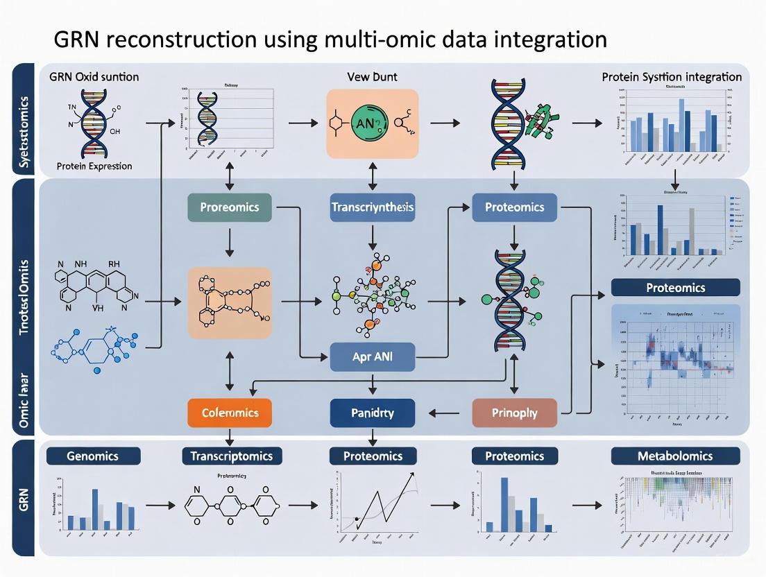
The following diagram illustrates the foundational workflow for generating multi-omics data and its primary application in GRN reconstruction, showcasing the flow from sample to biological insight.
The Omics Cascade: From Gene to Function
Each omics layer Interrogates a specific class of biological molecules, collectively providing a systems-level view. Their relationships and the central dogma of molecular biology are foundational to multi-omics integration.
Genomics
The genome is the complete sequence of DNA in a cell or organism, providing the fundamental, static blueprint of life [16] [17]. Genomics involves discovering and noting all sequences in an entire genome, studying the complete set of genes and their interactions [17]. With the exception of mutations, the genome of an organism remains essentially constant over time and across cell types [16].
Key Analytical Techniques:
- Whole Genome Sequencing (WGS): Provides the complete DNA sequence of an organism [16] [18].
- Genome-Wide Association Studies (GWAS): Identify associations between genomic variations (like Single Nucleotide Polymorphisms - SNPs) and complex traits or diseases [19] [20].
- Single Nucleotide Polymorphism (SNP) Chips: Arrays of oligonucleotide probes that hybridize to specific DNA sequences to assay known common variants [16].
Epigenomics
The epigenome consists of reversible chemical modifications to the DNA, or to the histones that bind DNA, which change gene expression without altering the underlying DNA base sequence [16] [20]. These modifications, which can be tissue-specific and respond to environmental factors, produce heritable changes in gene expression [16] [19]. The epigenome effectively determines the accessibility and packaging of the genomic blueprint.
Key Analytical Techniques:
- Bisulfite Sequencing: Measures DNA methylation status by treating DNA with bisulfite to convert unmethylated cytosines to uracils [20].
- ChIP-seq and CUT&Tag: Identify genome-wide protein-DNA interactions, such as histone modifications and transcription factor binding sites [3] [20].
- ATAC-seq: Assays for transposase-accessible chromatin to identify open, potentially regulatory regions of the genome [19] [3].
Transcriptomics
The transcriptome is the complete set of RNA transcripts (including mRNA, rRNA, tRNA, and non-coding RNA) from DNA in a cell or tissue at a specific point in time [16] [21]. It provides a dynamic snapshot of genomic potential, indicating which genes are actively being transcribed [20]. In humans, only 1.5 to 2 percent of the genome is represented in the transcriptome as protein-coding genes [16].
Key Analytical Techniques:
- RNA Sequencing (RNA-seq): Allows for direct sequencing of RNAs, providing a high-resolution view of the transcriptome, including novel transcripts and splice variants [16] [18].
- Microarrays: Oligonucleotide probes hybridize to specific RNA transcripts to measure their abundance [16].
- Single-Cell RNA-seq (scRNA-seq): Profiles the transcriptomes of individual cells, revealing cellular heterogeneity and identifying rare cell types [3] [18].
Proteomics
The proteome is the complete set of proteins expressed by a cell, tissue, or organism at a given time [16] [17]. Proteins are the functional effectors of cellular processes, and the proteome is highly complex due to post-translational modifications, different spatial configurations, and protein-protein interactions [16]. Unlike the relatively static genome, the proteome is highly dynamic and changes in response to environmental stimuli [17].
Key Analytical Techniques:
- Mass Spectrometry (MS): The dominant technology for high-throughput protein identification and quantification. Advances like the SRMAtlas enable targeted quantification of proteins [16] [18].
- Antibody-Based Arrays: Use antibodies or aptamers as capture agents to make quantitative measurements of proteins from complex mixtures like blood [16] [22].
- Western Blotting and ELISA: Used for targeted protein detection and quantification [22].
Metabolomics
The metabolome refers to the complete set of small molecule metabolites (e.g., sugars, lipids, amino acids, signaling molecules) within a biological sample [16] [20]. These compounds are the substrates and by-products of enzymatic reactions, making them the closest link to the phenotype of an organism [17]. The metabolome is highly dynamic and can vary due to diet, stress, drugs, and disease [16].
Key Analytical Techniques:
- Mass Spectrometry (MS) and Gas/Liquid Chromatography-MS (GC/LC-MS): Workhorse technologies for identifying and quantifying a vast number of metabolites [16] [18] [20].
- Nuclear Magnetic Resonance (NMR) Spectroscopy: Used for metabolic profiling and structural elucidation of metabolites without destroying the sample [16] [20].
Table 1: Summary of Key Omics Layers, Their Molecular Readouts, and Primary Technologies
| Omics Layer | Core Definition | Key Molecules Analyzed | Primary Analytical Technologies |
|---|---|---|---|
| Genomics | Study of the complete set of DNA (genome) [14] [17] | DNA sequence, genetic variants (SNPs, CNVs) [22] [20] | Next-Generation Sequencing (NGS), SNP microarrays [16] [22] |
| Epigenomics | Study of reversible, heritable chemical modifications to DNA and histones (epigenome) [16] [20] | DNA methylation, histone modifications, chromatin accessibility [16] [20] | Bisulfite sequencing, ChIP-seq, CUT&Tag, ATAC-seq [3] [20] |
| Transcriptomics | Study of the complete set of RNA transcripts (transcriptome) [14] [21] | mRNA, tRNA, rRNA, non-coding RNA [16] [21] | RNA-seq, scRNA-seq, microarrays [16] [3] |
| Proteomics | Study of the complete set of proteins (proteome) [14] [17] | Proteins, peptides, post-translational modifications [16] [22] | Mass spectrometry, antibody/aptamer arrays [16] [22] |
| Metabolomics | Study of the complete set of small-molecule metabolites (metabolome) [14] [20] | Sugars, lipids, amino acids, metabolic intermediates [16] [17] | Mass spectrometry, NMR spectroscopy [16] [20] |
Experimental Protocols for Multi-Omics Data Generation
Robust and reproducible experimental protocols are the bedrock of reliable multi-omics data. The following sections outline standard methodologies for generating data from each omics layer.
Protocol: Whole Genome Sequencing for Genomics
Objective: To determine the complete DNA sequence of an organism for variant discovery and genome assembly [16] [21].
Methodology:
- DNA Extraction: Isolate high-molecular-weight genomic DNA from tissue or cells using phenol-chloroform extraction or commercial kits.
- Library Preparation: Fragment DNA via sonication or enzymatic digestion. Repair ends, add 'A' bases, and ligate platform-specific adapter sequences. For long-read sequencing (PacBio, Oxford Nanopore), size selection is critical [19].
- Amplification: Amplify the adapter-ligated DNA library using PCR to generate sufficient material for sequencing.
- Sequencing: Load the library onto a sequencing platform (e.g., Illumina, PacBio, Oxford Nanopore). Illumina uses sequencing-by-synthesis, while PacBio (SMRT) and Nanopore provide long reads [16] [19].
- Data Analysis: Align generated reads to a reference genome using tools like BWA or Bowtie. Call variants (SNPs, indels) using GATK or similar software [19].
Protocol: ATAC-seq for Epigenomics
Objective: To map genome-wide chromatin accessibility and identify putative regulatory elements [3] [19].
Methodology:
- Nuclei Isolation: Gently lyse cells or tissue to isolate intact nuclei, keeping them cold to prevent artifact generation.
- Tagmentation: Treat nuclei with the Tn5 transposase enzyme. Tn5 simultaneously fragments DNA and inserts sequencing adapters into open, accessible regions of chromatin.
- DNA Purification: Purify the tagmented DNA using a standard column- or bead-based protocol.
- PCR Amplification: Amplify the purified DNA with primers containing full Illumina adapter sequences and barcodes.
- Sequencing & Analysis: Sequence the library on an Illumina platform. Analyze data by aligning reads (Bowtie2), calling peaks (MACS2) to identify accessible regions, and integrating with transcriptomic data to infer regulatory connections [3].
Protocol: Bulk RNA-seq for Transcriptomics
Objective: To quantify the abundance and sequence of RNA transcripts in a biological sample [16] [17].
Methodology:
- RNA Extraction: Isolate total RNA using a guanidinium thiocyanate-phenol-chloroform extraction (e.g., TRIzol) or silica-membrane columns. Assess RNA integrity (RIN > 8).
- rRNA Depletion / Poly-A Selection: Enrich for mRNA by removing ribosomal RNA (rRNA) using probe-based depletion or by selecting RNA molecules with poly-A tails.
- Library Preparation: Fragment RNA, synthesize complementary DNA (cDNA) using reverse transcriptase, ligate adapters, and PCR amplify.
- Sequencing: Perform sequencing on an Illumina platform to generate short reads (e.g., 150 bp paired-end).
- Data Analysis: Align reads to a reference genome/transcriptome (STAR, HISAT2). Quantify gene expression (e.g., as FPKM or TPM) using tools like featureCounts.
Protocol: Mass Spectrometry-Based Proteomics
Objective: To identify and quantify the proteins present in a complex biological sample [16] [15].
Methodology:
- Protein Extraction: Lyse cells or tissue in a denaturing buffer (e.g., containing SDS or urea) to solubilize the entire proteome.
- Digestion: Reduce disulfide bonds (DTT), alkylate cysteines (iodoacetamide), and digest proteins into peptides using a sequence-specific protease (typically trypsin).
- Desalting/Cleanup: Purify peptides using C18 solid-phase extraction tips or columns.
- Liquid Chromatography-Mass Spectrometry (LC-MS/MS):
- Separation: Load peptides onto a reverse-phase C18 LC column and separate them by hydrophobicity using a gradient of increasing organic solvent.
- Ionization & Mass Analysis: Ionize eluting peptides via electrospray ionization (ESI) and introduce them into the mass spectrometer. The instrument cycles between a full MS1 scan (to measure peptide abundance) and subsequent MS2 scans (to fragment and sequence selected peptides).
- Data Analysis: Search MS2 spectra against a protein sequence database using software (MaxQuant, Proteome Discoverer) for identification and quantification.
Protocol: Metabolite Profiling via LC-MS
Objective: To comprehensively profile and quantify small-molecule metabolites in a biological sample [16] [20].
Methodology:
- Metabolite Extraction: Use a solvent mixture (e.g., methanol:acetonitrile:water) to precipitate proteins and extract metabolites from biofluids (plasma, urine) or tissue homogenates. Keep samples cold to preserve labile metabolites.
- Liquid Chromatography (LC): Separate the extract using LC (e.g., HILIC for polar metabolites, reverse-phase C18 for lipids) to reduce complexity and ion suppression.
- Mass Spectrometry (MS):
- Data-Dependent Acquisition (DDA): For untargeted discovery, the MS instrument acquires MS1 and MS2 spectra for the most abundant ions to enable metabolite identification.
- Selected Reaction Monitoring (SRM)/Multiple Reaction Monitoring (MRM): For targeted, highly sensitive quantification of a pre-defined set of metabolites.
- Data Processing & Identification: Process raw data using software (XCMS, Progenesis QI) for peak picking, alignment, and normalization. Identify metabolites by matching MS1 and MS2 spectra against reference databases (e.g., METLIN [20], Human Metabolome Database).
Multi-Omics Data Integration for GRN Reconstruction
The ultimate goal of multi-omics in systems biology is often the reconstruction of Gene Regulatory Networks (GRNs)—the intricate interplay between transcription factors (TFs), cis-regulatory elements (CREs), and target genes that orchestrate cellular identity and function [3]. The following workflow details a standard computational pipeline for GRN inference from integrated multi-omics data.
Computational Workflow for GRN Reconstruction:
- Data Preprocessing & Quality Control: Each single-omics dataset (e.g., scRNA-seq, scATAC-seq) undergoes modality-specific preprocessing. This includes read alignment, filtering, normalization, and removal of technical artifacts. For single-cell data, cell clustering and annotation are performed to define cell types/states [3].
- Feature Linking: A critical step for integrating different data types. For example, scATAC-seq peaks are linked to potential target genes based on genomic proximity or by using chromatin conformation data. This creates a set of candidate regulatory relationships (e.g., TF -> CRE -> Gene) [3].
- GRN Inference: Matched multi-omic data is fed into computational methods that employ diverse statistical and machine learning approaches to infer the strength and direction of regulatory connections [3]. Key methodologies include:
- Correlation-Based Approaches: Identify co-expression between TFs and target genes or correlation between CRE accessibility and gene expression. Measures include Pearson/Spearman correlation and mutual information. While simple, they struggle to distinguish direct from indirect regulation [3].
- Regression Models: Model the expression of a target gene as a function of the expression/accessibility of multiple potential TFs/CREs. Penalized methods like LASSO regression are common to handle high dimensionality and prevent overfitting [3].
- Probabilistic Models: Use graphical models (e.g., Bayesian networks) to estimate the most probable regulatory relationships that explain the observed data, providing a measure of uncertainty [3].
- Deep Learning Models: Leverage architectures like graph convolutional networks (GCNs) or autoencoders to learn complex, non-linear relationships in an unsupervised or semi-supervised manner from large, integrated datasets [3] [18].
- Network Evaluation & Validation: The inferred GRN must be rigorously evaluated. This can involve benchmarking against curated gold-standard networks, testing for enrichment of known biological pathways, and, most importantly, experimental validation (e.g., CRISPR perturbations) of novel predicted interactions [3].
Successful execution of multi-omics protocols relies on high-quality, specific research reagents. The following table catalogs essential materials and their functions.
Table 2: Key Research Reagent Solutions for Multi-Omics Workflows
| Reagent / Tool Category | Specific Examples | Function in Multi-Omics Workflow |
|---|---|---|
| Nucleic Acid Enzymes | DNA Polymerases (PCR), Reverse Transcriptases (RT-PCR), Restriction Enzymes, Ligases [22] | Fundamental for library preparation (amplification, adapter ligation), cDNA synthesis, and targeted assays across genomics, epigenomics, and transcriptomics [22]. |
| PCR & Library Prep Kits | PCR Master Mixes, RT-PCR Kits, cDNA Synthesis Kits, Bisulfite Conversion Kits [22] | Provide optimized, ready-to-use reagents for efficient and reproducible amplification, reverse transcription, and specific library construction steps. |
| Oligonucleotides | PCR Primers, Sequencing Adapters, Barcoded Index Primers, Probes [22] | Enable targeted amplification, multiplexing of samples, and the attachment of sequences required for cluster generation and sequencing on NGS platforms. |
| Separation & Analysis | Electrophoresis Systems, DNA/RNA Stains and Ladders, HPLC/UPLC Systems [22] | Used for quality control (e.g., assessing DNA/RNA integrity, library fragment size) and separation of molecules (e.g., peptides, metabolites) prior to MS analysis. |
| Mass Spectrometry | Trypsin Protease, LC Columns (C18), Stable Isotope-Labeled Standards [16] | Critical for proteomics and metabolomics. Enzymes digest proteins; LC columns separate peptides/metabolites; labeled standards enable precise quantification. |
| Bioinformatics Tools | Alignment software (STAR, BWA), Peak Callers (MACS2), GRN tools (pySCENIC, CellOracle) [3] | Computational software and pipelines for analyzing raw sequencing/spectral data, identifying features, and performing advanced integrative analysis like GRN inference. |
Gene Regulatory Networks (GRNs) are collections of molecular regulators that interact with each other and with other substances in the cell to govern gene expression levels of mRNA and proteins, which in turn determine cellular function [1]. The classical view of combinatorial control often presumes coincident interactions between transcription factors (TFs). However, emerging research reveals that sequential molecular interactions rather than coincident ones primarily drive the specification of complex gene expression programs [23]. Understanding these temporal dynamics is crucial for accurate GRN reconstruction, especially when integrating multi-omic data. This application note elucidates the biological rationale for sequential interaction models and provides detailed protocols for their experimental validation and computational integration.
Key Concepts and Biological Principles
The Hierarchical Nature of GRNs
GRNs operate through a hierarchical structure where master transcriptional regulators control subordinate networks, creating layers of regulation that unfold over time [24]. This hierarchy enables:
- Temporal gating of gene expression during cellular differentiation
- Staged response to external stimuli such as pathogens or cytokines
- Integration of multiple signaling pathways through sequential TF activation
Feedback loops within these networks provide cellular memory and stability to gene expression states, ensuring maintenance of cellular identity through repeated cell divisions [1].
Sequential vs. Coincident Control Logic
Research on pathogen-responsive transcriptomes in murine fibroblasts and macrophages demonstrates that stimulus-responsive TFs typically function sequentially in logical OR gates or individually, rather than through coincident AND gates [23]. This represents a fundamental shift from traditional understandings of combinatorial control.
Key evidence for sequential control:
- AND gates occur between NFκB-responsive mRNA synthesis and MAPKp38-responsive control of mRNA half-life - processes that are temporally separated
- Logical OR gates are prevalent between sequentially acting NFκB and ISGF3 transcription factors
- Temporal coordination between nuclear transcription events and cytoplasmic mRNA stability mechanisms
Experimental Evidence and Data Presentation
Quantitative Analysis of Logical Gates in Immune Response
Table 1: Distribution of Logical Gate Types in Pathogen-Responsive Genes
| Gene Cluster | TF Logic Gate | Regulatory Mechanism | Frequency (%) | Primary TFs Involved |
|---|---|---|---|---|
| Inflammatory Early Responders | OR | Sequential TF activation | 42% | NFκB, AP1 |
| Antiviral Response | OR | Sequential TF activation | 38% | IRF, ISGF3 |
| Sustained Inflammatory | AND | mRNA synthesis + decay | 12% | NFκB, MAPKp38 |
| Cell Identity Maintainers | Single TF | Independent action | 8% | Cell-type specific TFs |
Data derived from mechanistic modeling of 714 endotoxin-inducible genes across 85 datasets measuring transcriptional responses of murine fibroblasts and macrophages to cytokines and pathogens [23].
Methodological Foundations for Inferring Sequential Interactions
Table 2: Computational Approaches for Sequential Interaction Detection
| Method Type | Key Capabilities | Limitations for Sequential Analysis |
|---|---|---|
| Dynamical Systems | Models time-evolving behavior of systems; captures synthesis and decay parameters | Requires prior domain knowledge; less scalable for large networks [3] |
| Boolean Networks | Logical operations with temporal ordering; can model sequential steps | Discretizes continuous expression data; may oversimplify [24] |
| Bayesian Networks | Probabilistic dependencies with directionality; infers causal relationships | Assumes specific distribution of gene expression [3] |
| Deep Learning (Enformer) | Integrates long-range interactions up to 100kb; uses attention mechanisms | Requires large training datasets; computationally intensive [25] |
Experimental Protocols
Protocol: Elucidating Sequential TF Control Logic
Objective: Determine whether combinations of transcription factors function sequentially or coincidentally in regulating target genes.
Materials:
- Wild-type primary mouse embryonic fibroblasts (MEFs)
- Recombinant cytokines: PDGFβ, TNF, IFNβ
- RNA extraction kit and qPCR reagents
- RNA-seq library preparation kit
Procedure:
Stimulus Panel Design:
- Prepare three stimulation conditions:
- PDGFβ (activates JNK pathway and AP1)
- TNF (activates AP1 and NFκB)
- IFNβ (activates IRF transcription factor ISGF3)
- Include unstimulated control
- Prepare three stimulation conditions:
Time-Course Experiment:
- Explicate MEF cultures to each stimulus for 0, 15, 30, 60, 120, and 240 minutes
- Include technical triplicates for each time point
TF Activity Measurement:
- Harvest cells at each time point for nuclear extraction
- Quantify TF activation through:
- Western blot for phospho-TF forms
- EMSA for DNA binding activity
- Immunofluorescence for nuclear localization
Transcriptome Profiling:
- Extract total RNA using column-based method
- Prepare RNA-seq libraries using poly-A selection
- Sequence on Illumina platform to minimum depth of 30M reads/sample
Data Integration:
- Cluster genes by expression patterns using K-means clustering
- Map TF activity profiles to gene expression clusters
- Assign logical gates based on stimulus-response patterns
Expected Results: The majority of pathogen-responsive genes will show expression patterns consistent with sequential OR gates rather than coincident AND gates [23].
Protocol: Validating mRNA Synthesis-Decay AND Gates
Objective: Experimentally confirm AND gates between nuclear transcription and cytoplasmic mRNA stability control.
Materials:
- NFκB inhibitor (e.g., BAY 11-7082)
- MAPKp38 inhibitor (e.g., SB203580)
- Metabolic labeling reagent (4-thiouridine)
- RT-qPCR reagents
Procedure:
Inhibitor Treatment:
- Pre-treat cells with DMSO (control), NFκB inhibitor, MAPKp38 inhibitor, or both for 1 hour
- Stimulate with TNF (10 ng/mL) for 2 hours
Transcriptional Pulse-Chase:
- Add 4-thiouridine to culture medium for 15 minutes to label newly synthesized RNA
- Replace with regular medium and harvest cells at 0, 15, 30, 60, 120 minutes post-labeling
Biotinylation and Separation:
- Biotinylate thiol-labeled RNA using EZ-Link Biotin-HPDP
- Separate labeled (newly synthesized) and unlabeled (pre-existing) RNA using streptavidin beads
Quantification:
- Quantify both newly synthesized and pre-existing RNA for target genes via RT-qPCR
- Calculate mRNA half-life from decay curves of pre-existing RNA
Data Analysis:
- Identify genes requiring both NFκB activity (for synthesis) and p38 activity (for stability)
- Confirm AND gate logic through synergistic inhibition with both inhibitors
Validation: Genes showing significantly reduced expression only when both pathways are inhibited demonstrate the AND gate between synthesis and decay mechanisms [23].
Visualization of Sequential Interactions
DOT Visualization: Sequential TF Logic Gates
DOT Visualization: Experimental Workflow for Sequential Analysis
The Scientist's Toolkit: Research Reagent Solutions
Table 3: Essential Research Reagents for Sequential Interaction Studies
| Reagent Category | Specific Examples | Function in Sequential Studies | Key Considerations |
|---|---|---|---|
| Pathway-Specific Agonists | TNF-α, IFN-β, PDGF-BB, LPS | Selective activation of specific TFs to map temporal hierarchies | Use at defined concentrations with precise timing |
| Kinase Inhibitors | BAY 11-7082 (NFκB), SB203580 (p38), SP600125 (JNK) | Dissect contribution of specific pathways to sequential logic | Validate specificity and use multiple inhibitors per pathway |
| Metabolic RNA Labels | 4-thiouridine, 5-ethynyl uridine | Distinguish newly synthesized vs. pre-existing mRNA for decay studies | Optimize labeling time for specific transcript half-lives |
| TF Activity Assays | Phospho-specific antibodies, EMSA kits, NanoBIT systems | Measure timing and magnitude of TF activation | Combine multiple methods for validation |
| Single-Cell Multi-omic Platforms | 10x Multiome, SHARE-seq | Simultaneously profile gene expression and chromatin accessibility | Ensure sufficient cell numbers for robust clustering |
| CRISPR Screening Tools | CRISPRi/a libraries for enhancer validation | Functionally test regulatory elements identified in models | Include multiple gRNAs per target for confidence |
Integration with Multi-omic GRN Reconstruction
The recognition of sequential molecular interactions necessitates specific computational approaches for accurate GRN reconstruction from multi-omic data:
Methodological Recommendations
Temporal Data Integration:
- Prioritize methods that incorporate time-course data rather than single time points
- Utilize dynamical systems models that capture synthesis and decay parameters [3]
- Implement ensemble approaches that combine multiple inference methods for improved stability and accuracy [26]
Multi-omic Feature Alignment:
- Align scRNA-seq and scATAC-seq data to connect TF expression, chromatin accessibility, and target gene expression
- Use deep learning architectures like Enformer that integrate long-range interactions (up to 100kb) to capture distal enhancer-promoter interactions [25]
- Leverage transformer-based models with attention mechanisms to identify relevant regulatory elements across extended genomic distances
Validation Strategies for Sequential Models
Experimental Validation:
- Perform targeted perturbations of identified sequential nodes
- Measure effects on both direct targets and downstream network components
- Use metabolic RNA labeling to distinguish transcriptional vs. post-transcriptional regulation
Computational Validation:
- Compare inferred networks to gold standard interactions from databases like KEGG and I2D [26]
- Assess biological consistency through enrichment of known biological pathways
- Evaluate temporal predictions against held-out time-course data
The paradigm of sequential rather than coincident molecular interactions represents a fundamental advance in understanding the biological rationale of gene regulatory networks. This perspective enables more accurate GRN reconstruction from multi-omic data by respecting the temporal hierarchy of regulatory events. The protocols and methodologies presented here provide researchers with practical tools to elucidate these sequential interactions and integrate them into predictive network models, ultimately enhancing our ability to understand cellular responses in development, homeostasis, and disease.
The Promise of Multi-Omic GRNs for Uncovering Disease Mechanisms and Identifying Therapeutic Targets
Gene Regulatory Networks (GRNs) are complex systems that determine the development, differentiation, and function of cells and organisms, as well as their response to environmental stimuli [27]. These networks consist of genes, transcription factors (TFs), microRNAs, and other regulatory molecules that interact to control gene expression [27]. The reconstruction of GRNs from multi-omic data represents a paradigm shift in biomedical research, enabling unprecedented insights into disease mechanisms and therapeutic targeting. Despite decades of cancer research, cancer ranks as the top cause of death and shortened life expectancy globally, with the global cancer burden estimated to increase by 47% from 2020 to 2040 [28]. Traditional single-omics approaches cannot fully capture the complex, multi-layered nature of disease mechanisms, as mutations that occur in DNA will affect the expression of proteins, but it is hard to tell the extent of the loss of function based on the genome alone [29]. Multi-omics integration provides a powerful framework to address these limitations by enabling researchers to filter out novel associations between biomolecules and disease phenotypes, identify relevant signaling pathways, and establish detailed biomarkers of disease [29]. The advent of high-throughput sequencing technologies has revolutionized our ability to profile various molecular features, including genomics, transcriptomics, proteomics, and metabolomics, providing the essential data layers for comprehensive GRN reconstruction [3]. This Application Note details standardized protocols for multi-omic GRN reconstruction and their applications in translational research, providing researchers with practical methodologies to advance precision medicine initiatives.
Computational Foundations of Multi-Omic GRN Inference
Methodological Approaches for GRN Reconstruction
GRN inference relies on diverse statistical and algorithmic principles to uncover regulatory connections between genes and their regulators. Table 1 summarizes the primary computational approaches used in GRN reconstruction, each with distinct strengths and applications.
Table 1: Computational Methods for Multi-Omic GRN Inference
| Method Category | Key Principles | Representative Algorithms | Best Use Cases |
|---|---|---|---|
| Correlation-based | Measures linear/non-linear associations between TFs and target genes using Pearson's/Spearman's correlation or mutual information [3] | ARACNE, CLR [27] | Initial network screening; hypothesis generation |
| Regression Models | Models gene expression as response variable regressed on TF expression/accessibility; handles high dimensionality via penalization [3] | LASSO [27] | Identifying direct regulatory relationships; sparse network inference |
| Probabilistic Models | Graphical models capturing dependence between variables; estimates most probable regulatory relationships [3] | Bayesian Networks [27] | Network inference with uncertainty quantification |
| Dynamical Systems | Models system behavior over time using differential equations; captures transcription, regulation, and stochasticity [3] | dynGENIE3 [27] | Time-course data; modeling network dynamics |
| Deep Learning | Neural networks (CNNs, VAEs, GNNs) that learn complex, non-linear relationships from large multi-omic datasets [3] [27] | GRN-VAE, DeepSEM, GRNFormer [27] | Large-scale multi-omic integration; capturing complex non-linearities |
Evolution of GRN Inference Methods
The field of GRN inference has evolved significantly from early approaches that leveraged microarray and RNA-sequencing data to identify co-expressed genes using measures of association [3]. The expansion from bulk transcriptomics to bulk multi-omics technologies such as ATAC-seq, Hi-C, and ChIP-seq enabled researchers to identify accessible regions of chromatin, capture structural changes and chromatin interactions, and profile protein-DNA interactions [3]. The advent of single-cell omics technologies has further revolutionized the field by enabling the inference of regulatory relationships at cell type, cell state, and single-cell resolution [3]. Recent sequencing platforms can simultaneously profile RNA and cis-regulatory element (CRE) accessibility within a single cell, leading to the development of novel GRN inference methods that exploit these matched multi-omic data to comprehensively recapitulate regulatory networks [3].
Figure 1: Evolution of GRN inference technologies, showing progression from early microarray-based approaches to modern AI-driven methods that leverage single-cell multi-omic data.
Protocols for Multi-Omic GRN Reconstruction
Data Processing and Harmonization
Protocol 3.1.1: Multi-Omic Data Preprocessing
- Genomic Variant Calling: Process raw sequencing data using the Genome Analysis Toolkit (GATK), the industry standard for identifying single nucleotides (SNPs) and indels, somatic short variants, copy number variations (CNV), and structural variations (SV) in germline DNA and RNAseq data [28].
- Copy Number Variation Analysis:
- Data Harmonization: Convert genome assembly versions to the latest reference (e.g., hg38) using tools like CruzDB to ensure compatibility across datasets generated from different platforms [28].
- Expression Data Categorization: Define up-regulation, normal, and down-regulation ranges by categorizing gene expression data using housekeeping genes as references [28].
Protocol 3.1.2: Single-Cell Multi-Omic Data Processing
- Quality Control: Filter cells based on quality metrics (mitochondrial read percentage, number of detected genes, doublet prediction).
- Normalization: Apply appropriate normalization methods for each modality (e.g., SCTransform for scRNA-seq, term frequency-inverse document frequency (TF-IDF) for scATAC-seq).
- Integration: Use mutual nearest neighbors (MNN) or other integration methods to align cells across modalities and correct for technical variation.
- Imputation: Address sparsity in single-cell data using deep learning methods like variational autoencoders (VAEs) for data imputation and augmentation [30].
GRN Inference Workflow
Protocol 3.2.1: Network Reconstruction Using PLBINs
- Data Input: Prepare normalized multi-omic matrices (expression, chromatin accessibility, methylation) for a patient cohort.
- Network Inference: Apply Prediction Logic Boolean Implication Networks (PLBINs), which have advantages over other methods in constructing genome-scale multi-omics networks in bulk tumors and single cells in terms of computational efficiency, scalability, and accuracy [28].
- Validation: Use external datasets or experimental validations to confirm high-confidence network edges.
- Hub Gene Identification: Apply graph theory network centrality metrics (betweenness, closeness, eigenvector centrality) to prioritize candidate genes for further investigation [28].
Protocol 3.2.2: Deep Learning-Based GRN Inference with Flexynesis
- Framework Setup: Install Flexynesis, available on PyPi, Guix, Bioconda, and Galaxy Server, which streamlines data processing, feature selection, hyperparameter tuning, and marker discovery [31].
- Architecture Selection: Choose from deep learning architectures or classical supervised machine learning methods with a standardized input interface for single/multi-task training and evaluation [31].
- Model Training:
- For classification tasks (e.g., cancer subtype prediction): Use multi-layer perceptron (MLP) with cross-entropy loss.
- For survival modeling: Employ supervisor MLP with Cox Proportional Hazards loss function [31].
- For multi-task learning: Attach multiple MLPs on top of sample encoding networks to shape the embedding space using multiple clinically relevant variables [31].
- Biomarker Discovery: Extract and interpret important features from the trained model to identify potential therapeutic targets.
Figure 2: Generalized workflow for multi-omic GRN reconstruction and therapeutic target identification.
Integration with Clinical Data and Therapeutic Applications
Connecting Multi-Omic GRNs to Clinical Outcomes
Protocol 4.1.1: Electronic Medical Record (EMR) Integration
- Data Mapping: Integrate patient multi-omic biomarkers with clinical, pathological, demographic, and comorbid factors from EMRs using standardized ontologies [28].
- Retrospective Analysis: Conduct retrospective analysis of EMRs to discover new drug targets and reposition existing drugs [28].
- Validation Framework: Implement extensive external validation using large-scale patient registries such as the SEER-Medicare cancer registry to identify biomarkers applicable to large patient populations [28].
Protocol 4.1.2: Survival Analysis and Risk Stratification
- Data Preparation: Prepare multi-omic data (e.g., gene expression and methylation profiles) with matched clinical outcome data (overall survival, progression-free survival).
- Risk Modeling: Use supervised learning approaches with Cox Proportional Hazards models to predict patient-specific risk scores based on input overall survival endpoints [31].
- Stratification: Split patients into high-risk and low-risk groups based on median risk scores and validate stratification using Kaplan-Meier survival analysis [31].
Applications in Precision Oncology
Multi-omic GRN analysis has demonstrated significant utility across various cancer types. Table 2 highlights key applications and findings from recent studies.
Table 2: Therapeutic Applications of Multi-Omic GRN Analysis in Precision Oncology
| Cancer Type | Multi-Omic Approach | Key Findings | Therapeutic Implications |
|---|---|---|---|
| Pan-Gastrointestinal and Gynecological Cancers | Gene expression + promoter methylation [31] | High accuracy classification of microsatellite instability (MSI) status (AUC = 0.981) without mutation data [31] | Identifies patients likely to respond to immune checkpoint blockade therapies |
| Lower Grade Glioma (LGG) and Glioblastoma (GBM) | Multi-omic integration with survival modeling [31] | Significant separation of patients by risk scores in embedding space and Kaplan-Meier plots [31] | Enables risk stratification and personalized treatment approaches |
| Non-Small-Cell Lung Cancer (NSCLC) | CNV analysis of immunotherapy targets [28] | CD20, CD27, PD1, PDL1 have more CNVs than SNVs in TCGA tumors [28] | Suggests CNV profiling could complement current biomarker strategies |
| Triple-Negative Breast Cancer | Multi-omics analysis [30] | Identification of therapeutic vulnerabilities in TNBC subtypes [30] | Reveals novel subtype-specific therapeutic targets |
| Serous Ovarian Cancer | Multi-omics molecular subtyping [30] | Identification of molecular subtypes with prognostic significance [30] | Enables subtype-specific treatment strategies |
Successful multi-omic GRN research requires specialized computational tools and data resources. Table 3 catalogues essential reagents and their applications in multi-omic GRN studies.
Table 3: Essential Research Reagents and Computational Tools for Multi-Omic GRN Studies
| Resource Category | Specific Tools/Resources | Application | Key Features |
|---|---|---|---|
| Data Resources | The Cancer Genome Atlas (TCGA) [31], Cancer Cell Line Encyclopedia (CCLE) [31], SEER-Medicare [28] | Provides large-scale multi-omic datasets with clinical annotations | Enables training and validation of GRN models across diverse patient populations |
| GRN Inference Software | PLBINs [28], Flexynesis [31], GRN-VAE [27], GRNFormer [27] | Reconstructs regulatory networks from multi-omic data | Various methodological approaches; specialized for different data types and research questions |
| Deep Learning Frameworks | PyTorch, TensorFlow (via Flexynesis) [31] | Provides architectures for multi-omic integration tasks | Supports single/multi-task learning for regression, classification, and survival modeling |
| Data Processing Tools | Genome Analysis Toolkit (GATK) [28], PennCNV-Affy [28], CGHcall [28] | Processes raw sequencing data into analyzable formats | Industry standards for variant calling, CNV analysis, and quality control |
| Validation Resources | DREAM challenges [27], external patient cohorts [28] | Benchmarks GRN inference performance | Provides gold-standard datasets and networks for method validation |
Multi-omic GRN reconstruction represents a powerful approach for elucidating disease mechanisms and identifying novel therapeutic targets. The methodologies outlined in this Application Note form a conceptually innovative framework to analyze various available information from research laboratories and healthcare systems, accelerating the discovery of biomarkers and therapeutic targets to ultimately improve patient survival outcomes [28]. As single-cell multi-omics technologies continue to advance and computational methods become more sophisticated, the precision and comprehensiveness of GRN models will further improve. Researchers are encouraged to adopt these standardized protocols to enhance reproducibility and accelerate translational applications in precision oncology and beyond.
A Guide to Computational Methods and Tools for Multi-Omic GRN Inference
Application Notes
The reconstruction of Gene Regulatory Networks (GRNs) from multi-omic data is a cornerstone of modern systems biology, critical for understanding cell identity, fate decisions, and disease mechanisms [3]. The integration of data from single-cell RNA-seq (scRNA-seq) and single-cell ATAC-seq (scATAC-seq) has revolutionized this field, enabling the inference of regulatory relationships at unprecedented resolution [3]. Several core computational frameworks have been developed to harness this data, each with distinct strengths, assumptions, and applications. Correlation-based methods offer a simple starting point for identifying potential associations, while regression models provide more robust inference of direct regulatory links. Bayesian approaches excel at incorporating prior knowledge and topology, and dynamical systems models uniquely capture the temporal evolution of gene expression, providing deep insights into the stability and dynamics of regulatory circuits [32] [33].
Selecting the appropriate methodological framework depends on the specific biological question, data type, and desired level of mechanistic insight. The following sections and accompanying tables detail the application, experimental protocols, and key reagents for each framework, providing a practical guide for researchers embarking on GRN reconstruction.
Quantitative Comparison of Core Methodological Frameworks
Table 1: Key characteristics and applications of the four core GRN inference frameworks.
| Framework | Primary Application in GRN | Key Strengths | Key Limitations | Suitable Data Types |
|---|---|---|---|---|
| Correlation Models | Initial screening for co-expressed genes and co-accessible regions [3]. | Simple, fast to compute, intuitive results; can capture non-linear relationships with Spearman correlation or Mutual Information [3]. | Cannot distinguish direct from indirect regulation; prone to false positives from confounders [3]. | scRNA-seq, scATAC-seq, bulk RNA-seq. |
| Regression Models | Inferring direct regulatory links by modeling a target gene's expression as a function of multiple potential regulators [3]. | More robust than correlation; can handle multiple regulators simultaneously; coefficients indicate interaction strength and direction [3]. | Can be unstable with highly correlated predictors; requires careful regularization (e.g., LASSO) to avoid overfitting [3]. | scRNA-seq, scATAC-seq (as potential regulators). |
| Bayesian Models | Incorporating prior knowledge (e.g., network topology, TF binding motifs) to refine network inference [34] [3]. | Naturally integrates diverse data types as priors; provides probabilistic measures of confidence for each edge [34]. | Computationally intensive; often assumes specific data distributions (e.g., Gaussian) which may not hold [3]. | scRNA-seq, plus prior data (e.g., protein-DNA interaction, known network topologies). |
| Dynamical Systems | Modeling the temporal dynamics of GRNs to understand stability, multistability, and response to perturbations [32] [33]. | Captures the dynamic and emergent properties of networks (e.g., oscillations, switches); highly interpretable parameters [3]. | Requires time-series data; model complexity increases rapidly with network size; can be difficult to parameterize [3]. | Time-series scRNA-seq or bulk RNA-seq. |
Table 2: Typical workflow outputs and validation strategies for each framework.
| Framework | Typical Output | Common Software/Packages | Suggested Validation Approaches |
|---|---|---|---|
| Correlation Models | A matrix of association scores (e.g., correlation coefficients, MI values) between all gene/feature pairs. | WGCNA, SCENIC, pySCENIC | Comparison with ChIP-seq validated interactions; functional enrichment of co-expression modules. |
| Regression Models | A list of regulator-target links with estimated coefficients; a sparse adjacency matrix for the network. | scLink, BSLIMs, LEAP | Knock-out/knock-down experiments; cross-validation on held-out data. |
| Bayesian Models | A posterior probability for each potential regulatory interaction; a confidence-weighted network. | Banjo, BGRMI, Bayesian Group Lasso [34] | Precision-recall analysis against gold-standard benchmarks (e.g., DREAM challenges) [34]. |
| Dynamical Systems | A set of equations (ODEs/Boolean rules) describing the system's evolution; parameters like degradation/rate constants. | BoolNet, GNA, Oscill8 | Testing predicted system responses (e.g., oscillation period, fate decisions) against new experimental data. |
Experimental Protocols
Protocol 1: Correlation-Based GRN Inference from scRNA-seq Data
Purpose: To identify potential regulatory relationships by measuring the association between gene expression patterns.
Background: This protocol uses the "guilt-by-association" principle, where the co-expression of a transcription factor (TF) and a putative target gene suggests a potential regulatory relationship [3]. Non-parametric measures like Spearman correlation are preferred for their ability to capture non-linear monotonic relationships.
Procedure:
- Data Preprocessing: Begin with a preprocessed scRNA-seq count matrix (cells x genes). Perform quality control, normalization, and log-transformation.
- Regulator Selection: Compile a list of potential regulator genes (e.g., all TFs).
- Association Calculation: For each target gene, calculate the Spearman correlation coefficient between its expression and the expression of every potential regulator across all cells.
- Network Construction: For each target gene, retain the top N regulators with the highest absolute correlation coefficients or apply a significance threshold (p-value < 0.05, adjusted for multiple testing).
- Multi-omic Integration (Optional): Filter potential regulatory links by requiring that the TF binding motif is present in an accessible chromatin region (from scATAC-seq data) near the target gene's promoter.
Key Reagent Solutions:
- TF Gene List: A curated list of transcription factors (e.g., from AnimalTFDB or MSigDB) is essential to define the set of potential regulators.
- Motif Database: A database of TF binding motifs (e.g., JASPAR, CIS-BP) is required for the optional scATAC-seq integration step.
Protocol 2: Bayesian GRN Inference with Topology Priors
Purpose: To reconstruct a GRN by incorporating prior knowledge about network structure, such as scale-free topology, to improve inference accuracy.
Background: This protocol uses a Bayesian framework to integrate gene expression data with the prior belief that biological networks often exhibit a scale-free or exponential in-degree distribution, where most genes are regulated by only a few TFs [34]. A Bayesian group lasso with spike and slab priors is used to perform gene selection and estimation for nonparametric models, effectively controlling model size and reducing false positives [34].
Procedure:
- Model Specification: Define a dynamic Bayesian network (DBN) model. For each gene g, model its expression at time t as a function of the expression of all other genes at time t-1 using B-spline basis functions to capture non-linearity [34].
- Prior Setting: Incorporate the topology information as a prior. Apply a prior distribution that restricts the maximum number of parents (regulators) for any target gene in the network, reflecting the biological observation of constrained in-degree distribution [34].
- Posterior Inference: Use Markov Chain Monte Carlo (MCMC) sampling methods to estimate the posterior distribution of the model parameters, which includes the probability of each regulatory link.
- Network Extraction: Extract the final network by including edges where the posterior probability of regulation exceeds a predefined threshold (e.g., 0.95).
Key Reagent Solutions:
- Benchmark Datasets: Gold-standard networks like DREAM3 and DREAM4 challenge datasets are critical for validating and tuning the model parameters [34].
- MCMC Sampling Software: Computational tools like JAGS, Stan, or custom implementations in R/Python are required for the Bayesian inference.
Protocol 3: Dynamical Systems Modeling using Boolean Networks
Purpose: To model GRNs as discrete dynamical systems to study their long-term behavior, including stable states (attractors) and their robustness to perturbations [33].
Background: Boolean networks provide a tractable framework to explore the mathematical principles of network stability, where gene expression is simplified to an ON (1) or OFF (0) state. A key mechanism conferring stability in these models is canalization, where a subset of inputs can determine the state of a node, making the network robust to other input variations [33].
Procedure:
- Network Structure Definition: Define the network structure (nodes and directed edges) based on prior knowledge (e.g., from literature or inferred through other methods).
- Boolean Rule Assignment: For each node (gene), define a Boolean update function (e.g., AND, OR) that determines its next state based on the states of its input nodes.
- System Simulation: Starting from random or specific initial states, simulate the network's trajectory over discrete time steps until it reaches a stable state (fixed point) or a set of repeating states (limit cycle).
- Stability Analysis: Quantify the robustness of the network's attractors by introducing random perturbations (flipping gene states) and observing the system's ability to return to the original attractor. This measures the degree of canalization [33].
Key Reagent Solutions:
- Boolean Network Simulation Software: Tools like BoolNet (R package) or CellCollective are essential for defining rules and running simulations.
- Perturbation Library (in silico): A defined set of in silico perturbations (e.g., gene knock-outs, sustained activation) is a key reagent for testing model predictions.
Mandatory Visualization
Diagram 1: GRN Inference Method Workflows
Diagram 2: Boolean Network Dynamics & Canalization
Research Reagent Solutions
Table 3: Essential reagents and computational tools for GRN inference.
| Category | Item | Function/Application |
|---|---|---|
| Data Generation | 10x Genomics Single Cell Multiome ATAC + Gene Expression | Simultaneously profiles gene expression and chromatin accessibility from the same single cell, providing matched multi-omic data for GRN inference [3]. |
| Data Generation | JASPAR Database | A curated, open-access database of transcription factor binding profiles (motifs) used to link accessible chromatin regions to potential regulators [3]. |
| Computational Tools | SCENIC (pySCENIC) | A widely-used computational tool that uses correlation (co-expression) and cis-regulatory motif analysis to infer GRNs and identify cellular states from scRNA-seq data. |
| Computational Tools | BoolNet | An R package that provides tools for the reconstruction, simulation, and analysis of Boolean networks, ideal for dynamical systems modeling of GRNs [33]. |
| Benchmarking | DREAM Network Inference Challenges | Community-standard in silico benchmark datasets (e.g., DREAM3, DREAM4) and in vivo networks for objectively evaluating and comparing the performance of GRN inference methods [34]. |
Harnessing Single-Cell Multi-Omics Data for Cell-Type-Specific Network Inference
Gene regulatory networks (GRNs) represent the complex circuitry of cellular identity and function, detailing the interactions between transcription factors (TFs), cis-regulatory elements (CREs), and their target genes. Inferring these networks is fundamental to understanding the molecular basis of development, cellular differentiation, and disease pathogenesis. The advent of single-cell multi-omics technologies has revolutionized this field by enabling the simultaneous measurement of multiple molecular layers—such as the transcriptome, epigenome, and proteome—within individual cells. This capability is crucial for dissecting cellular heterogeneity and reconstructing cell-type-specific regulatory maps that are often obscured in bulk analyses [35] [36].
The integration of these multi-omic data types addresses a critical limitation of single-modality studies. For instance, while single-cell RNA sequencing (scRNA-seq) reveals gene expression states, it cannot directly identify the accessible regulatory regions that control these expression patterns. Conversely, single-cell ATAC-seq (scATAC-seq) maps chromatin accessibility but does not directly link these regions to target gene expression. Multi-omics integration provides a more holistic and causal framework for GRN inference by functionally linking regulators to their targets [3] [13]. This review details the experimental and computational protocols essential for leveraging single-cell multi-omics data to infer accurate, cell-type-specific gene regulatory networks.
Methodological Foundations for Multi-Omics GRN Inference
The computational inference of GRNs from multi-omics data relies on a variety of statistical and algorithmic principles. Understanding these foundations is key to selecting and applying the appropriate tools.
Table 1: Core Methodological Approaches for GRN Inference
| Approach | Underlying Principle | Key Advantages | Common Tools/Examples |
|---|---|---|---|
| Correlation/Information-based | Identifies co-expression or co-variation between TFs and potential target genes. | Simple, intuitive, and computationally efficient. | LEAP, PIDC [3] [37] |
| Regression Models | Models the expression of a target gene as a linear/non-linear function of potential TF regulators. | Provides interpretable coefficients indicating interaction strength and direction. | GENIE3, SINCERITIES [3] [37] |
| Probabilistic Models | Uses graphical models to represent and infer the probabilistic dependencies between variables. | Allows for uncertainty quantification in predictions. | Methods in MAGICAL, scMTNI [3] [38] |
| Dynamical Systems | Utilizes differential equations to model the temporal dynamics of gene expression. | Captures causal, time-dependent relationships directly. | SCODE, GRISLI, MINIE [39] [3] [37] |
| Deep Learning | Employs neural networks (e.g., autoencoders, graph neural networks) to learn complex, non-linear relationships. | Highly flexible and can model intricate regulatory patterns. | GLUE, DeepMAPS [3] [40] [38] |
A significant challenge in multi-omics integration is the distinct feature spaces of different modalities (e.g., ATAC-seq peaks vs. RNA-seq genes). Frameworks like GLUE (Graph-Linked Unified Embedding) overcome this by using a prior knowledge-based "guidance graph" that explicitly links features across omics layers, such as connecting an accessible chromatin region to its putative target gene. This graph then guides the adversarial alignment of cells from different modalities into a shared latent space, enabling integrated analysis and regulatory inference [40]. Another advanced method, MINIE, integrates bulk metabolomics and single-cell transcriptomics through a Bayesian regression framework that explicitly models the timescale separation between molecular layers using a differential-algebraic equation model, providing a powerful tool for cross-layer network inference [39].
Experimental Protocols for Single-Cell Multi-Omics Data Generation
Generating high-quality single-cell multi-omics data is the first critical step. The following protocols outline the process from sample preparation to sequencing.
Sample Preparation and Cell Labeling
The goal is to obtain a viable single-cell suspension that preserves the integrity of multiple molecular types.
- Tissue Dissociation: Mechanically or enzymatically dissociate fresh tissue samples into single-cell suspensions. The choice of enzymes (e.g., collagenase, papain) and dissociation time must be optimized to minimize degradation of RNA, proteins, and epitopes [36].
- Cell Viability and Counting: Assess viability using trypan blue or other fluorescent dyes. Aim for >90% viability. Count cells to determine concentration.
- Cell Hashing and Multiplexing: To increase throughput and mitigate batch effects, label cells from different samples or conditions with unique oligonucleotide-barcoded antibodies (e.g., BD Single-Cell Multiplexing Kit). This allows multiple samples to be pooled before capture [41].
- Surface Protein Staining (CITE-seq): If profiling the surface proteome, stain the cell suspension with oligonucleotide-conjugated antibodies (BD AbSeq Ab-Oligos). Use a panel of antibodies targeting key surface markers relevant to the biological system [41].
- Nuclear vs. Cytoplasmic Separation (for specific assays): For methods like scG&T-seq or scMT-seq, gently lyse the cell membrane while keeping the nucleus intact. Separate the nucleus (for DNA/methylome sequencing) from the cytoplasm (for RNA sequencing) via micropipetting, centrifugation, or magnetic beads [36].
Single-Cell Capture and cDNA Synthesis
This protocol uses a droplet-based system (e.g., BD Rhapsody) for capturing single cells and preparing sequencing libraries.
- Cell Loading and Capture: Load the stained, single-cell suspension into a BD Rhapsody Cartridge. The instrument will isolate individual cells along with uniquely barcoded magnetic beads in microwells or droplets [41].
- Cell Lysis and Molecular Tagging: Lysing the cell releases its RNA. Poly-adenylated RNA molecules hybridize to the oligo-dT primers on the beads. Each primer contains a unique molecular identifier (UMI) and a cell barcode, tagging every mRNA molecule from a single cell.
- cDNA Synthesis: Perform reverse transcription on the beads to create cDNA libraries from the captured mRNA. If performing paired multi-omics (e.g., 10x Multiome), a transposase reaction is simultaneously performed on the same cell to fragment and tag accessible chromatin regions [41] [13].
Library Preparation and Sequencing
Generate separate but linked libraries for each omics modality from the same set of barcoded beads/cells.
- mRNA Library (WTA): Amplify the cDNA via PCR to create a Whole Transcriptome Analysis (WTA) library for Illumina sequencing [41].
- ATAC Library (if applicable): Amplify the transposed DNA fragments to create a separate ATAC-seq library [41].
- AbSeq Library (Protein): Amplify the antibody-derived tags to create the AbSeq (proteomics) library [41].
- Sample Tag Library (Multiplexing): Amplify the sample barcode reads from the multiplexing kit to enable sample demultiplexing post-sequencing [41].
- Library QC and Sequencing: Quantify and quality-check each library using a bioanalyzer. Pool libraries at appropriate molar ratios and sequence on an Illumina platform. Recommended sequencing depth is typically 20,000-50,000 reads per cell for scRNA-seq and 25,000-100,000 reads per cell for scATAC-seq.
Computational Workflow for Cell-Type-Specific Network Inference
Once multi-omics data is generated, the following computational protocol enables the inference of cell-type-specific GRNs.
Data Preprocessing and Integration
- Quality Control and Filtering:
- scRNA-seq: Filter out cells with low unique gene counts, high mitochondrial read percentage, and doublets. Filter genes detected in very few cells.
- scATAC-seq: Filter cells based on unique nuclear fragments and transcription start site (TSS) enrichment score. Remove peaks in blacklisted genomic regions.
- Normalization and Dimensionality Reduction:
- Normalize scRNA-seq data using methods like SCTransform. For scATAC-seq, perform term frequency-inverse document frequency (TF-IDF) normalization.
- Reduce dimensionality for each modality separately using Principal Component Analysis (PCA) on highly variable features.
- Multi-Omics Integration: Use tools like GLUE [40], Seurat CCA [37], or Harmony to integrate the scRNA-seq and scATAC-seq datasets. This aligns cells from different modalities into a shared low-dimensional space, allowing the identification of matched cellular states.
Cell-Type Identification and cis-Regulatory Analysis
- Clustering and Annotation: Perform graph-based clustering on the integrated cell embeddings. Annotate cell types using known marker genes from the scRNA-seq data.
- Linking cis-Regulatory Elements to Genes: A critical step for GRN inference. Tools like Signac or the functionality within SCENIC+ can connect distal enhancers and promoters (from scATAC-seq) to their target genes (from scRNA-seq) based on correlation and genomic distance, often leveraging chromatin co-accessibility. The method scSAGRN explicitly incorporates spatial association to compute correlations between gene expression and chromatin openness, effectively linking distal CREs to genes [13].
Regulatory Network Inference
This is the core step for building the GRN. The choice of tool depends on the data type and biological question.
For Paired scRNA-seq + scATAC-seq Data:
- Using scSAGRN:
- Input: The preprocessed and integrated paired data, along with a TF-motif database.
- Process: The algorithm uses weighted nearest neighbor (WNN) information and spatial association to compute robust correlations between TFs, CREs, and genes.
- Output: A GRN with signed (activating/repressing) TF-gene interactions and identified key TFs for each cell type [13].
- Using SCENIC+:
- Input: Similarly, paired data and a TF-motif database.
- Process: It first identifies candidate enhancer-to-gene links, then uses a regression framework to refine these links based on TF binding motifs, expression, and chromatin accessibility.
- Output: Cell-type-specific regulons (TF and its target genes) [38].
For Unpaired or Integrated Multi-Omics Data:
- Using GLUE:
- Input: Unpaired scRNA-seq and scATAC-seq data, plus a guidance graph of prior regulatory interactions.
- Process: It performs graph-linked variational autoencoder training and adversarial alignment to integrate the data and simultaneously infer the feature-feature graph.
- Output: Integrated cell embeddings and a refined regulatory interaction graph [40].
The Scientist's Toolkit: Essential Reagents and Computational Tools
Table 2: Research Reagent Solutions for Single-Cell Multi-Omics
| Category | Product/Kit | Function |
|---|---|---|
| Cell Multiplexing | BD Single-Cell Multiplexing Kit | Labels cells from different samples with unique DNA barcodes, enabling sample pooling and batch effect reduction. |
| Surface Protein Profiling | BD AbSeq Ab-Oligos | Oligonucleotide-conjugated antibodies for high-parameter surface protein quantification alongside transcriptomics. |
| Whole Transcriptome | BD Rhapsody WTA Kit | Generates cDNA libraries for whole transcriptome analysis from single cells. |
| Chromatin Accessibility | BD Rhapsody ATAC-Seq Assay | Generates libraries for profiling accessible chromatin regions in single cells. |
| Immune Profiling | BD Rhapsody TCR/BCR Assay | Enables sequencing of T-cell and B-cell receptor repertoires in single cells. |
| Multiome Kit | 10x Genomics Multiome ATAC + Gene Exp. | Allows for simultaneous scRNA-seq and scATAC-seq profiling from the same single nucleus. |
Table 3: Key Computational Tools for Multi-Omics GRN Inference
| Tool | Data Input | Core Methodology | Key Feature |
|---|---|---|---|
| GLUE [40] | Unpaired multi-omics | Graph-linked variational autoencoder | Integrates data and infers networks simultaneously; robust to noisy prior knowledge. |
| scSAGRN [13] | Paired scRNA+scATAC | Spatial association & WNN | Identifies activating/repressive TFs; superior in peak-gene linkage. |
| SCENIC+ [38] | Paired or integrated | Linear regression & motif enrichment | Extends SCENIC; infers enhancer-driven networks and cis-regulatory interactions. |
| MINIE [39] | scRNA-seq + bulk metabolomics | Bayesian regression & DAEs | Infers cross-omic interactions; models timescale separation between layers. |
| ScReNI [42] | Paired or unpaired scRNA+scATAC | Nearest neighbors & random forest | Infers cell-specific networks and identifies cell-enriched regulators. |
Concluding Remarks
The integration of single-cell multi-omics data represents a paradigm shift in our ability to infer accurate, cell-type-specific gene regulatory networks. By coupling experimental protocols that simultaneously profile the transcriptome, epigenome, and proteome with advanced computational methods that intelligently integrate these data, researchers can now move beyond correlation to uncover causal regulatory mechanisms. Frameworks like GLUE, which use biological knowledge to guide integration, and tools like scSAGRN and MINIE, which are designed to capture the unique dynamics of multi-omic data, are at the forefront of this advancement [39] [13] [40]. As these technologies and algorithms continue to mature, they will profoundly deepen our understanding of cellular identity in health and disease, ultimately accelerating drug discovery and the development of novel therapeutic strategies.
Advanced Machine Learning and Deep Learning Approaches for Network Reconstruction
Gene Regulatory Network (GRN) reconstruction is a fundamental challenge in computational biology, aiming to unravel the complex causal relationships between genes and their regulators that control cellular processes, development, and disease progression [3] [27]. The advent of high-throughput sequencing technologies has revolutionized this field, generating vast amounts of multi-omics data—encompassing genomics, transcriptomics, epigenomics, and metabolomics—that provide unprecedented opportunities for comprehensive network inference [39] [3].
Traditional GRN inference methods primarily focused on single-omic studies, particularly transcriptomics, overlooking the critical regulatory relationships across molecular layers [39]. However, biological phenotypes emerge from intricate interactions across these molecular layers, necessitating integrative approaches [39]. The emergence of single-cell multi-omics technologies now enables researchers to simultaneously profile multiple molecular features within individual cells, capturing cellular heterogeneity and revealing regulatory mechanisms at unprecedented resolution [3] [43].
This application note explores advanced machine learning and deep learning approaches for network reconstruction from multi-omic data, providing detailed methodologies, computational frameworks, and practical resources to empower researchers in drug development and systems biology to leverage these cutting-edge techniques.
Methodological Foundations for Multi-Omic Network Inference
Computational Approaches for Network Inference
Diverse mathematical and statistical methodologies have been developed to reconstruct GRNs from multi-omics data, each with distinct strengths and considerations for different data types and biological questions [3].
Correlation-based approaches operate on the "guilt by association" principle, where genes with similar expression patterns are assumed to be functionally related or co-regulated [3]. These methods utilize measures such as Pearson's correlation for linear relationships or Spearman's correlation and mutual information for nonlinear associations [3]. While computationally efficient and intuitive, correlation-based methods cannot easily distinguish direct from indirect regulatory relationships or establish causal directions [3] [44].
Regression models establish relationships between a response variable (e.g., gene expression) and multiple predictor variables (e.g., transcription factors or cis-regulatory elements) [3]. Regularization techniques like LASSO (Least Absolute Shrinkage and Selection Operator) are particularly valuable for handling the high-dimensionality of genomic data, where the number of potential predictors far exceeds sample sizes, by introducing penalty terms that shrink coefficients and reduce overfitting [3] [27].
Probabilistic models represent regulatory relationships as graphical models that capture dependencies between variables [3]. These approaches estimate the probability of regulatory relationships given observed data, allowing for filtering and prioritization of interactions for downstream validation [3]. However, they often assume specific distributions for gene expression that may not always hold true biologically [3].
Dynamical systems model the temporal evolution of gene expression using differential equations that incorporate regulatory effects, basal transcription rates, and stochasticity [3]. These models are particularly powerful for time-series data as they can capture the dynamic nature of regulatory processes [39] [3]. Methods like MINIE use differential-algebraic equations (DAEs) to explicitly model the timescale separation between different molecular layers, such as the faster metabolic processes versus slower transcriptional changes [39].
Deep learning models have recently gained significant attention for their ability to capture complex, nonlinear relationships in large-scale omics data [3] [27]. Architectures including convolutional neural networks (CNNs), autoencoders, graph neural networks (GNNs), and transformers can learn hierarchical representations of regulatory interactions [27]. While highly flexible, these approaches typically require substantial computational resources and training data, and their parameters can be challenging to interpret biologically [3].
Multi-Omic Integration Strategies
Integrating data across multiple omic layers presents both challenges and opportunities for network inference. Biological systems exhibit regulation across different timescales—from rapid metabolic changes (seconds) to slower transcriptional responses (hours)—which must be accounted for in integrative models [39]. Multi-omic data also often combines different measurement modalities (e.g., bulk metabolomics with single-cell transcriptomics) with significant sample heterogeneity [39].
Network-based integration approaches address these challenges by constructing hybrid multi-omics networks that combine both inferred and known relationships within and between omics layers [45]. These methods leverage prior knowledge from curated databases alongside data-driven inferences, enabling the identification of cross-layer regulatory mechanisms [45]. Propagation algorithms then allow researchers to explore these networks and identify functional modules and key regulators associated with specific phenotypes or experimental conditions [45].
Machine Learning Paradigms for GRN Inference
Machine learning approaches for GRN reconstruction can be broadly categorized into four learning paradigms, each with distinct methodological foundations and applications.
Table 1: Machine Learning Paradigms for GRN Inference
| Learning Paradigm | Key Characteristics | Representative Algorithms | Best-Suited Applications |
|---|---|---|---|
| Supervised Learning | Trained on labeled datasets with known regulatory interactions; predicts novel interactions based on learned patterns | GENIE3, DeepSEM, GRNFormer, SIRENE | Prediction of transcription factor targets; network inference with partial prior knowledge |
| Unsupervised Learning | Identifies patterns and structures from unlabeled data; does not require known regulatory interactions | ARACNE, LASSO, CLR, GRN-VAE, BiRGRN | De novo network inference; exploratory analysis of novel biological systems |
| Semi-Supervised Learning | Combines small amounts of labeled data with large unlabeled datasets; leverages both sources | GRGNN | Scenarios with limited validated interactions but abundant expression data |
| Contrastive Learning | Learns representations by contrasting positive and negative samples; identifies invariant features | GCLink, DeepMCL | Multi-condition networks; identifying conserved regulatory programs |
Supervised Learning Approaches
Supervised learning methods require labeled training datasets containing experimentally validated regulatory interactions [27]. These algorithms learn to recognize patterns associated with these known relationships, then generalize to predict novel interactions in new datasets [27]. GENIE3, an early supervised approach, uses Random Forests to infer regulatory relationships [27]. More recently, deep learning architectures have demonstrated superior performance in capturing complex regulatory patterns. DeepSEM employs structural equation modeling within a deep learning framework, while GRNFormer leverages transformer architectures adapted for graph-structured biological data [27].
Unsupervised and Semi-Supervised Approaches
Unsupervised methods identify regulatory relationships directly from expression data without pre-existing labels, making them particularly valuable for exploratory analysis of novel biological systems [27]. Classical approaches include ARACNE, which uses information theory and mutual information to identify likely interactions, and LASSO regression for sparse network inference [27]. Modern deep learning implementations include GRN-VAE, which uses variational autoencoders to model regulatory relationships, and BiRGRN, which employs bidirectional recurrent neural networks to capture temporal dependencies in expression data [27].
Semi-supervised approaches like GRGNN bridge the gap between supervised and unsupervised paradigms by combining limited labeled data with larger unlabeled datasets, leveraging graph neural networks to propagate information across the network [27]. This is particularly valuable when only a small subset of regulatory interactions has been experimentally validated.
Emerging Contrastive Learning Frameworks
Contrastive learning represents the cutting edge of GRN inference, focusing on learning representations by contrasting positive pairs (genuinely related genes) against negative pairs (unrelated genes) [27]. Methods like GCLink use graph contrastive learning for link prediction in regulatory networks, while DeepMCL employs convolutional networks to learn conserved regulatory patterns across different conditions or cell types [27]. These approaches excel at identifying invariant regulatory features across multiple experimental conditions or biological contexts.
Advanced Protocols for Multi-Omic Network Reconstruction
Protocol 1: Multi-Layer Network Inference with MINIE
The MINIE (Multi-omIc Network Inference from timE-series data) framework enables the reconstruction of regulatory networks integrating transcriptomic and metabolomic data through a Bayesian regression approach that explicitly models timescale separation between molecular layers [39].
Experimental Workflow:
Data Preparation and Preprocessing
- Collect time-series single-cell RNA sequencing (scRNA-seq) data and bulk metabolomics data from the same biological samples
- Perform quality control, normalization, and batch effect correction separately for each datatype
- Align measurements across timepoints and conditions
Transcriptome-Metabolome Mapping
- Formalize the metabolic dynamics using algebraic equations based on quasi-steady-state approximation:
- (0 \approx A{\rm mg}{\boldsymbol{g}} + A{\rm mm}{\boldsymbol{m}} + {\boldsymbol{b}}{\rm m})
- Where (A{\rm mg}) represents gene-metabolite interactions, (A{\rm mm}) represents metabolite-metabolite interactions, ({\boldsymbol{g}}) denotes gene expression, ({\boldsymbol{m}}) denotes metabolite concentrations, and ({\boldsymbol{b}}{\rm m}) represents baseline effects [39]
- Solve using sparse regression constrained by prior knowledge of metabolic reactions from curated databases (e.g., Human Metabolic Reactions database [39])
- Formalize the metabolic dynamics using algebraic equations based on quasi-steady-state approximation:
Regulatory Network Inference via Bayesian Regression
- Model the slow transcriptomic dynamics using differential equations:
- (\dot{{\boldsymbol{g}}} = {\boldsymbol{f}}({\boldsymbol{g}}, {\boldsymbol{m}}, {\boldsymbol{b}}_{\rm g}; {\boldsymbol{\theta}}) + {\boldsymbol{\rho}}({\boldsymbol{g}}, {\boldsymbol{m}}){\boldsymbol{w}}) [39]
- Integrate the algebraic metabolic constraints from step 2
- Infer parameters ({\boldsymbol{\theta}}) representing network topology using Bayesian regression with appropriate prior distributions
- Perform posterior sampling to obtain confidence estimates for inferred interactions
- Model the slow transcriptomic dynamics using differential equations:
Network Validation and Interpretation
- Validate inferred interactions against held-out timepoints or experimental conditions
- Compare high-confidence interactions with known pathways and literature evidence
- Perform functional enrichment analysis to identify biologically relevant modules
The following diagram illustrates the MINIE workflow:
Protocol 2: Hybrid Knowledge- and Data-Driven Network Integration
This protocol combines data-driven network inference with prior knowledge from curated databases to construct comprehensive multi-omics networks, as implemented in the netOmics framework [45].
Experimental Workflow:
Longitudinal Multi-Omics Data Preprocessing
- Process raw count tables from multiple omics assays (e.g., RNA-seq, proteomics, metabolomics)
- Apply data-type specific normalization and filter low-abundance features
- Retain molecules with highest expression fold change across the time course
Temporal Modeling and Clustering
- Model each molecule's expression profile over time using Linear Mixed Model Splines
- Accommodate non-regular experimental designs and interpolate missing timepoints
- Cluster profiles with similar temporal patterns using multivariate methods (e.g., multi-block PLS)
- Determine optimal cluster number by maximizing average silhouette coefficient
Multi-Layer Network Reconstruction
- Data-Driven Component: Apply inference algorithms (e.g., ARACNE for gene regulatory interactions) to expression profiles within each temporal cluster [45]
- Knowledge-Driven Component: Integrate experimentally determined interactions from curated databases:
- Construct cluster-specific sub-networks while maintaining cross-cluster connections
Network Propagation and Interpretation
- Apply random walk with restart algorithms to propagate signals from seed nodes through the multi-omics network
- Identify network modules enriched for specific biological functions or phenotypes
- Predict novel regulatory interactions based on network proximity and connectivity patterns
The protocol implementation is visualized below:
Protocol 3: Single-Cell Multi-Omic GRN Inference with Deep Learning
This protocol leverages deep learning architectures for GRN inference from paired single-cell multi-omics data (e.g., simultaneous scRNA-seq and scATAC-seq profiles) [3] [43].
Experimental Workflow:
Single-Cell Multi-Omic Data Processing
- Process raw single-cell multi-omics data (e.g., 10x Multiome, SHARE-seq)
- Perform quality control, removing low-quality cells and doublets
- Normalize counts across cells and features
- Impute missing values using appropriate methods
Feature Selection and Integration
- Select highly variable genes and accessible chromatin regions
- Reduce dimensionality using PCA or autoencoders for each modality
- Develop integrated representations linking genes to putative regulatory elements
- Define candidate regulator-target pairs based on genomic proximity and chromatin accessibility
Deep Learning Model Training
- Implement appropriate architecture based on data characteristics:
- Train models using appropriate loss functions and regularization
- Validate architecture choices through ablation studies
Network Construction and Biological Validation
- Extract regulatory scores or interaction probabilities from trained model
- Apply statistical thresholds to define high-confidence interactions
- Validate predictions using orthogonal data (e.g., ChIP-seq, CRISPR screens)
- Compare with gold-standard networks where available
Comparative Analysis of GRN Inference Methods
Table 2: Performance Comparison of GRN Inference Algorithms
| Algorithm | Learning Type | Deep Learning | Data Types | Key Technology | Scalability | Interpretability |
|---|---|---|---|---|---|---|
| MINIE | Unsupervised | No | Time-series, scRNA-seq, Metabolomics | Bayesian regression, DAEs | Medium | High |
| GENIE3 | Supervised | No | Bulk RNA-seq | Random Forest | High | Medium |
| DeepSEM | Supervised | Yes | Single-cell RNA-seq | Deep structural equation | Medium | Medium |
| GRN-VAE | Unsupervised | Yes | Single-cell RNA-seq | Variational autoencoder | Medium | Low |
| ARACNE | Unsupervised | No | Bulk RNA-seq | Information theory | High | High |
| GRNFormer | Supervised | Yes | Single-cell RNA-seq | Graph Transformer | Low | Low |
| GRGNN | Semi-supervised | Yes | Single-cell RNA-seq | Graph neural network | Medium | Low |
Table 3: Key Research Reagents and Computational Resources for Multi-Omic Network Inference
| Resource Category | Specific Tool/Database | Primary Function | Application Context |
|---|---|---|---|
| Multi-Omic Databases | BioGRID | Protein-protein and genetic interactions | Knowledge-driven network component [45] |
| KEGG Pathway | Metabolic pathways and reactions | Metabolic network reconstruction [45] | |
| GRN Inference Software | Inferelator | Regression-based network inference | Dynamical systems modeling [46] |
| netOmics | Multi-omics network integration | Longitudinal multi-omics studies [45] | |
| Single-Cell Platforms | 10x Multiome | Paired scRNA-seq + scATAC-seq | Single-cell multi-omic profiling [3] |
| SHARE-seq | Paired gene expression + chromatin accessibility | Single-cell regulatory mapping [3] | |
| Validation Resources | ChIP-seq | Transcription factor binding sites | Experimental validation of predictions [44] |
| Perturb-seq | Functional screening of regulatory elements | Causal validation of network edges [46] |
Concluding Remarks
The field of network reconstruction has evolved dramatically from correlation-based approaches applied to bulk transcriptomics to sophisticated deep learning frameworks that integrate diverse multi-omics data types [3] [27]. The methods and protocols outlined in this application note represent the current state-of-the-art in leveraging machine learning for deciphering complex regulatory networks.
Key challenges remain, including improving computational scalability for ever-increasing dataset sizes, enhancing model interpretability for biological insight, and developing robust benchmarks for method evaluation [3] [27]. Future directions will likely focus on incorporating three-dimensional genomic architecture, modeling spatial transcriptomics data, and developing personalized network models for precision medicine applications [3].
As multi-omics technologies continue to advance and generate increasingly complex datasets, the development and application of advanced machine learning approaches will be crucial for unlocking the comprehensive regulatory mechanisms underlying health and disease. The protocols provided here offer researchers practical roadmaps for implementing these powerful methods in their own systems biology and drug discovery research.
Gene Regulatory Network (GRN) reconstruction is fundamental to understanding the complex interactions that govern cellular identity, function, and response to disease. The advent of single-cell RNA sequencing (scRNA-seq) and other multi-omic technologies has provided unprecedented resolution for probing these networks, revealing cellular heterogeneity and dynamic regulatory processes. However, the analysis of such data introduces significant computational challenges, chief among them being the pervasive "dropout" effect in scRNA-seq data, where true gene expressions are erroneously measured as zero. This article presents two emerging computational tools, DAZZLE and MINIE, designed to address critical challenges in GRN inference from single-cell and time-series data, thereby advancing the broader thesis of robust multi-omic data integration.
DAZZLE: Enhanced GRN Inference from Single-Cell Data
Background and Conceptual Foundation
Single-cell RNA sequencing data is characterized by zero-inflation, where a significant proportion of observed zeros (57% to 92% across datasets) are "dropout" events—technical artifacts rather than biological absence [47]. These dropouts severely hamper downstream analyses, including GRN inference. Traditional approaches have focused on data imputation methods to replace missing values. In contrast, DAZZLE (Dropout Augmentation for Zero-inflated Learning Enhancement) introduces a novel paradigm of model regularization to improve resilience to zero-inflation [47] [48].
DAZZLE is built upon a autoencoder-based Structural Equation Modeling (SEM) framework, similar to its predecessor DeepSEM, but incorporates several key innovations, most notably Dropout Augmentation (DA). The core, counter-intuitive insight of DA is that augmenting the input data with additional, synthetically generated dropout noise during training can regularize the model, making it less likely to overfit the existing dropout noise in the real data [47].
Detailed Protocol: Implementing DAZZLE for GRN Inference
The following workflow outlines the primary steps for applying DAZZLE to infer gene regulatory networks from single-cell RNA-sequencing data.
Title: DAZZLE GRN Inference Workflow
Step 1: Data Preprocessing
- Input: Raw scRNA-seq count matrix (cells x genes).
- Transformation: Apply a log-transformation to the raw counts using ( \log(x + 1) ) to reduce variance and avoid taking the logarithm of zero. This transformed matrix serves as the primary input for the model [47].
Step 2: Model Training with Dropout Augmentation
- Dropout Augmentation (DA): During each training iteration, a small, randomly selected proportion of the non-zero expression values in the input matrix are artificially set to zero. This simulates additional dropout events, exposing the model to varied noise patterns and preventing overfitting to the specific dropout pattern in the original data [47] [48].
- Noise Classifier: A dedicated component within the neural network is trained simultaneously to predict whether a zero value is a result of this augmented dropout. This helps the model isolate and down-weight potentially unreliable data points during reconstruction [47].
- Sparsity Control: A critical modification over earlier models is the delayed application of the sparsity loss term on the adjacency matrix. This allows the model to stabilize somewhat before enforcing sparsity, improving the quality of the inferred network [47].
Step 3: GRN Extraction
- Output: After training, the weights of the trained, parameterized adjacency matrix (A) are extracted. This matrix represents the inferred GRN, where the connection strengths indicate the predicted regulatory relationships between genes (specifically, from TFs to their target genes) [47].
Key Advantages and Benchmark Performance
DAZZLE demonstrates significant improvements over existing methods like DeepSEM. It offers enhanced model stability, as its performance does not degrade rapidly with continued training. It also features a simplified model architecture and a closed-form prior, which collectively reduce the number of model parameters by 21.7% and decrease computational runtime by 50.8% on benchmark datasets [47].
Table 1: Key Reagent Solutions for DAZZLE GRN Inference
| Research Reagent / Resource | Function / Description | Source / Availability |
|---|---|---|
| scRNA-seq Dataset | Primary input data (cells x genes matrix) for inferring context-specific GRNs. | Public repositories (e.g., GEO, accession numbers like GSE121654) [48]. |
| DAZZLE Software | The core computational tool implementing Dropout Augmentation and the stabilized SEM. | GitHub: https://github.com/TuftsBCB/dazzle [48]. |
| BEELINE Benchmark Framework | A standardized platform and dataset for evaluating and comparing the performance of GRN inference methods. | GitHub: https://github.com/Murali-group/Beeline [47]. |
| Prior Network (Optional) | Existing, possibly incomplete, GRN knowledge that can be incorporated to guide inference (method dependent). | Databases like STRING, ENCODE, or literature-derived networks. |
| GPU Resources (e.g., H100) | Computational hardware to accelerate the training of the neural network model. | Standard high-performance computing (HPC) environments. |
MINIE: A Tool for Time-Series GRN Inference
The Need for Temporal Analysis in GRNs
Biological processes are dynamic. Capturing the temporal dependencies in gene expression is crucial for understanding the causal, directional relationships within GRNs, such as identifying master regulators during cell differentiation or disease progression. While the provided search results confirm MINIE as a tool for time-series data, specific methodological details were not available. Based on the general context of time-series GRN inference, tools like MINIE typically leverage pseudotime trajectories or direct time-course data to infer regulatory links.
General Protocol for Time-Series GRN Inference
The following protocol outlines a common computational approach for inferring GRNs from time-series single-cell data, a category to which MINIE belongs.
Title: Time-Series GRN Inference Logic
Step 1: Temporal Ordering of Cells
- Input: Single-cell data from a time-course experiment or a dynamic process.
- Pseudotime Inference: If explicit time stamps are unavailable, use computational tools (e.g., Monocle, PAGA) to order individual cells along a continuous trajectory representing the biological process, such as differentiation [47] [3].
Step 2: Model Formulation and Training
- Dynamical Systems Modeling: Methods like SCODE and SINGE, referenced alongside MINIE, use Ordinary Differential Equations (ODEs) to model the rate of change of gene expression as a function of regulator expression [47].
- Granger Causality: This statistical concept tests if past values of a potential regulator (TF) can predict the future expression of a target gene, helping to establish directionality [47].
- Model Fitting: The parameters of the chosen model (e.g., MINIE) are estimated from the temporally ordered data to infer the strength and direction of regulatory interactions.
Step 3: Network Validation
- Output: A dynamic GRN that may include information on the timing and strength of interactions.
- Validation: The inferred network should be validated against known regulatory interactions from literature or databases, and its predictive power should be tested on held-out temporal data.
Table 2: Comparison of DAZZLE and Time-Series Methods like MINIE
| Feature | DAZZLE | Time-Series Methods (e.g., MINIE, SCODE, SINGE) |
|---|---|---|
| Primary Data Input | Standard scRNA-seq count matrix (static snapshot). | Time-course scRNA-seq or pseudotime-ordered cells. |
| Core Innovation | Dropout Augmentation for robustness to technical zeros. | Modeling temporal dynamics/causality (ODEs, Granger causality). |
| Key Advantage | Handles high dropout rates; works with minimal gene filtration. | Infers directionality and causal relationships more effectively. |
| Inferred Network | Static, context-specific GRN. | Dynamic GRN, potentially showing progression of states. |
| Mathematical Foundation | Autoencoder-based Structural Equation Model (SEM). | Ordinary Differential Equations (ODEs), Granger Causality. |
Integrated Application in Multi-Omic Research
The future of accurate GRN reconstruction lies in the integration of diverse data modalities. While DAZZLE robustly handles transcriptomic dropout and time-series tools like MINIE extract dynamic information, a powerful strategy involves combining their strengths. For instance, a GRN inferred from single-cell data using DAZZLE can be refined and its dynamics validated using temporal inferences from MINIE applied to a separate time-course experiment. Furthermore, integrating these tools with epigenetic data (e.g., scATAC-seq) can provide mechanistic evidence for regulatory interactions, as the simultaneous accessibility of a cis-regulatory element and expression of a linked TF strongly suggests a direct regulatory relationship [3].
Table 3: Reagent Solutions for Multi-Omic GRN Integration
| Resource Category | Examples | Role in Integrated GRN Analysis |
|---|---|---|
| Multi-omic Single-Cell Platforms | 10x Genomics Multiome, SHARE-seq | Generate matched scRNA-seq and scATAC-seq data from the same cell [3]. |
| Epigenetic Data Sources | scATAC-seq, scChIP-seq | Identify accessible chromatin regions and TF binding sites to constrain and validate GRN connections [3]. |
| Prior Knowledge Databases | STRING, ENCODE, JASPAR | Provide known TF-target interactions and binding motifs for network priors [3]. |
| Unified GRN Inference Tools | Methods accepting multi-omic input | Leverage multiple data types simultaneously to build more comprehensive and accurate networks [3]. |
The challenges of GRN inference from single-cell data are multifaceted, requiring specialized tools for different aspects of the problem. DAZZLE addresses the critical issue of technical noise and data sparsity through its innovative Dropout Augmentation approach, offering a stable and practical solution for researchers. Meanwhile, tools like MINIE for time-series data are essential for unraveling the temporal dynamics of regulation. Framed within the broader objective of multi-omic data integration, these emerging tools represent vital components of a sophisticated toolkit. By selecting and combining these methods based on their complementary strengths—such as applying DAZZLE for robust initial network inference and MINIE for elucidating temporal dynamics—researchers and drug developers can construct more accurate and comprehensive models of gene regulation, ultimately accelerating discoveries in basic biology and therapeutic development.
Application Note: Multi-Omics Biomarker Discovery for Clinical Translation
The integration of multi-omics data has revolutionized biomarker discovery by providing a comprehensive view of the molecular architecture of disease. This approach moves beyond single-omics analyses to uncover complex, clinically actionable biomarkers that support cancer diagnosis, prognosis, and therapeutic decision-making [49]. The functional genomics context for this application note is Gene Regulatory Network (GRN) reconstruction, which utilizes integrated multi-omic data to model the complex regulatory relationships between genes and their products that drive disease phenotypes.
Key Applications and Quantitative Evidence
Multi-omics strategies have yielded validated biomarker panels across various cancer types, demonstrating significant clinical impact. The table below summarizes prominent examples of multi-omics biomarkers and their clinical applications.
Table 1: Clinically Validated Multi-Omics Biomarkers in Oncology
| Biomarker | Omics Layer | Cancer Type | Clinical Application | Trial/Validation Context |
|---|---|---|---|---|
| Tumor Mutational Burden (TMB) [49] | Genomics | Multiple Solid Tumors | Predicts response to pembrolizumab immunotherapy | KEYNOTE-158 trial, FDA-approved [49] |
| Oncotype DX (21-gene signature) [49] | Transcriptomics | Breast Cancer | Guides adjuvant chemotherapy decisions | TAILORx clinical trial [49] |
| MGMT Promoter Methylation [49] | Epigenomics | Glioblastoma | Predicts benefit from temozolomide chemotherapy | Standard clinical biomarker [49] |
| 2-hydroxyglutarate (2-HG) [49] | Metabolomics | IDH1/2-mutant Gliomas | Diagnostic and mechanistic biomarker | Functional characterization [49] |
| 10-metabolite Plasma Signature [49] | Metabolomics | Gastric Cancer | Diagnostic with superior accuracy vs. conventional markers | Development and validation study [49] |
Experimental Protocol: Multi-Omics Biomarker Discovery Workflow
Objective: To identify and validate a panel of biomarkers for cancer subtype classification and prognosis prediction by integrating genomics, transcriptomics, and proteomics data.
Materials and Reagents:
- Tissue samples (fresh frozen or FFPE)
- DNA/RNA/protein extraction kits (e.g., Qiagen, Thermo Fisher)
- Whole exome or genome sequencing library prep kits
- RNA sequencing library prep kits
- Mass spectrometry reagents for proteomics (trypsin, LC-MS grade solvents)
- Multiplex immunohistochemistry/immunofluorescence panels
Procedure:
Sample Preparation and Data Generation:
- Extract high-quality DNA, RNA, and protein from matched patient samples using standardized protocols.
- Perform Whole Exome Sequencing (WES) or Whole Genome Sequencing (WGS) to identify genetic variants (SNVs, CNVs) [49] [50].
- Conduct RNA Sequencing (RNA-seq) to profile gene expression, including mRNA and non-coding RNAs [49] [50].
- Analyze proteins using Liquid Chromatography-Mass Spectrometry (LC-MS) to quantify abundance and post-translational modifications [49].
Data Preprocessing and Quality Control:
- Process raw sequencing data through established pipelines (e.g., GATK for genomics, STAR for transcriptomics).
- Normalize omics data to account for technical variance (e.g., TPM/FPKM for RNA-seq, intensity-based normalization for proteomics) [51].
- Perform rigorous quality control to remove low-quality samples and correct for batch effects using methods like ComBat [51].
Multi-Omics Data Integration and Analysis:
- Employ an intermediate integration strategy to combine the different data types.
- Use Similarity Network Fusion (SNF) to construct patient similarity networks from each omics layer and fuse them into a single network to identify robust disease subtypes [51].
- Alternatively, apply unsupervised methods like Multi-Omics Factor Analysis (MOFA) to disentangle the sources of variation across omics layers.
- For GRN reconstruction, utilize the integrated data to infer regulatory interactions. Tools like SCENIC can be applied using the transcriptomics data as a proxy for regulatory activity, constrained by cis-regulatory information from epigenomics or genomics data.
Biomarker Identification and Validation:
- Perform differential analysis between the identified subtypes to define a multi-omics biomarker signature.
- Validate the biomarker panel in an independent patient cohort.
- Assess clinical utility by correlating the biomarker signature with patient outcomes (overall survival, progression-free survival) or treatment response.
Workflow Visualization
The following diagram illustrates the logical flow of the multi-omics biomarker discovery protocol, from sample collection to clinical application.
Application Note: AI-Driven Patient Stratification for Precision Oncology Trials
Patient stratification based on molecular profiles is fundamental to the success of modern clinical trials. Multi-omics data, when integrated with artificial intelligence (AI), enables the identification of distinct patient subgroups with unique disease drivers, prognoses, and treatment responses [52] [50]. This approach addresses the challenge of tumor heterogeneity, which often leads to drug resistance and trial failure [50]. The reconstruction of GRNs provides a biological framework for this stratification, as different patient subgroups often exhibit distinct network perturbations.
AI Fusion Strategies for Stratification
The integration of diverse data modalities requires sophisticated computational approaches. The table below compares the primary AI-based fusion strategies used for patient stratification.
Table 2: AI Data Fusion Strategies for Multi-Modal Patient Stratification
| Fusion Strategy | Description | Advantages | Disadvantages |
|---|---|---|---|
| Early Fusion [52] [51] | Concatenating raw features from all omics layers before model input. | Captures all potential cross-omics interactions. | High dimensionality; prone to overfitting; requires aligned data. |
| Intermediate Fusion [52] [51] | Transforming each data type then combining representations (e.g., using networks). | Reduces complexity; incorporates biological context. | May lose some raw information; requires careful design. |
| Late Fusion [52] [51] | Training separate models per modality and combining predictions. | Handles missing data well; computationally efficient. | May miss subtle cross-omics interactions. |
| Hybrid Fusion [52] | Combines early and late fusion at multiple levels. | Balances interaction capture with robustness. | Increased model complexity. |
Experimental Protocol: AI-Based Patient Stratification
Objective: To stratify patients into molecularly distinct subgroups for targeted therapy assignment using integrated multi-omics data and AI models.
Materials and Reagents:
- Multi-omics datasets (e.g., from public repositories like TCGA or in-house cohorts)
- High-performance computing infrastructure (cloud or cluster)
- AI/ML libraries (e.g., PyTorch, TensorFlow, scikit-learn)
- Pathway databases (e.g., Reactome, KEGG) in BioPAX or similar format [53] [54]
Procedure:
Data Collection and Curation:
- Assemble a cohort with genomics, transcriptomics, and clinical data. Proteomics and metabolomics data are included if available.
- Curate and preprocess each omics dataset as described in Protocol 1.1.
- Annotate data with clinical endpoints (e.g., treatment response, survival).
Model Training and Stratification:
- Select a fusion strategy based on data availability and alignment (see Table 2).
- For a late fusion approach:
- Train a separate classifier (e.g., a deep learning model) on each omics type to predict therapy response.
- Combine the predictions from all models using a meta-classifier (e.g., a logistic regression model) [51].
- For an intermediate fusion approach leveraging GRNs:
- Reconstruct condition-specific GRNs for patient subgroups using tools like PANDA or SCENIC.
- Use Graph Convolutional Networks (GCNs) to analyze the multi-omics data in the context of these biological networks. The GCN learns from the network structure to identify key regulatory nodes and pathways driving each subtype [51].
- Validate the model's stratification performance using cross-validation and hold-out test sets.
Biological Interpretation and Validation:
- Interpret the AI model to identify the key molecular features (e.g., genes, proteins, network modules) driving the stratification.
- Perform pathway enrichment analysis on these features to understand the underlying biology.
- Validate the stratification in preclinical models, such as Patient-Derived Xenografts (PDXs) or Organoids (PDOs), by testing if the predicted responder subgroup shows better treatment response [50].
Workflow Visualization
The following diagram outlines the process of AI-driven patient stratification, highlighting the fusion of multi-omics data and the role of GRN analysis.
Table 3: Key Research Reagent Solutions for Multi-Omics Integration Studies
| Item | Function/Application | Example Products/Tools |
|---|---|---|
| Nucleic Acid Extraction Kits [49] | Isolation of high-quality DNA and RNA from diverse sample types (tissue, blood). | Qiagen AllPrep, Thermo Fisher KingFisher. |
| Library Prep Kits [49] | Preparation of sequencing libraries for WGS, WES, and RNA-seq. | Illumina Nextera, NEBNext Ultra II. |
| Mass Spectrometry Systems [49] | High-throughput profiling of protein abundance and modifications. | Thermo Fisher Orbitrap, Bruker timSTOF. |
| Spatial Biology Platforms [49] [50] | Mapping RNA and protein expression within tissue architecture. | 10x Genomics Visium, NanoString GeoMx, Akoya Biosciences CODEX. |
| Pathway Analysis Databases [53] [54] | Providing curated biological pathways for network analysis and GRN validation. | Reactome, Pathway Interaction Database, Pathway Commons (BioPAX format). |
| Multi-Omics Integration Algorithms [49] [51] | Computational tools for combining and analyzing multiple omics datasets. | Similarity Network Fusion (SNF), MOFA, IntegrAO, Graph Convolutional Networks. |
| Preclinical Models [50] | Functional validation of biomarkers and therapeutic strategies. | Patient-Derived Xenografts (PDX), Patient-Derived Organoids (PDO). |
Navigating Pitfalls: A Practical Guide to Multi-Omic Data Integration Challenges
Gene Regulatory Network (GRN) reconstruction is a fundamental challenge in systems biology that aims to unravel the complex causal relationships between genes and their regulators. The advent of single-cell and multi-omic sequencing technologies has revolutionized this field by enabling researchers to probe regulatory interactions at unprecedented resolution across multiple molecular layers [3]. These technologies can simultaneously profile various molecular features within single cells, including RNA expression, chromatin accessibility (scATAC-seq), histone modifications (ChIP-seq), and chromatin conformation (Hi-C) [3] [55].
However, the integration of these diverse data types presents substantial computational and methodological challenges that must be addressed to accurately reconstruct comprehensive GRNs. This protocol examines four key integration hurdles—data heterogeneity, noise, batch effects, and timescale separation—and provides detailed application notes for mitigating these issues in multi-omic GRN reconstruction studies. Effectively addressing these challenges is critical for understanding the regulatory crosstalk that drives cellular processes, cell fate decisions, and disease mechanisms [3].
The table below summarizes the core integration challenges, their impact on GRN reconstruction, and the primary strategies for their mitigation.
Table 1: Key Integration Hurdles in Multi-omic GRN Reconstruction
| Challenge | Primary Cause | Impact on GRN Inference | Principal Mitigation Strategies |
|---|---|---|---|
| Data Heterogeneity | Different data modalities (e.g., scRNA-seq, scATAC-seq), scales, and distributions [3] | Reduces power to detect true regulatory relationships; obscures cross-omic interactions [3] [39] | Multi-view learning; Dimension reduction; Cross-modal alignment [3] [39] |
| Technical Noise | Single-cell protocols with low RNA input, high dropout rates, cell-to-cell variation [56] | Introduces spurious correlations; masks true biological signals [56] | Imputation methods; Probabilistic modeling; Deep learning architectures [3] |
| Batch Effects | Technical variations from different labs, reagents, equipment, or processing times [56] [57] | Skews differential expression analysis; reduces reproducibility; leads to false conclusions [56] [57] | Ratio-based scaling with reference materials; Harmony; ComBat [57] |
| Timescale Separation | Different turnover rates across omic layers (e.g., metabolites: minutes, mRNA: hours) [39] | Misalignment of causal relationships; inaccurate dynamical models [39] | Differential-Algebraic Equations (DAEs); Multi-timescale modeling [39] |
Experimental Protocols for Addressing Integration Challenges
Protocol 1: Batch Effect Correction Using Reference Materials
Purpose: To remove technical batch effects in multi-omics studies using a ratio-based scaling approach with reference materials, enabling robust integration of datasets across different batches, platforms, and laboratories [57].
Materials and Reagents:
- Quartet multi-omics reference materials (DNA, RNA, protein, metabolite) derived from B-lymphoblastoid cell lines [57]
- Study samples of interest
- Standard laboratory equipment for multi-omics profiling (e.g., sequencing platforms, mass spectrometers)
Procedure:
- Experimental Design: Concurrently profile one or more technical reference materials (e.g., Quartet D6 sample) alongside your study samples in each batch [57].
- Data Generation: Generate transcriptomics, proteomics, and/or metabolomics data for both reference materials and study samples across all batches using consistent protocols.
- Ratio Calculation: For each feature (gene, protein, metabolite) in every study sample, calculate a ratio-based value using the formula:
Ratio = Study_sample_value / Reference_value[57]. - Data Integration: Use the resulting ratio-scaled data matrices for all downstream integrative analyses, including differential expression analysis and GRN inference.
- Quality Assessment: Validate batch effect removal by performing PCA and checking that samples cluster by biological group rather than by batch.
Notes: This approach is particularly effective in confounded scenarios where biological factors of interest are completely aligned with batch factors, a situation where most other batch correction methods fail [57]. The method has been validated across transcriptomics, proteomics, and metabolomics data types.
Protocol 2: Multi-omic Network Inference with Timescale Separation (MINIE)
Purpose: To infer causal regulatory networks across omic layers while explicitly accounting for the different timescales at which various molecular layers operate [39].
Materials and Reagents:
- Time-series single-cell RNA-seq data
- Time-series bulk metabolomics data
- Prior knowledge database of molecular interactions (e.g., human metabolic reactions)
Procedure:
- Data Collection: Collect coordinated time-series measurements of transcriptomic (preferably single-cell) and metabolomic (typically bulk) data [39].
- Timescale Modeling: Formalize the system using Differential-Algebraic Equations (DAEs), where slow transcriptomic dynamics are modeled with differential equations and fast metabolic dynamics are modeled with algebraic equations assuming instantaneous equilibration [39].
- Transcriptome-Metabolome Mapping: Infer gene-metabolite interactions using sparse regression constrained by prior knowledge of human metabolic reactions [39].
- Regulatory Network Inference: Apply Bayesian regression to infer intra-layer and cross-layer regulatory interactions within a unified mathematical framework [39].
- Network Validation: Validate inferred interactions using curated network databases and targeted experimental follow-up.
Notes: The DAE framework is essential for managing the substantial timescale separation in biological systems, where metabolite turnover occurs in minutes while mRNA turnover occurs over hours. This approach has been successfully applied to Parkinson's disease data, identifying both known and novel regulatory interactions [39].
Visualization of Multi-omic Data Integration Workflow
Figure 1: Multi-omic data integration workflow for GRN reconstruction, showing key processing steps (blue) and where major challenges (red) are addressed.
Visualization of Timescale Separation Modeling
Figure 2: Modeling timescale separation between omic layers using a Differential-Algebraic Equation (DAE) framework, which handles fast metabolic dynamics as algebraic constraints and slow transcriptomic dynamics as differential equations.
The Scientist's Toolkit: Research Reagent Solutions
Table 2: Essential Research Reagents and Resources for Multi-omic GRN Studies
| Resource | Type | Primary Function | Application Context |
|---|---|---|---|
| Quartet Reference Materials [57] | Reference Materials | Provides multi-omics benchmark for batch effect correction | Enables ratio-based scaling across transcriptomics, proteomics, and metabolomics datasets |
| Chromatin State Maps [55] | Data Resource | Defines regulatory elements (promoters, enhancers) across cell types | Provides prior knowledge for linking regulatory elements to target genes |
| Barcoded Reporter Clones [58] | Experimental Tool | Systematically measures position effects on gene expression | Identifies chromatin features that influence expression mean and variability |
| MINIE Software [39] | Computational Tool | Infers multi-omic networks from time-series data | Models timescale separation between transcriptomic and metabolomic layers |
| BioTapestry [59] | Visualization Software | Specialized GRN modeling and visualization | Represents regulatory networks at cis-regulatory level with hierarchical views |
| v3c-viz [60] | Visualization Tool | Implements Voronoi diagrams for chromatin contact data | Enables adaptive-binning visualization of Hi-C/micro-C data at moderate sequencing depth |
The reconstruction of Gene Regulatory Networks (GRNs) from multi-omic data is a fundamental challenge in systems biology, aiming to unravel the complex causal relationships between genes and their regulators [3]. The success of these efforts depends critically on the rigorous pre-processing of raw data from diverse omic technologies. Multi-omic studies integrate measurements from genomics, transcriptomics, proteomics, metabolomics, and epigenomics to build comprehensive models of cellular systems [61]. However, this data originates from various technologies, each with unique noise profiles, detection limits, statistical distributions, and batch effects [62] [63]. Without careful pre-processing, these technical heterogeneities can obscure biological signals and lead to spurious regulatory inferences.
Pre-processing multi-omic data for GRN reconstruction involves three critical steps: standardization, which establishes consistent data formats and annotations; normalization, which removes technical variations to make measurements comparable; and harmonization, which integrates the disparate data types into a unified analytical framework [63]. The importance of these steps is magnified in GRN studies because most inference algorithms—whether correlation-based, regression models, probabilistic methods, dynamical systems, or deep learning approaches—are highly sensitive to data quality and consistency [3]. Proper pre-processing ensures that the inferred regulatory relationships reflect biology rather than technical artifacts, enabling more accurate reconstruction of the complex regulatory crosstalk that drives cellular processes and diseases.
Methodologies for Multi-Omic Data Pre-processing
Standardization of Raw Data
Standardization establishes consistent data formats, quality controls, and annotation systems across different omic platforms, creating the foundation for subsequent integration. This process begins with platform-specific quality assessment and data formatting to ensure compatibility with analytical pipelines.
Table 1: Standardization Procedures for Major Omic Technologies
| Omic Technology | Primary Standardization Steps | Key Quality Metrics | Common File Formats |
|---|---|---|---|
| DNA/RNA Sequencing | Adapter trimming, quality scoring, sequence alignment, format conversion | Base call quality, GC content, alignment rates, duplication rates | FASTQ, BAM, VCF [61] |
| Mass Spectrometry | Peak detection, chromatogram alignment, feature identification | Signal-to-noise ratio, peak resolution, retention time stability | mzML, mzXML, .raw [61] |
| Nuclear Magnetic Resonance | Phasing, baseline correction, chemical shift referencing, solvent filtering | Signal strength, spectral resolution, line shape, signal-to-noise | FID, 1R, NV [61] |
For sequencing-based technologies (e.g., RNA-seq, ATAC-seq, ChIP-seq), standardization includes quality control using tools like FastQC to assess sequence quality, adapter content, and GC distribution [62]. Sequence alignment to reference genomes converts raw reads (FASTQ) to mapped reads (BAM), enabling subsequent feature counting. For mass spectrometry-based proteomics and metabolomics, standardization involves peak detection, chromatogram alignment, and compound identification using reference libraries. Nuclear Magnetic Resonance (NMR) data requires phasing, baseline correction, and chemical shift referencing to ensure consistent spectral interpretation [61].
Normalization Techniques
Normalization removes non-biological technical variations arising from differences in sample handling, sequencing depth, library preparation, or instrument sensitivity, enabling meaningful biological comparisons. The appropriate normalization strategy depends on the data type and its specific technical characteristics.
Table 2: Normalization Methods for Different Omic Data Types
| Data Type | Recommended Methods | Application Context | Key Assumptions |
|---|---|---|---|
| RNA-seq | TPM, FPKM, DESeq2 median ratio, TMM | Gene expression quantification | Most genes are not differentially expressed |
| Proteomics | Total ion current, reference protein normalization, quantile | LC-MS/MS quantification | Total protein content similar across samples |
| Metabolomics | Probabilistic quotient normalization, total ion count, internal standards | MS-based metabolomics | Overall metabolic concentration profiles are similar |
| Methylation arrays | Background correction, dye bias correction, subset quantile normalization | Illumina Infinium arrays | Most probes not differentially methylated |
| Single-cell RNA-seq | SCTransform, deconvolution size factors, downsampling | UMI-based single-cell data | Captures technical noise model |
For sequencing-based transcriptomics, normalization addresses differences in sequencing depth and library composition. The DESeq2 median ratio method assumes most genes are not differentially expressed and computes size factors based on the geometric mean across samples [3]. The TMM (Trimmed Mean of M-values) method is similarly robust to composition biases. For mass spectrometry-based proteomics and metabolomics, total ion current normalization assumes the overall abundance of proteins or metabolites is similar across samples, while quantile normalization forces the empirical distributions to be identical [62]. NMR-based metabolomics often uses probabilistic quotient normalization, which references each spectrum to a dilution-invariant reference [61].
In single-cell multi-omics for GRN reconstruction, specialized normalization is critical. Methods like SCTransform model technical noise using generalized linear models or regularized negative binomial regression to account for varying sequencing depth, amplification efficiency, and dropout events [3]. These approaches are particularly important when integrating scRNA-seq with scATAC-seq data for GRN inference, as they ensure that technical variations do not confound the relationships between chromatin accessibility and gene expression.
Data Harmonization and Integration
Harmonization transforms normalized data from different omic platforms into a unified framework for integrated analysis, addressing the challenges of different scales, distributions, and missing value patterns that characterize multi-omic datasets.
Batch effect correction is a critical harmonization step that removes systematic technical variations between experimental batches. Combat uses empirical Bayes methods to adjust for batch effects while preserving biological signals [62]. Harmony iteratively clusters cells and corrects embeddings, particularly effective for single-cell multi-omic data integration. Remove Unwanted Variation (RUV) methods utilize control genes or factors to remove technical noise.
Cross-omic alignment ensures proper correspondence between features across different data types. For GRN reconstruction integrating scRNA-seq and scATAC-seq, this may involve linking genomic regions to potential target genes based on chromosomal proximity, chromatin conformation data, or correlation patterns [3]. In matched multi-omics, "vertical integration" maintains the biological context from the same samples, while in unmatched data, "diagonal integration" combines omics from different technologies, cells, and studies [63].
Integration methods include similarity-based approaches like Similarity Network Fusion (SNF), which constructs sample-similarity networks for each data type and fuses them into a combined network [63]. Factorization methods like Multi-Omics Factor Analysis (MOFA) infer latent factors that capture shared and specific sources of variation across omics modalities [63]. Supervised integration methods like DIABLO (Data Integration Analysis for Biomarker discovery using Latent Components) use known phenotype labels to identify integrative components maximally associated with outcomes [63].
Experimental Protocols for Quality Assessment
Protocol 1: Quality Control for Sequencing-Based Omics
This protocol establishes quality assessment for sequencing data used in GRN reconstruction, particularly RNA-seq and ATAC-seq.
Materials:
- Raw sequencing reads in FASTQ format
- Reference genome and annotation files
- Computing resources with adequate storage and memory
Procedure:
- Quality Assessment: Run FastQC on raw FASTQ files to evaluate per-base sequence quality, adapter contamination, and overrepresented sequences [62].
- Adapter Trimming: Use Trimmomatic or Cutadapt to remove adapter sequences and low-quality bases.
- Alignment: Map reads to reference genome using appropriate aligners (STAR for RNA-seq, BWA for DNA-seq).
- Alignment QC: Assess mapping quality using Qualimap or SAMstat, including mapping rates, insert sizes, and coverage uniformity [62].
- Feature Quantification: Generate count matrices for features (genes for RNA-seq, peaks for ATAC-seq) using featureCounts or HTSeq.
- Sample-level QC: Calculate quality metrics including total reads, aligned reads, duplicate rates, and for RNA-seq, rRNA alignment rates and 3' bias.
Quality Assessment:
- Minimum sequencing depth: >20 million reads per sample for bulk RNA-seq
- Minimum alignment rate: >70% for RNA-seq, >80% for DNA-seq
- Identify outliers using principal component analysis of quality metrics
Protocol 2: Technical Validation for Mass Spectrometry-Based Omics
This protocol validates data quality for proteomics and metabolomics data integrated with transcriptomics in GRN studies.
Materials:
- Raw mass spectrometry files (.raw, .d, mzML)
- Quality control samples (pooled samples, internal standards)
- Compound identification databases
Procedure:
- Peak Detection: Extract chromatographic peaks and their intensities using platform-specific software (MaxQuant for proteomics, XCMS for metabolomics).
- Retention Time Alignment: Correct for retention time drift across samples using alignment algorithms.
- Feature Identification: Match MS/MS spectra to reference libraries for compound identification.
- Quality Control Samples: Analyze QC samples to monitor instrument performance and reproducibility [62].
- Batch Effect Detection: Use principal component analysis to visualize batch associations and identify drift.
- Reproducibility Assessment: Calculate coefficients of variation for technical replicates and quality control samples.
Quality Assessment:
- Retention time stability: <0.5 minute drift across sequence
- Peak intensity correlation >0.9 between technical replicates
- Coefficient of variation <20% in quality control samples
Protocol 3: Multi-Omic Data Integration Quality Assessment
This protocol evaluates the success of multi-omic integration for GRN reconstruction applications.
Materials:
- Normalized and harmonized matrices from multiple omic platforms
- Sample metadata including experimental conditions and batches
- Computing resources with statistical software (R, Python)
Procedure:
- Integration Method Application: Apply selected integration method (MOFA, SNF, DIABLO) to harmonized data matrices.
- Variance Exploration: Examine variance explained by integration components and their association with biological and technical factors.
- Concordance Assessment: Evaluate biological concordance by measuring correlation between connected features across platforms (e.g., transcription factor RNA and protein levels).
- Stability Testing: Assess robustness through cross-validation or bootstrap resampling.
- Network Validation: Compare GRN predictions with known regulatory interactions from reference databases.
Quality Assessment:
- Successful removal of batch effects while preserving biological signal
- Identification of shared multi-omic factors explaining substantial variance
- Higher correlation between connected features across platforms after integration
The Scientist's Toolkit: Essential Research Reagents and Materials
Table 3: Key Research Reagent Solutions for Multi-Omic Pre-processing
| Reagent/Material | Function | Application Context |
|---|---|---|
| Illumina Nextera DNA Flex Library Prep | Automated high-throughput DNA library preparation | Genomics, transcriptomics, epigenomics [61] |
| Qiagen QIAseq FX Library Kit | Flexible library preparation protocols compatible with multiple sequencing platforms | Cross-platform sequencing studies [61] |
| High-field NMR systems (>800 MHz) | Provides heightened signal strength and resolution for molecular structure analysis | Metabolomics, structural biology [61] |
| Orbitrap mass analyzers | High-resolution mass spectrometry for precise mass measurement | Proteomics, metabolomics [61] |
| Quality control reference materials | Standardized samples for monitoring technical performance | Cross-platform quality assessment [62] |
| Internal standard compounds | Isotope-labeled compounds for retention time alignment and quantification | Mass spectrometry-based metabolomics and proteomics [62] |
| Cross-linking reagents | Protein-DNA interaction preservation for chromatin studies | ChIP-seq, GRN reconstruction [3] |
| Single-cell multi-ome kits | Simultaneous profiling of RNA and chromatin accessibility from single cells | Single-cell GRN reconstruction [3] |
Workflow Visualization
Multi-Omic Pre-processing Workflow
Multi-Omic Integration Methods for GRN Reconstruction
Gene Regulatory Network (GRN) reconstruction is a fundamental goal in modern biology, essential for understanding the complex mechanisms that govern cellular identity, function, and disease pathogenesis. The advent of multi-omics technologies, which enable the concurrent measurement of genomic, transcriptomic, epigenomic, proteomic, and metabolomic layers from the same biological sample, has provided unprecedented data for this task [13]. However, the high-dimensionality, heterogeneity, and technical noise inherent in these datasets pose significant analytical challenges. Success hinges on selecting an appropriate data integration strategy.
This Application Note provides a comparative analysis of three widely used multi-omics integration methods—MOFA, SNF, and DIABLO—framed within the specific context of GRN reconstruction research. We detail their underlying algorithms, present structured comparisons, and offer explicit protocols to guide researchers and drug development professionals in applying these methods to uncover the regulatory logic of biological systems.
The choice of integration method is dictated by the biological question, data structure, and desired outcome. The table below summarizes the core characteristics of MOFA, SNF, and DIABLO.
Table 1: Core Characteristics of MOFA, SNF, and DIABLO
| Feature | MOFA (Multi-Omics Factor Analysis) | SNF (Similarity Network Fusion) | DIABLO (Data Integration Analysis for Biomarker discovery using Latent cOmponents) |
|---|---|---|---|
| Core Approach | Unsupervised Bayesian factorization into latent factors [64] [63] | Unsupervised network fusion of sample-similarity networks [63] | Supervised multivariate analysis to maximize separation between pre-defined classes [64] [11] [63] |
| Learning Type | Unsupervised | Unsupervised | Supervised |
| Primary Objective | Identify latent sources of variation across multiple data modalities [63] | Fuse data types to construct a holistic sample network for clustering [63] | Identify a small, correlated set of multi-omics features predictive of a phenotype [64] [63] |
| Ideal Use Case in GRNs | Exploratory analysis to discover major axes of variation (e.g., developmental trajectories, unknown subtypes) driving coordinated molecular changes. | Identifying distinct cellular states or patient subgroups based on integrated molecular profiles, without using labels. | Building predictive models of a specific condition (e.g., disease vs. healthy) and extracting biomarker panels across omics layers. |
| Key Outputs | Factors capturing shared/unique variance; factor loadings (features); factor values (samples) [63] | A fused sample-similarity network [63] | Latent components; selected feature set across omics correlated with the outcome [64] [63] |
| Handling Missing Data | Yes, inherent in the probabilistic framework [11] | Requires complete cases or imputation | Designed for matched samples; can be extended with method-specific tricks |
Table 2: Technical Considerations and Applications
| Aspect | MOFA | SNF | DIABLO |
|---|---|---|---|
| Strengths | Interpretable factors; quantifies variance per factor per view; handles missing data naturally [64] [63] | Captures complex, non-linear relationships; robust to noise and data scale [63] | Directly addresses classification/prediction; provides a shortlist of multi-omics biomarkers [64] [11] |
| Limitations | Linear assumptions; factors can be biologically abstract [11] | Limited interpretability of features driving fusion; no direct feature selection [63] | Requires a categorical outcome; risk of overfitting without careful validation [11] |
| GRN Application Example | Uncovering co-regulated gene/protein modules associated with CKD progression, highlighting pathways like JAK-STAT signaling [64]. | Clustering patients into molecular subtypes based on integrated transcriptomic, proteomic, and metabolomic data for stratified analysis [63]. | Identifying a minimal set of mRNA, protein, and metabolite biomarkers that distinguish AD patients from controls [65]. |
The following workflow diagram illustrates the decision process for selecting the most appropriate method based on the research objective.
Experimental Protocols
Protocol 1: Unsupervised Exploration of Molecular Variation with MOFA for GRN Hypothesis Generation
This protocol uses MOFA to decompose multi-omics data into factors representing key sources of biological variation, which can inform upstream regulators in GRNs.
1. Input Data Preparation
- Data Types: Matched multi-omics data (e.g., transcriptomics, proteomics, metabolomics) from the same set of samples or cells [64].
- Preprocessing: Normalize and scale each omics dataset individually using platform-specific methods (e.g., DESeq2 for RNA-seq, quantile normalization for proteomics) [66]. Filter for highly variable features to reduce dimensionality and noise [64].
- Format: Create a
Talldata frame or a list of matrices where rows are samples and columns are features for each omics view.
2. Model Training and Factor Selection
- Initialization: Use the
create_mofafunction to structure the data. Standard options are typically sufficient for the model setup. - Training: Run the
run_mofafunction to train the model. The number of factors (K) can be set automatically or specified by the user based on model diagnostics (e.g., the proportion of variance explained) [64] [63]. - Factor Inspection: Use
plot_variance_explainedto assess the variance contributed by each factor to each data view. Prioritize factors that explain variance across multiple omics types for downstream analysis [64].
3. Downstream Analysis and Integration with GRN Inference
- Biological Interpretation: Correlate factor values with sample metadata (e.g., clinical traits, cell lineage). For a factor of interest, extract the top feature loadings (genes, proteins) from each view using
get_weights[64]. - Pathway & GRN Integration: Perform pathway enrichment analysis (e.g., with Gene Ontology, KEGG) on the high-weight features from a factor. This identifies biological processes and pathways driven by that factor. These features and pathways serve as prime candidates for input into GRN inference tools (e.g., SCENIC, CellChat) to reconstruct the underlying regulatory architecture [64] [67].
Protocol 2: Supervised Biomarker Discovery with DIABLO for Targeted GRN Analysis
This protocol uses DIABLO to identify a core set of multi-omics features that discriminate between predefined phenotypic classes, enabling focused investigation on a dysregulated GRN.
1. Input Data and Design Setup
- Data Types: Matched multi-omics data from the same samples.
- Phenotype: A categorical outcome variable (e.g., Disease vs. Control, different treatment responses) [64] [63].
- Preprocessing: Normalize and log-transform data as needed. The design matrix must be specified to define the connections between the different omics datasets, typically with values of 0 (no connection) or 1 (full connection) [11].
2. Model Tuning and Feature Selection
- Parameter Tuning: The critical step is to use
tune.block.splsdato perform cross-validation and select the number of components and the number of features to select per component and per omics type. This prevents overfitting [11]. - Model Training: Train the final DIABLO model using
block.splsdawith the tuned parameters. The model will find latent components that are highly correlated across omics datasets and maximally separated with respect to the phenotype classes [64] [63].
3. Biomarker Validation and Network Analysis
- Biomarker Extraction: Use the
selectVarfunction to extract the multi-omics features selected by the model. This yields a compact, cross-validated biomarker signature. - Network Visualization: Plot the relationships between the selected features from different omics layers using
plotDiabloandcircosPlotto visualize their correlations and co-regulation patterns. - GRN Contextualization: Input the shortlisted biomarker genes and proteins into network analysis tools. Overlaying these discriminative features onto a prior knowledge GRN or using them to seed a new network inference can reveal the core regulatory modules perturbed in the disease or condition under study [65].
Successful multi-omics integration and GRN reconstruction rely on a suite of computational tools and curated biological databases.
Table 3: Key Resources for Multi-Omics Integration and GRN Analysis
| Resource Name | Type | Function in Analysis |
|---|---|---|
| MOFA+ [63] | R/Python Package | Implements the MOFA model for unsupervised integration of multi-omics data. |
| mixOmics [11] | R Package | Provides the DIABLO framework for supervised multi-omics integration and biomarker discovery. |
| Similarity Network Fusion (SNF) | R/Python Tool | Constructs fused sample networks from multiple omics data types for clustering. |
| CellChat [67] | R Package | Infers and analyzes intercellular communication networks from single-cell or spatial data. |
| pySCENIC [67] | Python Tool | Infers transcription factor regulatory networks from single-cell RNA-seq data. |
| Pathway Commons [65] | Biological Database | A comprehensive resource of publicly available pathway and interaction data for prior knowledge. |
| CellMarker [67] | Database | Provides marker genes for various cell types, aiding in the annotation of single-cell data. |
| Omics Playground [63] | Commercial Platform | An integrated, code-free platform for analyzing and visualizing multi-omics data, including MOFA and DIABLO. |
Workflow Visualization for GRN Reconstruction
The following diagram outlines a generalized computational workflow for reconstructing Gene Regulatory Networks from multi-omics data, highlighting where integration methods like MOFA, SNF, and DIABLO fit into the pipeline.
MOFA, SNF, and DIABLO are powerful yet distinct tools for multi-omics data integration. MOFA excels in unsupervised exploration of coordinated biological variation, SNF in identifying robust sample subgroups based on complex data fusion, and DIABLO in supervised biomarker discovery for phenotypic prediction. The choice among them should be driven by the specific research objective. By following the structured protocols and utilizing the provided toolkit, researchers can effectively leverage these methods to distill meaningful biological insights from complex multi-omics datasets, ultimately advancing the reconstruction of accurate and informative Gene Regulatory Networks for basic research and drug development.
Single-cell RNA sequencing (scRNA-seq) has revolutionized biological research by enabling the transcriptomic profiling of individual cells, thereby uncovering cellular heterogeneity and dynamic processes within tissues. However, the analysis of scRNA-seq data presents unique challenges, including pervasive data sparsity, technical dropout events, and profound cellular heterogeneity. These challenges are particularly critical in the context of gene regulatory network (GRN) reconstruction, as they can obscure true biological signals and lead to inaccurate inference of regulatory relationships. Data sparsity in scRNA-seq arises from both biological factors, where genes may be genuinely unexpressed in certain cell types or states, and technical factors, including low mRNA capture efficiency and stochastic sampling effects. This sparsity is further compounded by cellular heterogeneity, where diverse cell populations with distinct transcriptional programs coexist within the same sample. Addressing these intertwined challenges requires sophisticated computational approaches that can distinguish technical artifacts from biological reality while preserving the rich diversity of cell states. This Application Note provides detailed protocols and frameworks for overcoming these challenges, with particular emphasis on their implications for GRN reconstruction using multi-omic data integration.
Understanding the Challenges
Nature and Origins of Data Sparsity and Dropouts
Single-cell RNA-seq data are characterized by an exceptionally high proportion of zero values, typically exceeding 95% of the data matrix. These zeros originate from two distinct sources: technical dropouts, where transcripts are present but not detected due to limitations in sequencing depth or capture efficiency, and biological zeros, representing genuine absence of expression. The distinction is critical, as misclassification can lead to erroneous biological conclusions. Dropout events occur more frequently for genes with low to moderate expression levels and can exhibit gene- and cell-type-specific patterns, further complicating analysis.
The fundamental nature of scRNA-seq data is compositional, meaning the data convey relative rather than absolute abundance information. This compositional characteristic necessitates specialized statistical approaches, as conventional methods assuming Euclidean geometry may yield misleading results. The high dimensionality of scRNA-seq data (~20,000 genes across thousands to millions of cells) further exacerbates these challenges, requiring scalable computational solutions [68].
Cellular Heterogeneity as Biological Reality and Analytical Challenge
Cellular heterogeneity represents both a primary motivation for single-cell studies and a significant analytical challenge. In complex tissues, multiple cell types and states coexist, each with distinct transcriptional programs and regulatory networks. For example, a recent single-cell atlas of human ureteral scar stricture tissue identified 11 major cell types, including epithelial, stromal, endothelial, and immune cells, each comprising distinct subpopulations with specialized functions [69]. This heterogeneity manifests as complex mixture distributions in transcriptional space that can confound conventional clustering and analysis approaches.
When reconstructing GRNs, cellular heterogeneity presents a particular challenge because regulatory relationships may be cell-type-specific. Pooling diverse cell types during analysis can obscure these specific interactions and lead to inferred networks that do not accurately represent biology in any specific cell type. Thus, accounting for heterogeneity is not merely a preprocessing step but a fundamental consideration throughout the analytical workflow.
Computational Frameworks and Methodologies
Strategies for Handling Data Sparsity and Dropouts
Table 1: Comparison of scRNA-seq Imputation Methods
| Method | Category | Underlying Approach | Strengths | Limitations |
|---|---|---|---|---|
| SCR-MF [70] | Hybrid | Combines scRecover dropout detection with random forest imputation | Preserves biological zeros, robust performance | Moderate computational demand |
| scRecover [70] | Model-based | Zero-inflated negative binomial model | Accurately identifies technical zeros | Requires high-quality initial clustering |
| MAGIC [70] | Smoothing | Graph diffusion on cell-cell affinity graph | Effective for trajectory inference | Can over-smooth and blur cell-type boundaries |
| SAVER [70] | Model-based | Borrows information across genes using priors | Gene-specific uncertainty estimates | Computationally intensive for large datasets |
| DeepImpute [70] | Deep learning | Neural network with dropout layer | Scalable to large datasets | Black-box nature limits interpretability |
| ALRA [70] | Low-rank approximation | Adaptively-thresholded low rank approximation | Computationally efficient | May miss nonlinear relationships |
An alternative perspective suggests embracing dropouts as useful signals rather than treating them as problems to be fixed. The binary dropout pattern (zero vs. non-zero) itself contains information about cellular identity, as genes in the same pathway tend to exhibit similar dropout patterns across cell types. Co-occurrence clustering algorithms that leverage these patterns have demonstrated effectiveness comparable to approaches using quantitative expression of highly variable genes for identifying cell populations [71].
Normalization Approaches Accounting for Biological Variability
Conventional normalization methods like CP10K (counts per 10,000) assume constant transcriptome size across all cells, but this assumption is biologically unrealistic. Different cell types exhibit substantial variation in total mRNA content, with transcriptome size varying by multiple folds across cell types. These differences reflect biological reality rather than technical artifacts [72].
ReDeconv introduces an innovative normalization approach called CLTS (Count based on Linearized Transcriptome Size) that preserves biological variation in transcriptome size while removing technology-derived effects. This approach corrects for scaling effects that distort differentially expressed gene identification and improves accuracy in downstream analyses like bulk deconvolution [72].
Compositional Data Analysis (CoDA) provides another framework for handling scRNA-seq data through log-ratio transformations. The centered-log-ratio (CLR) transformation has shown advantages in dimension reduction visualization, clustering, and trajectory inference compared to conventional methods. Specialized count addition schemes enable application of CoDA to high-dimensional sparse scRNA-seq data [68].
Integrated Analytical Workflow
The following diagram illustrates a comprehensive workflow for addressing single-cell challenges in GRN reconstruction:
Figure 1: Comprehensive workflow for addressing single-cell challenges in GRN reconstruction, integrating quality control, normalization, imputation, and heterogeneity analysis.
Experimental Protocols
Protocol 1: Comprehensive Quality Control and Preprocessing
Purpose: To ensure data quality and remove technical artifacts while preserving biological signals.
Materials:
- Raw scRNA-seq count matrix (genes × cells)
- Computing environment with R/Python and appropriate packages
Procedure:
Initial Quality Assessment
- Generate quality metrics using Cell Ranger
web_summary.htmlor equivalent - Examine mapping rates, sequencing saturation, and cell calling metrics
- Verify expected number of cells recovered and median genes per cell [73]
- Generate quality metrics using Cell Ranger
Cell-level Filtering
- Filter cells with unusually high or low UMI counts (potential multiplets or empty droplets)
- Remove cells with extreme number of detected genes
- Exclude cells with high mitochondrial percentage (typically >10% for PBMCs, though cell-type-dependent) [73]
- Apply these thresholds consistently but adjust based on cell type expectations
Gene-level Filtering
- Remove genes detected in fewer than a specified number of cells (e.g., <10 cells)
- Consider retaining mitochondrial and ribosomal genes for specialized analyses
Ambient RNA Correction (Optional but Recommended)
- Estimate background RNA profile using SoupX or CellBender
- Subtract contaminating transcripts from genuine cells
- Particularly important for detecting rare cell types or subtle expression patterns [73]
Troubleshooting Tips:
- If cell number seems excessively high after filtering, check for overly liberal cell calling thresholds
- If mitochondrial percentage is uniformly high, consider sample quality issues
- If UMI/gene distributions show unusual bimodality, investigate potential batch effects
Protocol 2: Handling Dropouts via SCR-MF Imputation
Purpose: To accurately distinguish technical dropouts from biological zeros and perform targeted imputation.
Materials:
- Quality-filtered scRNA-seq count matrix
- R environment with
scRecoverandmissForestpackages installed
Procedure:
Dropout Detection with scRecover
- Input quality-filtered count matrix
- For each gene (optionally stratified by preliminary cell subpopulations), fit a zero-inflated negative binomial (ZINB) model
- Calculate posterior probability that each observed zero is a technical dropout
- Estimate the number of dropouts per cell using species-accumulation style estimation [70]
Hyperparameter Tuning
- Use 5-fold cross-validation on 20% of training data
- Optimize parameters to minimize out-of-bag (OOB) error
- Typical optimal configuration: ntree=10, mtry=√p, maxiter=2 [70]
Random Forest Imputation
- Apply missForest algorithm only to entries identified as technical dropouts
- Preserve putative biological zeros unchanged
- Use normalized mean squared error (NME) to assess imputation quality
Validation
- Compare cluster separation metrics (ARI, NMI) before and after imputation
- Validate biological fidelity through known marker gene expression
- Assess preservation of rare cell populations
Technical Notes:
- The SCR-MF approach explicitly separates dropout detection from value recovery
- This modular design reduces oversmoothing and preserves heterogeneity across cell states
- Computational efficiency makes it suitable for mid-scale single-cell datasets [70]
Protocol 3: Multi-omic GRN Reconstruction with Foundation Models
Purpose: To infer gene regulatory networks by integrating single-cell multi-omic data.
Materials:
- Paired scRNA-seq and scATAC-seq data
- Computing resources capable of running foundation models (GPU recommended)
- Pre-trained models (scGPT, scPlantFormer, etc.)
Procedure:
Data Preprocessing and Integration
- Process each modality separately through appropriate normalization pipelines
- Integrate datasets using multimodal integration methods (StabMap, TMO-Net)
- Perform batch correction while preserving biological variation [74]
Foundation Model Application
- Load pre-trained model (e.g., scGPT pretrained on 33 million cells)
- Fine-tune on target dataset if necessary
- Extract cell embeddings that capture multimodal information [74]
GRN Inference
- Select appropriate GRN reconstruction method based on data characteristics:
- Correlation-based approaches for initial hypothesis generation
- Regression models for interpretable network inference
- Deep learning models for capturing complex nonlinear relationships [3]
- Incorporate epigenetic information (chromatin accessibility, TF binding) to constrain potential regulatory relationships
- Validate networks using orthogonal data (CRISPR screens, known pathways)
- Select appropriate GRN reconstruction method based on data characteristics:
Cell-Type-Specific Network Analysis
- Subset cells by type or state based on clustering results
- Reconstruct GRNs for individual cell types
- Identify conserved vs. cell-type-specific regulatory interactions
Validation Approaches:
- Compare with gold standard networks (e.g., from literature-curated databases)
- Perform in silico perturbation predictions and compare with experimental results
- Validate key predictions using multiplex immunofluorescence or immunohistochemistry [69]
The Scientist's Toolkit
Table 2: Essential Research Reagent Solutions for Single-Cell Multi-omic Studies
| Category | Item | Function/Application | Examples/Notes |
|---|---|---|---|
| Wet-lab Reagents | Chromium GEM-X Single Cell 3' Reagent Kits | Single-cell partitioning and barcoding | 10x Genomics platform; enables high-throughput scRNA-seq [73] |
| MobiCube High-throughput Single Cell 3' Transcriptome Set | Library preparation for scRNA-seq | Used with MobiNova-100 microfluidic platform [69] | |
| Enzyme digestion solution | Tissue dissociation to single-cell suspension | Critical step requiring optimization for different tissue types [69] | |
| Computational Tools | Seurat R package | Comprehensive scRNA-seq analysis | Industry standard for QC, clustering, and differential expression [69] [75] |
| scGPT foundation model | Cross-species annotation and perturbation modeling | Pretrained on 33M+ cells; enables zero-shot transfer learning [74] | |
| SCR-MF framework | Dropout detection and imputation | Combines scRecover and random forests for robust performance [70] | |
| CellChat | Cell-cell communication analysis | Infers signaling networks from scRNA-seq data [69] | |
| ReDeconv toolkit | scRNA-seq normalization and bulk deconvolution | Incorporates transcriptome size variation for accurate normalization [72] | |
| Reference Databases | DISCO and CZ CELLxGENE | Curated single-cell data repositories | Aggregate >100 million cells for comparative analysis [74] |
| Human Cell Atlas | Reference cell profiles | Global initiative to map all human cells [74] |
Signaling Pathways and Biological Insights
Recent applications of these methodologies have revealed novel biological insights, particularly in disease contexts. In ureteral scar stricture tissue, single-cell analysis uncovered expanded S100A8+ and MT1E+ basal epithelial cells with pro-inflammatory characteristics, heterogeneous fibroblast populations including inflammatory fibroblasts, mixed M1/M2 macrophage polarization, and elevated Th17, Treg, and CD8+ T cell populations. Cell-cell communication analysis revealed enhanced signaling via PERIOSTIN, collagen, and laminin pathways among fibroblasts, endothelial cells, and immune subsets [69].
The following diagram illustrates the cell-cell communication network identified in fibrotic microenvironments:
Figure 2: Cell-cell communication network in fibrotic microenvironment showing enhanced signaling via PERIOSTIN, collagen, and laminin pathways.
Addressing the unique challenges of single-cell data—sparsity, dropouts, and heterogeneity—requires specialized computational approaches that respect the biological complexity and technical limitations of these datasets. The frameworks and protocols presented here provide a roadmap for robust analysis, particularly in the context of GRN reconstruction. By implementing appropriate normalization strategies that account for transcriptome size variation, employing targeted imputation that preserves biological zeros, and leveraging foundation models for multi-omic integration, researchers can extract more biologically meaningful insights from their single-cell data. As these methodologies continue to evolve, they promise to further bridge the gap between cellular omics and actionable biological understanding, ultimately advancing both basic research and therapeutic development.
Ten Quick Tips for Avoiding Common Mistakes in Multi-Omics Integration Analyses
The integration of multi-omics data has become a cornerstone of modern computational biology, offering unprecedented opportunities for reconstructing gene regulatory networks (GRNs) and unraveling complex biological mechanisms. This process, which harmonizes diverse molecular data layers such as the genome, epigenome, transcriptome, and proteome, enables researchers to uncover regulatory relationships that remain invisible when analyzing individual omics layers in isolation [63]. However, the path to robust multi-omics integration is fraught with methodological pitfalls that can compromise analytical outcomes and biological interpretations. These challenges stem from the inherent heterogeneity of data structures, varying statistical distributions across platforms, differing noise profiles, and the high-dimensional nature of omics datasets where variables often dramatically outnumber samples [63] [76] [77]. For researchers focused on GRN reconstruction, these complexities are further compounded by the need to establish causal regulatory relationships rather than mere associations. This application note presents ten quick tips distilled from current best practices to help researchers, scientists, and drug development professionals navigate these challenges effectively, avoid common mistakes, and implement robust multi-omics integration strategies that yield biologically meaningful insights for gene regulatory network inference.
Multi-omics integration aims to harmonize multiple layers of biological data, including epigenomics, transcriptomics, proteomics, and metabolomics, to provide a holistic understanding of cellular processes [63]. Emerging research demonstrates that complex phenotypes, including multi-factorial diseases, are associated with concurrent alterations across these molecular layers. The integration of distinct molecular measurements can uncover relationships not detectable when analyzing each omics layer in isolation, making it uniquely powerful for uncovering disease mechanisms, identifying molecular biomarkers and novel drug targets, and aiding the development of precision medicine approaches [63].
In the specific context of GRN reconstruction, multi-omics data integration plays a particularly crucial role. Gene regulatory networks are mathematical representations of how gene regulators interact, typically presented in graphical format where genes are nodes connected by edges representing regulatory relationships [38]. These networks can be used to understand cell fate by mapping the regulatory programmes that trigger cells to shift to another cell type or cell state, with applications in both developmental research and disease settings [38]. While early GRN inference methods leveraged single-omics data (primarily transcriptomics), the integration of multi-omics data—particularly combining transcriptomic and epigenomic data—provides more robust information about the accessibility of transcription factor binding sites and adds critical context to networks drawn from transcriptomics alone [3] [38].
However, harmonizing multiple omics data presents significant bioinformatics and statistical challenges that can stall discovery efforts, especially for those without computational expertise [63]. Biologists and bioinformaticians often struggle with these analyses due to the fragmented and heterogeneous nature of such data. Distinct data types exhibit different statistical distributions and noise profiles, requiring tailored pre-processing and normalization approaches [63]. Furthermore, the lack of standardized preprocessing protocols, the specialized bioinformatics expertise required, the difficult choice of appropriate integration methods, and the challenging interpretation of biologically meaningful profiles represent key bottlenecks in the biomedical community [63]. The following ten tips provide a structured framework to avoid common mistakes in this complex analytical process.
Tip 1: Define Your Biological Question and Integration Strategy Before Data Collection
Rationale
The foundation of a successful multi-omics study lies in careful planning that begins before any data generation occurs. Many failed multi-omics projects suffer from inadequate upfront planning, where researchers collect data first and only later consider how to integrate it and what questions to ask. This approach often leads to fundamental mismatches between the data structure and the analytical goals, incompatible sample types across omics layers, or insufficient statistical power [77] [78].
Protocol for Implementation
Precisely frame your research question and define clear hypotheses before designing your study [77]. Determine whether your study aims to discover biomarkers, reconstruct regulatory networks, identify therapeutic targets, or characterize novel cell states.
Determine the appropriate integration approach based on your biological question:
Consider practical experimental factors during study design:
- "Will my samples be measured all at once, or over different time points?"
- "Will my samples be measured individually, or pooled?"
- "What is the control for my intervention of interest?"
- "What type of sample(s) am I using?" [77]
Engage bioinformatic expertise early in the process to ensure proper experimental design and power analysis [77]. The answers to these design questions impact the robustness, power, and necessary statistical tests to answer your overarching research question.
Tip 2: Select Appropriate Sequencing Platforms with Your End Goal in Mind
Rationale
The selection of appropriate sequencing platforms is critical for generating high-quality multi-omics data suitable for integration and GRN inference. Different omics technologies balance various performance characteristics such as error rates, read lengths, sensitivity, and throughput. Choosing incompatible platforms or technologies unsuited to your specific biological question can severely limit downstream integration potential and analytical outcomes [77].
Protocol for Implementation
Match technology selection to your biological question rather than simply choosing the most advanced or available platform [77]. Consider:
- For genomics: Balance error rates and read lengths based on your needs. Illumina short-read sequencing offers low error rates (~0.25% per base) but limited read length (~600 bases), while long-read technologies like PacBio and Oxford Nanopore offer much longer reads (up to 30 kb or more) but higher error rates (15-20% per base) [77].
- For proteomics: Consider protein abundance levels in your samples. For low-abundance proteins, you may need to remove high-abundance proteins from your sample beforehand [77].
- For GRN reconstruction: Prioritize technologies that provide complementary regulatory information. scRNA-seq combined with scATAC-seq offers transcriptome and chromatin accessibility information from single cells, enabling more accurate inference of transcription factor-target gene relationships [3] [38].
Ensure platform compatibility across omics layers. When designing a multi-omics study, verify that the sample preparation requirements, spatial resolution, and cellular coverage of your chosen technologies are compatible.
Consider computational requirements when selecting platforms. Some technologies generate substantially more data or require specialized computational approaches for processing and integration.
Table 1: Sequencing Platform Considerations for Multi-Omics Studies
| Technology Type | Key Performance Metrics | Strengths | Limitations | Best Suited For |
|---|---|---|---|---|
| Short-read Sequencing (Illumina) | Error rate: ~0.25%, Read length: ≤600 bases | High accuracy, cost-effective | Limited read length, sensitive to low diversity libraries | Variant calling, expression quantification |
| Long-read Sequencing (PacBio, Nanopore) | Error rate: 15-20%, Read length: up to 30kb+ | Resolves complex regions, detects structural variants | Higher error rates, more expensive | Genome assembly, isoform sequencing |
| scRNA-seq | Cells per run: 10,000+, Genes per cell: 1,000-5,000 | Cellular resolution, identifies heterogeneity | Sparse data, technical noise | Cell typing, differential expression |
| scATAC-seq | Cells per run: 10,000+, Peaks per cell: varies | Maps chromatin accessibility, infers TF binding | Very sparse data, complex analysis | GRN inference, regulatory element identification |
Tip 3: Implement Rigorous Quality Control and Preprocessing for Each Data Type
Rationale
Quality control is a fundamental step in multi-omics data analysis that cannot be overlooked. Different omics platforms have different signal-to-noise ratios and confer differing statistical powers, and there's always a possibility of confounding and technical artifacts leaking into your data [77]. Without careful quality control, these technical artifacts can propagate through the integration process and lead to spurious biological conclusions. This is particularly critical when leveraging public data, which can sometimes be of poor quality [77].
Protocol for Implementation
Perform modality-specific quality control for each omics dataset before integration:
- For transcriptomics: Filter cells/genes with low counts, high mitochondrial percentage, or evidence of doublets.
- For epigenomics: Remove low-quality cells based on transcription start site enrichment, fragment size distribution, and total fragments.
- For proteomics: Implement appropriate normalization and remove proteins with excessive missing values.
Address the missing value problem that commonly plagues multi-omics datasets. Use appropriate imputation methods tailored to each data type, but document all imputation steps and consider performing sensitivity analyses to ensure imputation isn't driving key results [76].
Apply appropriate normalization to account for technical variation within each omics modality. Different omics data types require different normalization approaches (e.g., counts per million for RNA-seq, vs. variance stabilization for proteomics).
Document all quality control steps thoroughly, including parameters used for filtering and normalization. This ensures reproducibility and helps identify potential sources of technical bias in downstream analyses.
The diagram below illustrates a recommended quality control and preprocessing workflow for multi-omics data:
Tip 4: Standardize and Harmonize Data Formats Before Integration
Rationale
Standardization and harmonization of data and metadata are key steps in multi-omics data integration because they ensure that data can be accurately and consistently interpreted and analyzed [78]. Data formats of multi-omics can vary widely, even within the same study, creating significant barriers to integration. Without proper standardization, technical differences between platforms and processing pipelines can masquerade as biological signals and lead to incorrect conclusions.
Protocol for Implementation
Convert all datasets to compatible formats. For compatibility with machine learning or statistical analysis methods, further processing is often needed to unify the format, for example, to an n-by-k samples-by-feature matrix [78].
Implement batch effect correction to account for technical variations between different processing batches, sequencing runs, or laboratory conditions. Methods such as ComBat, Harmony, or mutual nearest neighbors can be effective, but should be chosen based on your data structure and integration approach.
Use established ontologies and metadata standards to annotate your datasets. Harmonization involves mapping data from different sources onto a common scale or reference and may involve the use of domain-specific ontologies or other standardized data formats [78].
Document all processing steps thoroughly, including software versions, parameters, and any transformations applied to the data. This documentation is essential for reproducibility and for understanding potential sources of bias in your analysis.
Tip 5: Choose Integration Methods Matched to Your Data Structure and Biological Question
Rationale
The choice of integration method should be driven by both your data structure (matched vs. unmatched samples) and your specific biological question. Distinct multi-omics integration methods have been developed with different strengths, limitations, and underlying assumptions [63]. Using an inappropriate integration method for your data structure or research question can lead to loss of biological signal or identification of spurious relationships.
Protocol for Implementation
Characterize your data structure:
- Matched multi-omics: Multi-omics profiles are acquired concurrently from the same set of samples. This enables "vertical integration" and allows for more refined associations between often non-linear molecular modalities [63].
- Unmatched multi-omics: Data generated from different, unpaired samples. This may require more complex computational analyses involving "diagonal integration" to combine omics from different technologies, cells, and studies [63].
Select an integration strategy aligned with your analytical goals:
- Early integration: Concatenates all omics datasets into a single large matrix before analysis. This approach is simple but results in a complex, noisy, high-dimensional matrix [76].
- Mixed integration: Separately transforms each omics dataset into a new representation before combining them for analysis, reducing noise and dimensionality [76].
- Intermediate integration: Simultaneously integrates multi-omics datasets to output multiple representations (one common and some omics-specific) [76].
- Late integration: Analyzes each omics separately and combines the final predictions. This approach does not capture inter-omics interactions [76].
- Hierarchical integration: Focuses on inclusion of prior regulatory relationships between different omics layers. This truly embodies trans-omics analysis but is still a nascent field [76].
Choose specific algorithms based on your data and question:
- MOFA (Multi-Omics Factor Analysis): Unsupervised factorization method in a probabilistic Bayesian framework that infers latent factors capturing principal sources of variation across data types [63].
- DIABLO (Data Integration Analysis for Biomarker discovery using Latent Components): Supervised integration method that uses known phenotype labels to achieve integration and feature selection [63].
- SNF (Similarity Network Fusion): Network-based method that constructs sample-similarity networks for each omics dataset and fuses them to capture shared cross-sample similarity patterns [63].
Table 2: Multi-Omics Integration Methods for GRN Reconstruction
| Method | Data Type | Integration Strategy | Mathematical Framework | GRN Applications |
|---|---|---|---|---|
| MOFA | Matched or unmatched | Intermediate | Bayesian factorization | Identifies coordinated variation across omics layers |
| DIABLO | Matched | Supervised integration | Multiblock sPLS-DA | Biomarker discovery for phenotypic groups |
| SNF | Matched | Network fusion | Similarity networks | Patient stratification, cancer subtyping |
| SCENIC+ | Single-cell multi-omics | Multi-step GRN inference | Linear models + motif analysis | Direct GRN inference from scMulti-omics |
| CellOracle | Single-cell | Unpaired data integration | Linear models | Simulates network perturbations |
| Pando | Single-cell multi-omics | Paired or integrated | Linear/non-linear models | Infers TF-target relationships |
Tip 6: Address the High-Dimensionality Challenge in Multi-Omics Data
Rationale
Multi-omics data spans many dimensions across both samples and features of interest (genes, proteins, CpG sites, etc.) [77]. This high dimensionality, often called the "curse of dimensionality," presents significant statistical challenges. In multi-omics studies, a dataset encompassing hundreds of samples might include not only thousands of genes per sample but also numerous epigenomic modification sites and differentially expressed transcripts associated with each gene [77]. This can lead to overfitting, decreased generalizability, and reduced statistical power if not properly addressed.
Protocol for Implementation
Implement dimensionality reduction techniques appropriate for your data type and integration approach:
- Principal component analysis (PCA) for linear dimensionality reduction
- Uniform Manifold Approximation and Projection (UMAP) or t-distributed Stochastic Neighbor Embedding (t-SNE) for visualization
- Autoencoders for non-linear dimensionality reduction in deep learning approaches [3]
Use feature selection methods to identify the most informative variables before integration:
- Variance-based filtering to remove uninformative features
- Model-based feature selection (e.g., using linear models or random forests)
- Domain knowledge-driven selection of biologically relevant features
Apply regularization techniques in statistical models to prevent overfitting. Methods like LASSO (Least Absolute Shrinkage and Selection Operator) introduce penalty terms that effectively shrink coefficients toward zero, reducing model complexity [3].
Validate findings using independent datasets or resampling techniques like cross-validation to ensure that identified patterns generalize beyond your specific dataset.
Tip 7: Leverage Multiple Integration Methods to Validate Findings
Rationale
At present, no universal framework exists for multi-omics integration [63]. Current methods and algorithms may perform differently depending on data types and data characteristics, with no one-size-fits-all solution. Relying on a single integration method risks building conclusions on methodological artifacts rather than true biological signals. Using multiple, complementary integration approaches provides a more robust foundation for biological insights.
Protocol for Implementation
Apply multiple integration methods to your dataset. For example, combine:
- A factorization approach (e.g., MOFA)
- A network-based method (e.g., SNF)
- A supervised integration method (e.g., DIABLO) if you have phenotype labels [63]
Compare results across methods to identify consistent patterns. Regulatory relationships or biomarkers identified by multiple independent methods are more likely to represent true biological signals rather than methodological artifacts.
Use method disagreement to identify sensitive or uncertain relationships. Inconsistencies between methods can highlight areas where additional experimental validation is needed or where biological complexity may require more sophisticated modeling approaches.
Benchmark new methodologies against established methods using trusted datasets [77]. This is a key task for ensuring the fundamental pillar of science: repeatability.
Tip 8: Prioritize Biological Interpretation Throughout the Analytical Process
Rationale
Translating the outputs of multi-omics integration algorithms into actionable biological insight remains a significant bottleneck [63]. While statistical and machine learning models can effectively integrate omics datasets to uncover novel clusters, patterns, or features, the results can be challenging to interpret biologically. There is a risk of drawing spurious conclusions if the complexity of integration models, missing data, and lack of functional annotation are not properly considered [63].
Protocol for Implementation
Incorporate prior biological knowledge throughout the analysis process. Use established pathway databases, protein-protein interaction networks, and regulatory databases to contextualize your findings.
Implement functional enrichment analysis on features identified as important in your integrated models. Tools like GSEA, Enrichr, or clusterProfiler can help identify biological processes, pathways, and functions associated with your multi-omics signatures.
Use network analysis approaches to visualize and interpret complex multi-omics relationships. Consider:
Validate key findings experimentally when possible. While computational validation is important, ultimately, biological insights should be confirmed through targeted experiments such as CRISPR perturbations, reporter assays, or targeted proteomics.
Tip 9: Implement Effective Visualization Strategies for Multi-Omics Data
Rationale
Effective visualization of multi-omics data and integration results is crucial for interpretation and communication of findings. However, visualizing high-dimensional, multi-modal data presents unique challenges. Poor visualization choices can obscure important patterns or mislead interpretation. This is particularly important for GRN reconstruction, where the structure and dynamics of networks need to be communicated clearly [79].
Protocol for Implementation
Select appropriate visualization types for different aspects of your multi-omics data:
- Heatmaps for pattern visualization across samples and features
- Network diagrams for relationship structures [79]
- Dimensionality reduction plots (UMAP, t-SNE) for sample clustering
- Upset plots or Venn diagrams for set relationships across omics layers
- Sankey diagrams for flow relationships between molecular layers
Follow visualization best practices:
- Consider color blindness when choosing color palettes [79]
- Use shapes (trapezoids, triangles, rectangles) to encode additional information [79]
- Increase values (font size, node size, edge width) of data of interest rather than reducing the size of other data [79]
- Ensure sufficient contrast between elements and backgrounds
Use specialized multi-omics visualization tools such as:
The diagram below illustrates a recommended workflow for multi-omics data integration specifically for GRN inference:
Tip 10: Ensure Reproducibility and Documentation at Every Stage
Rationale
Reproducibility is a cornerstone of scientific research, yet it remains particularly challenging in complex multi-omics analyses where numerous preprocessing steps, parameter choices, and analytical decisions can dramatically impact results. Comprehensive documentation ensures that analyses can be understood, verified, and built upon by other researchers, increasing the impact and credibility of your work [78].
Protocol for Implementation
Document all analytical steps including software versions, parameters, and processing decisions. Use tools like R Markdown, Jupyter notebooks, or workflow management systems to create executable documentation that combines code, results, and explanations.
Make code and data publicly available whenever possible. When you have authorization to release data, we recommend releasing both the raw data and the preprocessed data in public repositories [78]. For data that cannot be shared publicly, provide detailed descriptions of access procedures.
Use version control systems like Git to track changes in analytical code and documentation. This creates an audit trail of your analytical decisions and facilitates collaboration.
Report negative results and methodological challenges encountered during your analysis. This transparency helps other researchers avoid similar pitfalls and contributes to methodological improvements in the field.
Table 3: Research Reagent Solutions for Multi-Omics Integration and GRN Inference
| Resource Category | Specific Tools/Platforms | Function | Application in GRN Research | |
|---|---|---|---|---|
| Integration Frameworks | mixOmics (R), INTEGRATE (Python) | Provide unified environments for multi-omics data integration | Statistical integration of diverse omics data types for network inference | |
| GRN-Specific Tools | SCENIC+, CellOracle, Pando | Specialized in gene regulatory network inference from multi-omics data | Direct reconstruction of regulatory networks from integrated data | |
| Visualization Platforms | Bio | Mx, Cytoscape, Omics Playground | Interactive visualization and exploration of multi-omics data | Network visualization, pattern identification, and result interpretation |
| Data Resources | TCGA, ENCODE, SignaLink | Provide curated multi-omics datasets and prior knowledge | Benchmarking, validation, and incorporation of existing biological knowledge | |
| Workflow Management | Nextflow, Snakemake | Orchestrate complex multi-omics analysis pipelines | Ensure reproducibility and scalability of analytical workflows |
Multi-omics data integration represents a powerful approach for reconstructing gene regulatory networks and understanding complex biological systems, but it requires careful attention to methodological details to avoid common pitfalls. By following these ten quick tips—from careful experimental design through appropriate method selection to rigorous validation and interpretation—researchers can navigate the challenges of multi-omics integration more effectively. The field continues to evolve rapidly, with new computational methods and experimental technologies emerging regularly. However, the fundamental principles of careful planning, methodological rigor, biological contextualization, and reproducibility will remain essential for extracting meaningful biological insights from integrated multi-omics data and advancing our understanding of gene regulatory networks in health and disease.
Benchmarking and Validation: Ensuring Biological Relevance in Reconstructed GRNs
Gene regulatory network (GRN) inference from multi-omic data represents a cornerstone of modern systems biology, promising to unravel the complex interactions between genes and their regulators. The computational methods to reconstruct these networks have grown increasingly sophisticated, leveraging diverse mathematical approaches from correlation analysis to deep learning [3]. However, the mere construction of a network is insufficient; its biological interpretation and utility hinge upon rigorous validation strategies. This application note examines the necessity of validation in GRN research, providing detailed protocols and frameworks to bridge the gap between computational predictions and biologically meaningful insights, with particular emphasis on multi-omic data integration.
The Validation Imperative: Confronting Inferential Challenges
GRN inference methods inherently make simplifying assumptions about complex biological systems, and their outputs must be critically evaluated against empirical evidence. Without proper validation, computational predictions remain speculative and risk leading research astray.
Methodological Limitations and Assumptions
Different GRN inference approaches carry distinct limitations that validation helps mitigate:
- Correlation-based methods (e.g., WGCNA) cannot readily distinguish direct from indirect regulatory relationships or infer causal directions [3] [81].
- Regression-based approaches (e.g., LASSO) struggle with correlated predictors and may become unstable when TFs regulate one another [3].
- Probabilistic models often assume specific distributions for gene expression that may not hold true across all biological contexts [3].
- Deep learning models, while flexible, require large training datasets and offer limited interpretability without specialized explainability frameworks [3] [65].
These methodological constraints underscore why validation is not merely a supplementary step but an essential component of credible GRN research.
Single-Cell Specific Challenges
Single-cell sequencing technologies introduce additional complications for GRN inference, primarily through zero-inflation or "dropout" events, where transcripts fail to be detected despite being present [47]. This phenomenon can severely distort network inferences. The DAZZLE model addresses this through Dropout Augmentation (DA), a regularization technique that improves model robustness by artificially introducing dropout noise during training [47]. Such specialized solutions still require validation to confirm their effectiveness in specific biological contexts.
Diagram 1: The DAZZLE framework addresses single-cell data challenges through dropout augmentation, requiring validation to confirm biological relevance.
Quantitative Benchmarking: Establishing Performance Baselines
Systematic benchmarking provides the foundational validation for any GRN inference method, enabling direct comparison against established approaches and ground truth data.
The PEREGGRN Benchmarking Framework
The PEREGGRN platform offers a comprehensive solution for expression forecasting evaluation, incorporating 11 large-scale perturbation datasets and configurable benchmarking software [82]. Its key innovation lies in a nonstandard data split where no perturbation condition appears in both training and test sets, ensuring models are evaluated on truly novel interventions rather than memorized patterns.
Protocol: Implementing PEREGGRN Benchmarking
Dataset Preparation: Collect and quality-control perturbation transcriptomics datasets, removing samples where targeted genes do not show expected expression changes [82].
Data Splitting: Allocate distinct perturbation conditions to training and test sets, ensuring no overlap.
Baseline Establishment: Implement simple dummy predictors (mean/median expression) as performance baselines [82].
Multi-Metric Evaluation: Calculate diverse performance metrics to capture different aspects of predictive accuracy (Table 1).
Table 1: Key Performance Metrics for GRN Validation in PEREGGRN
| Metric Category | Specific Metrics | Biological Interpretation | Strengths |
|---|---|---|---|
| Overall Accuracy | Mean Absolute Error (MAE), Mean Squared Error (MSE) | Average deviation from actual expression values | Comprehensive assessment of prediction error |
| Rank-Based | Spearman Correlation | Preservation of expression value ordering | Less sensitive to outliers |
| Directional Change | Proportion of genes with correct direction change | Accuracy in predicting up/down regulation | Particularly relevant for intervention studies |
| Classification Focus | Cell type classification accuracy | Success in predicting phenotypic outcomes | Relevant for developmental biology applications |
| Top-Effects Focus | Metrics on top 100 differentially expressed genes | Accuracy for most biologically relevant changes | Emphasizes signal over noise |
Cross-Method Comparative Validation
Recent benchmarking studies reveal that performance varies substantially across methods and biological contexts. The GNNRAI framework, which integrates multi-omics data with biological priors using graph neural networks, demonstrated a 2.2% average increase in validation accuracy across 16 Alzheimer's disease biodomains compared to MOGONET [65]. Such comparative validation is essential for selecting appropriate methods for specific research contexts.
Multi-Omic Integration Validation: The GNNRAI Framework
Integrating multiple omics layers presents unique validation challenges, as predictions must be consistent across molecular modalities and prior biological knowledge.
Explainable AI for Biomarker Identification
The GNNRAI framework incorporates integrated gradients as an explainability method to elucidate informative biomarkers from trained models [65]. This approach assigns importance scores to input features based on gradients of the model prediction, allowing researchers to prioritize predicted regulatory relationships for experimental validation.
Protocol: Validation via Explainable AI
Model Training: Train GNN models on multi-omic data integrated with biological knowledge graphs [65].
Importance Scoring: Apply integrated gradients to compute feature importance scores for genes, proteins, and network interactions.
Biomarker Prioritization: Rank features by their importance scores and filter based on established biological knowledge.
Cross-Validation: Assess biomarker consistency across multiple training iterations and data splits.
Literature Mining: Compare identified biomarkers against known disease-associated genes and pathways.
In Alzheimer's disease applications, this approach successfully identified nine well-known and eleven novel AD-related biomarkers among the top twenty predictions, demonstrating the value of explainable AI for validation [65].
Biological Prior Integration Validation
Methods that incorporate prior knowledge, such as GNNRAI's use of Alzheimer's biodomains, require validation to ensure biological plausibility rather than computational convenience.
Table 2: Research Reagent Solutions for Multi-Omic GRN Validation
| Reagent/Resource | Type | Function in Validation | Example Sources |
|---|---|---|---|
| SHARE-seq/10x Multiome | Experimental Platform | Generates paired scRNA-seq and scATAC-seq data | [3] |
| Pathway Commons | Knowledge Database | Provides prior biological knowledge for network topology | [65] |
| AD Biodomains | Curated Gene Sets | Functional units reflecting AD-associated endophenotypes | [65] |
| ROSMAP Cohort Data | Multi-omics Dataset | Provides transcriptomic/proteomic data for neurological disorders | [65] |
| BEELINE Benchmarks | Evaluation Framework | Standardized platform for GRN method comparison | [47] |
Experimental Validation Protocols: From In Silico to In Vitro
Computational predictions must ultimately be tested through experimental assays that provide direct evidence for regulatory relationships.
Priority Ranking for Experimental Testing
Given the cost and throughput limitations of experimental validation, predictions should be strategically prioritized:
High-Confidence Novel Predictions: Interactions strongly predicted by multiple methods or supported by orthogonal computational evidence.
Contextually Relevant Predictions: Interactions involving genes known to be important in the biological context of interest.
Therapeutically Relevant Predictions: Interactions involving druggable targets or pathways with therapeutic implications.
Technically Feasible Predictions: Interactions that can be tested with available experimental systems and assays.
Multi-Assay Validation Framework
No single experimental method can fully validate GRN predictions; a combination of approaches is necessary to establish different aspects of regulatory relationships.
Protocol: Experimental Validation Cascade
Phase 1: Binding Validation
- Method: Chromatin Immunoprecipitation Sequencing (ChIP-seq), DNA Affinity Purification Sequencing (DAP-seq)
- Objective: Confirm physical binding of transcription factors to predicted genomic regions
- Duration: 2-4 weeks per transcription factor
- Key Controls: IgG controls, input DNA, motif disruption mutants
Phase 2: Functional Validation
- Method: CRISPR Knockout/Knockdown, siRNA Screening
- Objective: Test whether perturbation of predicted regulators alters target gene expression
- Duration: 3-6 weeks including expression analysis
- Key Controls: Non-targeting guides, rescue experiments
Phase 3: Causal Validation
- Method: Perturb-seq (CRISPR screens with single-cell RNA sequencing)
- Objective: Establish causal relationships between regulators and targets at scale
- Duration: 4-8 weeks including library preparation and sequencing
- Key Controls: Non-targeting guides, housekeeping genes
Diagram 2: Multi-phase experimental validation cascade for GRN predictions, incorporating feedback loops for model refinement.
Cross-Species and Transfer Learning Validation
Transfer learning approaches that apply models trained on data-rich species to less-characterized organisms require specialized validation strategies to ensure regulatory conservation.
Orthology-Based Validation
When using transfer learning for cross-species GRN inference, predictions should be validated through:
Conservation Analysis: Assessing whether predicted regulatory relationships involve genes with conserved functions across species.
Expression Pattern Concordance: Verifying that predicted target genes show similar expression patterns in the target species.
Limited Experimental Validation: Conducting focused experimental testing of high-value predictions in the target species.
In plant studies, models trained on Arabidopsis thaliana have been successfully transferred to poplar and maize, with validation showing that hybrid machine learning/deep learning approaches achieved over 95% accuracy on holdout test datasets [7].
Validation must be recognized not as an afterthought but as an integral component of GRN inference research. The frameworks, protocols, and resources outlined herein provide a roadmap for establishing rigorous validation practices that transform computational predictions into biologically meaningful insights. As GRN inference methods continue to evolve—incorporating increasingly diverse omic data types and sophisticated algorithmic approaches—parallel advances in validation methodologies will be equally crucial. By adopting a validation-first mindset, researchers can ensure their network models genuinely illuminate biological mechanisms rather than merely reflecting computational artifacts, ultimately accelerating the translation of systems biology discoveries into therapeutic applications.
The reconstruction of Gene Regulatory Networks (GRNs) from multi-omic data represents a fundamental challenge in systems biology, with significant implications for understanding cellular mechanisms and advancing drug discovery [3]. As a plethora of computational methods has emerged to infer regulatory relationships from high-throughput biological data, the development of robust benchmarking platforms has become equally critical for validating these approaches under realistic conditions [83] [84]. Benchmarking GRN inference methods faces a unique double-bind: true biological networks are never fully known, and performance evaluation must therefore rely on carefully constructed gold standards that balance biological realism with computational tractability [83].
The evolution from bulk to single-cell multi-omics technologies has further complicated this landscape, introducing new dimensions of cellular heterogeneity, data sparsity, and technical noise that benchmarking frameworks must adequately capture [3] [83]. This protocol details comprehensive strategies for leveraging both simulated and curated biological networks to establish rigorous evaluation standards, enabling researchers to objectively compare GRN reconstruction methods and select the most appropriate approaches for their specific research contexts in multi-omics integration.
Conceptual Framework for GRN Benchmarking
Gene regulatory networks are defined as sets of directed regulatory interactions between gene pairs, where a source gene directly regulates the expression or function of a target gene [83]. In benchmarking contexts, it is essential to distinguish GRNs from related network types: Gene Co-expression Networks (GCNs) represent undirected correlation relationships without regulatory directionality; Transcriptional Regulatory Networks (TRNs) form a specialized subcategory of GRNs that exclusively model control orchestrated by transcription factors; and Gene Regulatory Circuits focus on specific functional modules within broader networks [83].
Table 1: Classification of Network Types in GRN Benchmarking
| Network Type | Edge Directionality | Node Types | Primary Application |
|---|---|---|---|
| Gene Regulatory Network (GRN) | Directed | All genes | Comprehensive regulatory mapping |
| Transcriptional Regulatory Network (TRN) | Directed | Transcription factors and targets | TF-specific regulation |
| Gene Co-expression Network (GCN) | Undirected | All genes | Correlation-based association |
| Gene Regulatory Circuit | Directed | Subset of genes | Specific pathway analysis |
A fundamental challenge in GRN benchmarking is establishing reliable ground truth networks for method validation. Current approaches utilize several complementary strategies:
2.2.1 Experimentally Curated Databases Well-studied model organisms provide practical foundations for ground truth construction. RegulonDB offers comprehensive information about transcriptional regulation in Escherichia coli, including validated TF-gene interactions [83]. The DREAM (Dialogue on Reverse Engineering Assessment and Methods) challenges have established standardized network inference benchmarks using both synthetic and biological data [83]. These resources typically derive from painstaking manual curation of experimental results from the scientific literature, providing high-confidence regulatory relationships.
2.2.2 Genetic Perturbation Datasets Recent advances in single-cell perturbation technologies, particularly CRISPR-based interventions, have enabled the generation of large-scale datasets that provide direct evidence for causal gene-gene interactions [84]. The CausalBench platform incorporates two large-scale perturbation datasets from RPE1 and K562 cell lines, containing over 200,000 interventional data points measuring gene expression in individual cells under both control and perturbed conditions [84]. These datasets provide a more dynamic perspective on regulatory relationships by capturing system responses to targeted interventions.
2.2.3 Protein-Protein Interaction Networks While not directly capturing transcriptional regulation, protein interaction networks provide valuable complementary information for benchmarking, particularly for methods that infer post-transcriptional regulatory mechanisms [83]. However, these networks often lack tissue specificity and may not accurately represent condition-specific regulatory relationships [83].
Benchmarking Platforms and Performance Metrics
Table 2: Major Benchmarking Platforms for GRN Inference
| Platform Name | Data Types | Key Features | Methods Evaluated |
|---|---|---|---|
| CausalBench | Single-cell perturbation data | Biology-driven metrics, distribution-based measures | Observational: PC, GES, NOTEARS; Interventional: GIES, DCDI; Challenge methods: Mean Difference, Guanlab |
| DREAM Challenges | Synthetic and biological networks | Community-wide blind assessment | Multiple network inference approaches |
| GRNBench | Single-cell multi-omics | Focus on scalability and robustness | Methods exploiting paired RNA-seq and ATAC-seq data |
The CausalBench platform represents a significant advancement in GRN benchmarking by utilizing real-world large-scale single-cell perturbation data rather than synthetic networks [84]. This platform employs two complementary evaluation frameworks: a biology-driven approximation of ground truth based on known biological mechanisms, and quantitative statistical evaluations that leverage comparisons between control and treated cells to empirically estimate causal effects [84].
Performance Metrics for GRN Assessment
Benchmarking GRN inference methods requires multiple performance dimensions to be evaluated simultaneously:
3.2.1 Accuracy Metrics Traditional accuracy metrics include precision (the fraction of correctly identified interactions among all predicted interactions) and recall (the fraction of true interactions correctly identified by the method) [84]. The F1 score, representing the harmonic mean of precision and recall, provides a balanced measure of both concerns [84]. In perturbation-based benchmarks, the False Omission Rate (FOR) measures the rate at which existing causal interactions are omitted by a model [84].
3.2.2 Statistical and Causal Metrics The Mean Wasserstein distance quantifies the extent to which predicted interactions correspond to strong causal effects by measuring the distributional shifts between control and perturbed conditions [84]. This metric is particularly valuable in perturbation-based benchmarks where the magnitude of regulatory effects provides additional validation beyond mere interaction existence.
3.2.3 Scalability and Robustness As single-cell datasets continue to grow in size and complexity, benchmarking must evaluate computational efficiency and method stability across diverse data conditions [83]. This includes assessing performance on networks of varying sizes, under different noise levels, and across multiple cell types or states.
Experimental Protocols for Benchmarking GRN Methods
Protocol 1: Benchmarking with CausalBench
4.1.1 Data Preparation
- Download the CausalBench dataset from https://github.com/causalbench/causalbench, which includes perturbational single-cell RNA sequencing data from RPE1 and K562 cell lines [84].
- Preprocess the data according to platform specifications, including normalization, quality control, and filtering of low-quality cells or genes.
- Split the data into training and validation sets, maintaining the balance between control and perturbed conditions.
4.1.2 Method Implementation
- Implement the GRN inference method of interest, ensuring compatibility with CausalBench's data format and evaluation framework.
- For observational methods, use only the control data. For interventional methods, incorporate both control and perturbation data [84].
- Execute the method on the training data to infer regulatory networks.
4.1.3 Evaluation
- Apply CausalBench's biology-driven evaluation by comparing predicted interactions to known biological mechanisms specific to the cell lines under study.
- Perform statistical evaluation using the Mean Wasserstein distance and False Omission Rate metrics provided by the platform.
- Compare performance against baseline methods included in CausalBench, such as NOTEARS, GRNBoost, and GIES [84].
4.1.4 Interpretation
- Analyze the trade-off between precision and recall, noting that methods like Mean Difference and Guanlab have demonstrated strong performance across both dimensions [84].
- Assess the method's ability to leverage interventional information, which has been a challenge for many existing approaches [84].
- Consider scalability limitations, as poor scalability has been identified as a key factor limiting performance on large-scale datasets [84].
Protocol 2: Evaluation with Database-Derived Gold Standards
4.2.1 Gold Standard Network Construction
- Select appropriate database sources based on the biological context of interest (e.g., RegulonDB for prokaryotic systems, organism-specific databases for eukaryotic systems) [83].
- Extract known regulatory interactions, applying appropriate filters for evidence quality and experimental validation.
- Construct a directed network representation, distinguishing between different types of regulatory relationships (activation, repression, etc.).
4.2.2 Experimental Data Integration
- Obtain single-cell multi-omics data (e.g., paired scRNA-seq and scATAC-seq) relevant to the biological context of the gold standard.
- Preprocess the data according to established best practices for the specific data types, including normalization for scRNA-seq and peak calling for scATAC-seq.
4.2.3 Network Inference and Comparison
- Apply the GRN inference method to the experimental data to predict regulatory interactions.
- Compare predicted interactions against the gold standard network using precision, recall, and F1 score.
- Perform enrichment analysis to determine whether the method shows preferential performance for specific types of regulators (e.g., transcription factors versus other regulators) or regulatory motifs.
4.2.4 Specificity Assessment
- Generate random networks with similar topological properties to the gold standard.
- Evaluate the method's performance on these random networks to establish a baseline for comparison.
- Calculate specificity metrics to ensure that observed performance reflects true biological insight rather than general network properties.
Protocol 3: Using Synthetic Networks with Realistic Properties
4.3.1 Network Simulation
- Utilize tools like RACIPE (Random Circuit Perturbation) to generate ensembles of mathematical models for a network topology of interest [85].
- Sample parameters from biologically plausible ranges, including production rates, degradation rates, and interaction strengths.
- Simulate network behavior across multiple parameter sets to capture diverse dynamical regimes.
4.3.2 Data Generation
- For each parameter set, generate synthetic single-cell data that reflects the technical characteristics of real sequencing data, including sparsity, dropout events, and technical noise [83].
- Incorporate multi-omic features by simulating both gene expression and chromatin accessibility data based on the regulatory relationships in the underlying network.
4.3.3 Method Validation
- Apply GRN inference methods to the synthetic data and compare reconstructed networks to the known ground truth.
- Evaluate method performance across different network topologies, dynamical regimes, and data quality conditions.
- Assess robustness to data sparsity by systematically varying dropout rates and noise levels in the synthetic data.
Visualization of Benchmarking Workflows
Diagram 1: Comprehensive GRN Benchmarking Workflow illustrating the key stages in evaluating gene regulatory network inference methods, from gold standard selection through final interpretation.
Table 3: Essential Research Reagents and Computational Tools for GRN Benchmarking
| Resource Category | Specific Tools/Platforms | Primary Function | Key Features |
|---|---|---|---|
| Benchmarking Platforms | CausalBench, DREAM Challenges | Standardized evaluation of GRN methods | Real perturbation data, multiple metrics, baseline methods |
| Gold Standard Databases | RegulonDB, STRING, IMEx Consortium | Source of validated interactions | Experimentally supported, manually curated |
| Network Inference Methods | scTFBridge, SCENIC, GRNBoost | GRN reconstruction from multi-omic data | Multi-omics integration, TF activity inference |
| Data Sources | Single-cell perturbation datasets, 10x Multiome, SHARE-seq | Experimental data for validation | Paired RNA-seq and ATAC-seq, genetic perturbations |
| Analysis Environments | Python/R ecosystems, Cytoscape | Network visualization and analysis | Interactive exploration, publication-ready graphics |
Robust benchmarking of GRN inference methods requires a multi-faceted approach that combines simulated networks, curated biological databases, and large-scale perturbation data. The protocols outlined here provide a comprehensive framework for evaluating method performance across multiple dimensions, including accuracy, scalability, and biological relevance. As single-cell multi-omics technologies continue to evolve, benchmarking platforms must similarly advance to incorporate new data types, more sophisticated evaluation metrics, and increasingly realistic biological scenarios. The recent development of platforms like CausalBench represents a significant step forward in this direction, enabling more principled assessment of method performance on real-world interventional data and accelerating progress toward more accurate and biologically meaningful GRN reconstruction.
Gene Regulatory Network (GRN) inference is a fundamental process in systems biology that aims to map the complex regulatory interactions between transcription factors (TFs) and their target genes. The reconstruction of accurate GRNs provides critical insights into cellular mechanisms, disease pathogenesis, and potential therapeutic targets. With the advent of high-throughput sequencing technologies, computational methods for GRN inference have evolved from traditional statistical approaches to sophisticated machine learning (ML) and deep learning (DL) algorithms capable of integrating multi-omic data. However, researchers face significant challenges in selecting appropriate methods given variations in performance, scalability to large datasets, and accuracy across different biological contexts. This review provides a comprehensive comparative analysis of contemporary GRN inference methods, highlighting their performance characteristics, scalability limitations, and accuracy under various experimental conditions, with particular emphasis on their application within multi-omic data integration frameworks.
Method Classifications and Performance Characteristics
GRN inference methods can be broadly categorized into several computational approaches, each with distinct strengths and limitations for specific data types and biological questions. The table below summarizes the key characteristics of major method categories.
Table 1: Comparative Performance of GRN Inference Method Categories
| Method Category | Representative Methods | Key Strengths | Key Limitations | Optimal Data Context |
|---|---|---|---|---|
| Traditional ML | GENIE3, GRNBoost2 | High interpretability, performs well on bulk data [7] [47] | Struggles with high-dimensional, noisy data; may miss nonlinear relationships [7] | Bulk transcriptomics, data with limited samples |
| Deep Learning | DeepSEM, DeepBind | Captures nonlinear, hierarchical relationships; excels with large datasets [7] [47] | High computational demand; requires large training datasets [7] | Large-scale single-cell data, sequence-based features |
| Hybrid Approaches | Hybrid CNN-ML | Combines feature learning of DL with classification strength of ML; achieves >95% accuracy in benchmarks [7] | Complex model architecture; potential overfitting on small datasets [7] | Multi-omic integration, cross-species inference |
| Autoencoder-based | DAZZLE, HyperG-VAE | Improved stability over predecessors; handles zero-inflation in scRNA-seq [47] [48] [86] | May degrade if over-fitted to dropout noise without regularization [47] | Single-cell data with high dropout rates |
| Multi-omic Integration | MINIE, MODA | Integrates temporal and cross-omic regulatory relationships; superior performance in curated networks [39] [87] | Requires careful handling of timescale separation between molecular layers [39] | Time-series multi-omics, metabolomics-transcriptomics integration |
Quantitative Performance Benchmarks
Recent large-scale benchmarking efforts provide critical insights into the actual performance of GRN inference methods under standardized conditions. The CausalBench study, which evaluated methods on large-scale single-cell perturbation data, revealed important trade-offs between precision and recall across different approaches [84].
Table 2: Performance Metrics from the CausalBench Benchmarking Study [84]
| Method | Type | Precision | Recall | F1 Score | Scalability to Large Networks |
|---|---|---|---|---|---|
| Mean Difference | Interventional | High | High | 0.89 | Excellent |
| Guanlab | Interventional | High | High | 0.87 | Excellent |
| GRNBoost2 | Observational | Low | Very High | 0.72 | Good |
| NOTEARS variants | Observational | Medium | Low | 0.61 | Moderate |
| PC | Observational | Medium | Low | 0.58 | Poor |
| GES/GIES | Observational/Interventional | Medium | Low | 0.59-0.63 | Poor |
The benchmark demonstrated that methods specifically designed to leverage interventional data, such as Mean Difference and Guanlab, generally outperformed those using only observational data [84]. Interestingly, simple interventional methods surpassed more complex approaches in many metrics, highlighting how scalability limitations can constrain performance in realistic biological contexts with thousands of genes.
Experimental Protocols for GRN Inference
Protocol 1: Hybrid Machine Learning/Deep Learning Pipeline for Cross-Species GRN Inference
This protocol outlines the methodology for implementing hybrid ML/DL approaches that have demonstrated >95% accuracy in plant species and enabled cross-species transfer learning [7].
Data Collection and Preprocessing
- RNA-seq Data Retrieval: Download raw FASTQ files from Sequence Read Archive (SRA) using SRA-Toolkit (v2.11+)
- Quality Control: Process raw reads with Trimmomatic (v0.38+) to remove adapters and low-quality bases; assess quality with FastQC
- Read Alignment and Quantification: Align trimmed reads to reference genome using STAR (v2.7.3a+); obtain gene-level raw counts with CoverageBed
- Normalization: Normalize raw counts using weighted trimmed mean of M-values (TMM) method in edgeR
Feature Engineering and Model Training
- Positive/Negative Pair Definition: Curate known regulatory interactions from validated databases (e.g., TRRUST, PlantRegMap) for positive pairs; generate negative pairs through random sampling of non-interacting TF-gene pairs
- Feature Integration: Incorporate sequence-based features (motifs, conservation), epigenetic marks, and expression correlation metrics
- Hybrid Model Architecture:
- Implement convolutional neural network (CNN) for feature extraction from integrated data
- Feed learned features into traditional ML classifiers (Random Forest, SVM)
- Optimize hyperparameters through cross-validation
- Transfer Learning Implementation:
- Train initial model on data-rich species (e.g., Arabidopsis thaliana)
- Fine-tune final layers on target species with limited data (e.g., poplar, maize)
- Validate transfer performance on holdout datasets
Validation and Interpretation
- Holdout Testing: Evaluate model performance on completely withheld experimental datasets
- Biological Validation: Compare predicted regulators with known pathway master regulators (e.g., MYB46, MYB83 for lignin biosynthesis)
- Cross-Species Validation: Assess conservation of predicted interactions across evolutionary distances
Protocol 2: DAZZLE Framework for Single-Cell RNA-seq Data with High Dropout Rates
This protocol details the implementation of DAZZLE, which addresses zero-inflation in single-cell data through dropout augmentation rather than imputation [47] [48].
Data Preprocessing and Transformation
- Data Loading: Input single-cell gene expression matrix (cells × genes)
- Count Transformation: Apply log(x+1) transformation to raw counts to reduce variance and avoid log(0)
- Quality Filtering: Remove cells with excessive mitochondrial content or low gene detection; filter genes detected in very few cells
Dropout Augmentation Implementation
- Synthetic Dropout Injection: At each training iteration, randomly select a small proportion of expression values (typically 5-15%) and set them to zero
- Noise Classifier Training: Simultaneously train a classifier to predict the probability that each zero represents augmented dropout versus biological absence
- Latent Space Regularization: Use classifier outputs to guide the encoder to cluster likely dropout events in latent space
Model Architecture and Training
- Structural Equation Model Framework: Parameterize adjacency matrix A within autoencoder architecture
- Sparsity Control: Implement delayed introduction of sparse loss term to improve training stability
- Optimization: Use single optimizer for entire model (unlike alternating optimization in DeepSEM)
- Prior Specification: Employ closed-form Normal distribution rather than separate latent variable estimation
Validation and Network Extraction
- Convergence Monitoring: Track reconstruction loss and network sparsity across epochs
- Early Stopping: Implement based on stability of inferred adjacency matrix
- Network Pruning: Apply thresholding to remove weak edges from final adjacency matrix
- Benchmarking: Compare against ground truth networks where available (e.g., BEELINE benchmarks)
Figure 1: DAZZLE workflow for GRN inference from single-cell data with dropout augmentation.
Protocol 3: MINIE Framework for Multi-omic Time-Series Data Integration
This protocol describes the MINIE methodology for inferring cross-omic regulatory networks from time-series transcriptomic and metabolomic data [39].
Data Integration and Timescale Modeling
- Timescale Separation: Model slow transcriptomic dynamics with differential equations and fast metabolic dynamics with algebraic constraints
- Differential-Algebraic Equation System:
- Formalize transcriptomic dynamics: ġ = f(g,m,b₉;θ) + ρ(g,m)w
- Formalize metabolic dynamics: ṁ = h(g,m,bₘ;θ) ≈ 0 (quasi-steady-state assumption)
- Multi-omic Data Alignment: Align single-cell transcriptomic and bulk metabolomic measurements across matched timepoints
Network Inference via Bayesian Regression
- Transcriptome-Metabolome Mapping:
- Assume linear approximation for metabolic function: 0 ≈ Aₘ₉g + Aₘₘm + bₘ
- Solve for metabolites: m ≈ -Aₘₘ⁻¹Aₘ₉g - Aₘₘ⁻¹bₘ
- Sparse Regression Implementation:
- Incorporate curated metabolic reaction networks as constraints
- Apply Bayesian regression with sparsity-promoting priors
- Leverage time-series structure to infer causal directions
Validation and Interpretation
- Synthetic Data Validation: Test inference accuracy on simulated networks with known topology
- Biological Prior Incorporation: Compare predictions against known pathways and regulatory relationships
- Experimental Validation: Design perturbation experiments to test high-confidence novel predictions
Figure 2: MINIE workflow for multi-omic network inference from time-series data.
Essential Research Reagent Solutions
The following table compiles key computational tools and resources essential for implementing the GRN inference methods discussed in this review.
Table 3: Essential Research Reagents and Computational Tools for GRN Inference
| Resource/Tool | Type | Primary Function | Application Context |
|---|---|---|---|
| SRA Toolkit | Data Access | Retrieval of sequencing data from NCBI SRA | Initial data acquisition for transcriptomic analysis [7] |
| STAR Aligner | Computational Tool | Spliced alignment of RNA-seq reads to reference genomes | Read mapping and quantification [7] |
| Trimmomatic | Computational Tool | Removal of adapter sequences and quality trimming | Preprocessing of raw sequencing data [7] |
| BEELINE Benchmarks | Benchmarking Framework | Standardized evaluation of GRN inference methods | Method comparison and performance validation [47] [84] |
| CausalBench | Benchmarking Suite | Evaluation on real-world single-cell perturbation data | Scalability testing and causal inference validation [84] |
| KEGG/STRING Databases | Knowledge Base | Curated molecular interactions and pathways | Prior knowledge integration and validation [39] [87] |
| TRRUST | Knowledge Base | Experimentally validated transcriptional regulatory networks | Ground truth for validation and positive pairs [7] [87] |
| COBRA Toolbox | Computational Tool | Constraint-based reconstruction and analysis of metabolic networks | Metabolic network integration and simulation [87] |
The comparative analysis of GRN inference methods reveals a complex landscape where method performance is highly dependent on data type, scale, and biological context. Hybrid approaches that combine ML and DL demonstrate superior accuracy (>95% in benchmarks) while addressing limitations of individual method categories. For single-cell data with high dropout rates, DAZZLE's dropout augmentation strategy provides enhanced robustness compared to traditional imputation approaches. In multi-omic integration, MINIE's explicit modeling of timescale separation enables more accurate inference of cross-omic regulatory relationships. Benchmarking studies consistently highlight the critical importance of scalability, with simpler methods often outperforming complex alternatives on large-scale real-world datasets due to computational constraints. As GRN inference continues to evolve, methods that effectively balance computational efficiency with biological interpretability while leveraging multi-omic data integration will be essential for advancing our understanding of complex regulatory mechanisms in health and disease.
Reconstructing Gene Regulatory Networks (GRNs) from multi-omic data represents a cornerstone of modern systems biology, enabling researchers to unravel the complex regulatory interactions that govern cellular identity, function, and disease mechanisms [3] [88]. However, the computational inference of these networks presents significant challenges, primarily due to the high-dimensional nature of omics data where the number of potential regulatory features vastly exceeds the number of observed cellular samples [3] [89]. This inherent complexity necessitates robust biological validation strategies to distinguish true regulatory relationships from spurious correlations and computational artifacts.
The validation paradigm for GRN research has evolved from simple correlation-based assessments toward integrated frameworks that leverage multiple lines of biological evidence. Two complementary approaches have emerged as fundamental to this process: (1) the integration of prior biological knowledge from curated databases and literature, and (2) functional enrichment analysis that evaluates whether inferred networks recapitulate established biological pathways and functions [90] [91]. These techniques provide essential biological context that transforms computationally inferred networks into biologically meaningful models with predictive power, ultimately building confidence in network predictions and facilitating their application in basic research and drug development.
This application note provides detailed protocols for implementing these validation strategies, specifically designed for researchers working with multi-omic data integration in GRN reconstruction. The protocols address common challenges in the field, including managing data heterogeneity, accounting for platform-specific noise, and addressing biases in functional analysis methods [92] [89].
Integrating Prior Knowledge into GRN Validation
Conceptual Foundation and Biological Rationale
The integration of prior knowledge leverages the vast repository of previously established biological facts to constrain and validate computationally inferred networks. This approach operates on the principle that genuine regulatory relationships are more likely to have supporting evidence in existing literature or databases, while entirely novel interactions require stronger computational evidence and experimental validation [91]. Prior knowledge typically encompasses transcription factor-binding motifs, known protein-DNA interactions from ChIP-seq experiments, curated pathway databases, and experimentally validated regulatory interactions from literature-mined resources.
The biological rationale for this approach stems from the evolutionary conservation of regulatory mechanisms and the modular nature of biological systems. Transcription factors often regulate specific sets of target genes across multiple cell types and conditions, forming recognizable regulatory modules that recur across different biological contexts [3] [38]. By incorporating these established relationships as prior information, researchers can significantly improve the biological plausibility of inferred networks while reducing false positive rates.
Protocol: Prior Knowledge Integration with Priori
Purpose: To validate transcription factor activity predictions in GRNs using literature-supported regulatory information.
Experimental Principle: This method applies linear models to determine the impact of transcription factor regulation on the expression of its target genes, using previously established regulatory relationships from curated biological databases [91].
Materials and Reagents:
- RNA-seq data (bulk or single-cell)
- Prior regulatory information database (e.g., literature-curated TF-target interactions)
- Computational environment with R or Python installed
- Priori software package
Step-by-Step Procedure:
Data Preparation:
- Format gene expression matrix with genes as rows and samples as cells.
- Ensure proper normalization of expression data appropriate for your experimental design.
- Import prior knowledge database of TF-target gene relationships.
Software Implementation:
- Install Priori following documentation from the original publication [91].
- Configure analysis parameters including organism specification and statistical thresholds.
Execution:
- Run Priori to apply linear models that assess the impact of each transcription factor on its predefined set of target genes.
- Generate transcription factor activity scores for each sample or cell in your dataset.
Interpretation:
- Identify transcription factors with statistically significant activity scores (p-value < 0.05, FDR-corrected).
- Compare activity patterns across experimental conditions or cell types.
- Correlate transcription factor activity with relevant phenotypic measurements.
Expected Results and Troubleshooting:
- Successful execution should identify key transcription factors driving expression patterns in your data.
- If no significant transcription factors are detected, verify compatibility between your expression data and the prior knowledge database (e.g., gene identifier matching, organism compatibility).
- For weak signals, consider increasing sample size or integrating additional prior knowledge sources.
Table 1: Computational Tools for Prior Knowledge Integration in GRN Validation
| Tool Name | Type of Prior Knowledge | Statistical Framework | Key Advantages | Applicable Data Types |
|---|---|---|---|---|
| Priori | Literature-curated TF-target interactions | Linear models | Superior detection of perturbed TFs; identified determinants in cancer survival | RNA-seq (bulk and single-cell) |
| SCENIC+ | cis-regulatory motifs + co-expression | Linear | Identifies key drivers in cell fate decisions; works on trajectories | Paired scRNA-seq + scATAC-seq |
| Pando | TF-binding motifs + regulatory regions | Linear/Non-linear | Integrates multimodal data; Frequentist or Bayesian framework | Multimodal single-cell data |
| BiologicalNetworks | Multiple curated databases + interactions | Network-based | Integrates heterogeneous data types; finds common regulators | Multi-omics, PPI, genetic interactions |
Application Case Study: Predicting Survival Determinants in Breast Cancer
In a recent application, researchers applied Priori to predict transcription factor activity from RNA sequencing data of breast cancer patient samples [91]. The analysis uniquely identified FOXA1 activity as a significant determinant of survival in breast invasive ductal carcinoma (BIDC), a finding that was not detected by 11 other benchmarked methods. This demonstrates how prior knowledge integration can reveal biologically and clinically relevant regulators that might otherwise be missed by purely data-driven approaches.
The validation workflow involved:
- Processing RNA-seq data from TCGA breast cancer samples
- Applying Priori with a comprehensive TF-target gene database
- Calculating activity scores for each transcription factor across patients
- Performing survival analysis correlating TF activity with patient outcomes
- Independent validation using experimental models
This case study highlights the translational potential of prior knowledge integration in nominating therapeutic targets and biomarkers from multi-omic data.
Functional Enrichment Analysis for GRN Validation
Methodological Foundations
Functional enrichment analysis provides a systems-level validation approach by testing whether genes comprising inferred regulatory modules show statistically significant enrichment for specific biological functions, pathways, or disease associations [93]. The Gene Set Enrichment Analysis (GSEA) methodology represents a cornerstone approach that evaluates the distribution of predefined gene sets across a ranked list of genes, typically ordered by their differential expression or association with a particular regulatory factor [93].
The statistical foundation of GSEA involves three key steps: (1) calculation of an enrichment score (ES) that reflects the degree to which a gene set is overrepresented at the extremes of the ranked list; (2) estimation of the statistical significance of the ES through permutation testing; and (3) adjustment for multiple hypothesis testing to control false discovery rates [93]. This approach offers significant advantages over single-gene analyses by detecting modest but coordinated changes across multiple genes in a pathway, thereby enhancing statistical power and biological interpretability.
Protocol: Gene Set Enrichment Analysis for Regulatory Modules
Purpose: To determine whether genes regulated by specific transcription factors or network modules are enriched for specific biological functions, pathways, or disease signatures.
Experimental Principle: This method tests whether members of a predefined gene set S (representing a biological pathway or function) tend to occur toward the top or bottom of a ranked list L of genes, where the ranking is based on association with a regulatory factor of interest [93].
Materials and Reagents:
- Ranked list of genes based on regulatory association
- Gene set database (e.g., MSigDB, GO, KEGG)
- GSEA software (available as gsea-p implementation)
- Computational resources for permutation testing
Step-by-Step Procedure:
Gene Ranking:
- Generate a ranked list L of genes based on their association with your transcription factor or regulatory module of interest.
- Use appropriate metrics such as correlation coefficient, regression weights, or mutual information.
Gene Set Selection:
- Select appropriate gene set database based on your biological question.
- Common choices include canonical pathways, Gene Ontology terms, or custom gene sets.
Enrichment Analysis:
- Calculate enrichment score using weighted Kolmogorov-Smirnov-like running sum statistic.
- Generate null distribution through phenotype-based permutation (recommended) or gene-based permutation.
- Compute normalized enrichment score (NES) accounting for gene set size.
- Calculate false discovery rate (FDR) to correct for multiple hypothesis testing.
Result Interpretation:
- Identify significantly enriched gene sets (typically FDR < 0.25 as original authors suggest).
- Examine leading-edge subset - the core genes that account for the enrichment signal.
- Cluster gene sets based on overlapping leading-edge subsets to identify overarching biological themes.
Troubleshooting and Optimization:
- For small sample sizes where phenotype permutation is not feasible, use gene set permutation with caution, recognizing potential inflation of false positives.
- If results show minimal enrichment, consider less stringent FDR thresholds or investigate whether the ranking metric appropriately captures regulatory relationships.
- Address background bias by ensuring the background gene set appropriately represents the experimental context [92].
Table 2: Functional Enrichment Tools for GRN Validation
| Tool Name | Enrichment Methodology | Gene Set Databases | Key Features | Integration with GRN Tools |
|---|---|---|---|---|
| GSEA | Kolmogorov-Smirnov running sum statistic | MSigDB (1,325+ sets initially) | Leading-edge analysis; phenotype permutation | Compatible with any GRN tool output |
| clusterProfiler | Over-representation analysis + GSEA | GO, KEGG, MSigDB, custom | Handles multi-omics; addresses background bias | Works with differential expression results |
| BiologicalNetworks | Fisher's exact test + network visualization | GO, KEGG, custom imports | Integrated network visualization; multi-omics data | Direct integration with network analysis |
Addressing Background Bias in Enrichment Analysis
Background bias represents a significant challenge in functional enrichment analysis, particularly when validating GRNs inferred from multi-omic data [92]. This bias arises when the background gene set used for statistical testing does not appropriately represent the experimental context, leading to skewed results. For example, using a general background of all genes in the genome when analyzing a cell-type specific regulatory network might miss important contextual signals.
Protocol Extension: Mitigating Background Bias
Background Selection:
- Define background gene set based on detection thresholds in your experimental data.
- For single-cell data, include only genes detected above minimum expression thresholds.
- Consider cell-type specific backgrounds when analyzing heterogeneous data.
Implementation in clusterProfiler:
- Use the 'background' parameter to specify custom gene universe.
- Compare results between general and context-specific backgrounds to assess robustness.
- Utilize the package's capabilities for comparing enrichment patterns across different backgrounds.
Interpretation Adjustments:
- Report both standard and background-adjusted results for transparency.
- Focus on enrichment patterns consistent across multiple background definitions.
- Use conservative statistical thresholds when background adjustments substantially change results.
Integrated Validation Workflow for Multi-omic GRNs
Comprehensive Validation Strategy
A robust validation strategy for GRNs reconstructed from multi-omic data integrates both prior knowledge and functional enrichment within a unified framework. This integrated approach leverages the complementary strengths of both methods: prior knowledge provides direct mechanistic support for specific regulatory interactions, while functional enrichment offers systems-level validation of the biological coherence of network modules.
The workflow begins with network inference using appropriate computational methods (e.g., GENIE3, SCENIC+, CellOracle) applied to multi-omic data [3] [38]. The inferred network is then decomposed into regulatory modules centered on specific transcription factors, and each module undergoes parallel validation through prior knowledge integration and functional enrichment analysis. Convergence of evidence from both approaches provides high-confidence validation, while discrepancies identify areas requiring additional experimental investigation or computational refinement.
Diagram: Integrated GRN validation workflow combining prior knowledge and functional enrichment approaches.
Protocol: Multi-tiered GRN Validation Framework
Purpose: To implement a comprehensive validation strategy for GRNs that integrates both prior knowledge and functional enrichment evidence.
Materials and Reagents:
- Inferred GRN from multi-omic data
- Prior knowledge databases (e.g., TRRUST, ENCODE, ReMap)
- Functional gene set collections (e.g., MSigDB, GO, KEGG)
- Computational environment for integrated analysis
Procedure:
Network Decomposition:
- Extract regulatory modules centered on transcription factors.
- For each TF module, identify directly regulated target genes.
- Apply confidence thresholds based on computational scores.
Parallel Validation Tracks:
Prior Knowledge Track:
- Query databases for known interactions between TFs and their targets.
- Calculate precision and recall metrics for recovered known interactions.
- Identify novel regulatory relationships lacking prior support.
Functional Enrichment Track:
- Rank target genes based on association strength with regulating TF.
- Perform GSEA using biological process and pathway gene sets.
- Identify significantly enriched functions (FDR < 0.25).
- Extract leading-edge genes for each enriched function.
Evidence Integration:
- Create evidence matrix categorizing regulatory interactions as:
- High-confidence (prior knowledge + functional support)
- Medium-confidence (one validation line + strong computational evidence)
- Low-confidence (computational evidence only)
- Calculate confidence scores combining computational and validation evidence.
- Create evidence matrix categorizing regulatory interactions as:
Iterative Refinement:
- Use validation results to refine network inference parameters.
- Prioritize high-confidence modules for experimental follow-up.
- Generate hypotheses about novel regulators based on functional coherence.
Expected Outcomes:
- Quantifiable validation metrics (e.g., precision, recall, enrichment FDR)
- Confidence-ranked regulatory interactions
- Functionally annotated network modules
- Specific hypotheses for experimental testing
Research Reagent Solutions for Experimental Validation
Table 3: Essential Research Reagents for GRN Experimental Validation
| Reagent/Category | Specific Examples | Function in Validation | Application Notes |
|---|---|---|---|
| Antibodies for TF Detection | Anti-FOXA1, Anti-P53, Anti-STAT1 | Chromatin immunoprecipitation; protein localization | Validate TF expression and binding; requires antibody specificity validation |
| Chromatin Accessibility Assays | ATAC-seq, DNase-seq, MNase-seq | Map accessible regulatory regions | Correlate with TF binding predictions; requires fresh nuclei for optimal results |
| Protein-DNA Interaction Methods | ChIP-seq, CUT&Tag, CUT&RUN | Direct validation of TF binding sites | CUT&Tag recommended for low cell numbers; requires specific antibodies |
| CRISPR Screening Tools | sgRNA libraries, Cas9 variants | Functional validation of regulatory predictions | Pooled screens assess phenotypic impact of perturbing network components |
| Reporter Assays | Luciferase, GFP constructs | Test enhancer activity of predicted regions | Clone predicted regulatory elements into reporter vectors |
| Perturbation Reagents | siRNA, shRNA, Small molecules | Experimental perturbation of network nodes | Assess network robustness and identify druggable regulators |
| Multi-omic Platforms | 10x Multiome, SHARE-seq | Simultaneous measurement of transcriptome and epigenome | Generate validation data with matched modalities |
The integration of prior knowledge and functional enrichment analysis provides a robust framework for biologically validating GRNs reconstructed from multi-omic data. These complementary approaches address the fundamental challenge of distinguishing true regulatory relationships from computational artifacts, thereby increasing confidence in network predictions and facilitating their application in basic research and drug development.
As the field advances, several emerging trends promise to enhance validation capabilities. First, the expanding availability of high-quality, cell-type specific regulatory annotations will improve the precision of prior knowledge integration. Second, single-cell multi-omic technologies are enabling validation at unprecedented resolution, capturing cellular heterogeneity that was previously obscured in bulk measurements. Third, machine learning approaches are increasingly capable of integrating diverse validation evidence to generate confidence scores that accurately predict experimental validation success.
For researchers and drug development professionals, implementing the protocols described in this application note will provide a systematic approach to GRN validation. By rigorously applying these techniques and maintaining awareness of their limitations—including database completeness, background biases, and contextual specificity—the research community can continue to advance toward more accurate, predictive models of gene regulation that ultimately inform therapeutic development across diverse disease contexts.
The reconstruction of Gene Regulatory Networks (GRNs) from multi-omic data represents a powerful approach to deciphering the complex molecular mechanisms underlying Parkinson's disease (PD). While single-omic analyses have provided valuable insights, they often overlook the complex, cross-layer regulatory interactions that define cellular homeostasis and disease pathogenesis [39]. This case study details the validation of a PD-associated GRN, focusing on the integrated stress response leader, Inositol-Requiring Enzyme 1 (IRE1). We demonstrate a structured workflow from multi-omic network inference through to experimental validation, providing a reproducible template for GRN reconstruction in neurodegenerative disease research.
Multi-Omic Inference of the PD Network
Computational Network Inference
The initial GRN was inferred using MINIE (Multi-omIc Network Inference from timE-series data), a computational method specifically designed for multi-omic time-series data [39]. MINIE addresses a critical challenge in multi-omic integration: the significant timescale separation between molecular layers (e.g., fast metabolic turnover versus slow transcriptional changes) [39].
- Data Input: The model integrated two common data modalities:
- Mathematical Framework: MINIE employs a Differential-Algebraic Equation (DAE) model, formalized as:
ĝ = f(g, m, b_g; θ) + ρ(g, m)w(for slow transcriptomic dynamics)ṁ = h(g, m, b_m; θ) ≈ 0(for fast metabolic dynamics, using a quasi-steady-state approximation) [39] Here,grepresents gene expression,mrepresents metabolite concentrations, and other terms model external influences and noise.
- Network Output: The inference procedure generated a directed network topology encompassing both intra-layer (e.g., gene-gene) and inter-layer (e.g., gene-metabolite) causal interactions, pinpointing IRE1 signaling as a key dysregulated pathway in PD [94].
Key Network Predictions
The inferred GRN highlighted several significant findings, with IRE1 emerging as a network hub. Key predictions are summarized in Table 1.
Table 1: Key Dysregulated Features in PD from Multi-Omic Integration
| Feature Category | Specific Feature | Observation in PD | Biological Implication |
|---|---|---|---|
| Alternative Splicing | XBP1 Splicing | Increased XBP1s/XBP1u ratio [94] | Indicator of IRE1 RNase activity |
| 3' UTR Length (A3) | 13% of affected genes showed 3' UTR gain [94] | Potential altered mRNA stability & localization | |
| 5' UTR Length (A5) | 24% of affected genes showed 5' UTR gain [94] | Potential altered translational regulation | |
| Protein Domain Integrity | Domain Loss | >75% of affected genes showed domain loss [94] | Potential loss of protein function |
| Non-Coding Isoforms | Non-coding Upregulation | >75% of affected genes showed upregulation [94] | Potential competitive inhibition or regulation |
| Cross-Omic Dysregulation | OSBPL3, TJP2, ANLN | Significant changes in transcriptomics, proteomics, and splicing [94] | Multi-level disruption in key cellular processes |
Experimental Validation of the IRE1 Subnetwork
The computational prediction of altered IRE1 signaling required direct experimental confirmation. The following protocols were executed to validate its activity and downstream targets.
Protocol 1: Assessing IRE1 Activity and Splicing Validation
This protocol quantifies IRE1 activation by measuring the splicing of its canonical target, XBP1 mRNA.
1. RNA Extraction and cDNA Synthesis * Isolate Total RNA: From flash-frozen post-mortem PD patient and control brain samples (e.g., substantia nigra) or relevant cellular models (e.g., neuronal PC12 cells treated with PD-mimetics like 6-OHDA) using a phenol-chloroform method (e.g., TRIzol Reagent). Quantify RNA purity and concentration via spectrophotometry (A260/A280 ratio ~2.0) [94]. * Synthesize cDNA: Using 1 µg of total RNA, a reverse transcriptase kit (e.g., SuperScript IV), and oligo(dT) or random hexamer primers in a 20 µL reaction volume. Use the following thermal cycler protocol: 25°C for 5 min, 50°C for 45 min, 80°C for 5 min.
2. Detect XBP1 Splicing via RT-qPCR * Primer Design: Design primers that flank the IRE1 cleavage site in human XBP1. * XBP1s Forward: 5'-CTGGAACAGCAAGTGGTAGA-3' * XBP1s Reverse: 5'-CTGGATCAGACTGCATGG-3' * XBP1u Forward: 5'-CCTTGTAGTTGAGAACCAGG-3' * XBP1u Reverse: 5'-GGGGCTTGGTATATATGTGG-3' * qPCR Reaction: Prepare a 10 µL reaction mix containing 1X SYBR Green Master Mix, 250 nM of each forward and reverse primer, and 10 ng of cDNA template. * Thermocycling Conditions: * UDG activation: 50°C for 2 min * Polymerase activation: 95°C for 2 min * 40 cycles of: Denature at 95°C for 15 sec, Anneal/Extend at 60°C for 1 min. * Data Analysis: Calculate the relative expression of XBP1s and XBP1u using the 2^(-ΔΔCt) method, normalizing to a housekeeping gene (e.g., GAPDH or ACTB). An increased XBP1s/XBP1u ratio in PD samples confirms elevated IRE1 RNase activity [94].
Protocol 2: In Vitro mRNA Cleavage Assay for RIDD Target Validation
This protocol biochemically validates direct cleavage of predicted RIDD targets by IRE1's RNase domain.
1. Generate RNA Substrates * Template Preparation: PCR-amplify DNA fragments containing the putative RIDD cleavage site (a consensus XBP1-like motif) from genes of interest (e.g., OSBPL3, C16orf74, SLC6A1) [94]. Clone the fragments into a plasmid vector under a T7 promoter. * In Vitro Transcription: Linearize the plasmid and transcribe RNA in vitro using the T7 RiboMAX Express Large Scale RNA Production System. Purify the RNA transcripts using spin-column based clean-up kits.
2. Execute Cleavage Assay * Prepare IRE1 Protein: Obtain the active, recombinant human IRE1 cytosolic domain (comprising the kinase and RNase domains) from a commercial supplier or purify it from an overexpression system (e.g., HEK293T cells). * Cleavage Reaction: Assemble a 20 µL reaction containing: * 1 µg of purified target RNA substrate * 100 nM of active IRE1 protein * Reaction Buffer: 20 mM HEPES (pH 7.4), 50 mM Potassium Acetate, 1 mM MnCl₂, 1 mM DTT. * Incubate: Conduct the reaction at 37°C for 60 minutes. * Negative Control: Run a parallel reaction without the IRE1 protein to account for non-specific RNA degradation.
3. Analyze Cleavage Products * Terminate Reaction: Add 20 µL of Formamide Loading Buffer (containing 95% formamide and EDTA) to stop the reaction. * Visualize Products: Denature the samples at 95°C for 5 min and resolve the RNA fragments by Denaturing Urea-PAGE (e.g., 8% polyacrylamide gel containing 8M urea). * Staining and Detection: Stain the gel with SYBR Gold nucleic acid gel stain for 15 min and visualize the RNA bands using a gel documentation system. The appearance of smaller, specific RNA fragments in the IRE1+ reaction, but not in the negative control, confirms direct cleavage [94].
The Scientist's Toolkit: Research Reagent Solutions
Table 2: Essential Reagents and Kits for GRN Validation
| Item Name | Supplier Examples | Function in Protocol |
|---|---|---|
| TRIzol Reagent | Thermo Fisher Scientific | Monophasic phenol solution for simultaneous dissociation of biological samples and isolation of high-quality total RNA [94]. |
| SuperScript IV First-Strand Synthesis System | Thermo Fisher Scientific | Reverse transcriptase kit for robust synthesis of cDNA from RNA templates, even with challenging GC-rich or structured RNA [94]. |
| SYBR Green PCR Master Mix | Thermo Fisher Scientific, Bio-Rad | Optimized mix for quantitative real-time PCR, containing HotStart Taq DNA Polymerase, dNTPs, and the fluorescent SYBR Green dye [94]. |
| T7 RiboMAX Express Large Scale RNA Production System | Promega | For high-yield in vitro synthesis of large amounts of RNA for use in cleavage assays and other biochemical studies [94]. |
| Recombinant Human IRE1α Protein (active) | R&D Systems, Abcam | Source of purified, active IRE1 enzyme essential for performing in vitro cleavage assays to validate RIDD targets [94]. |
Integrated Signaling Pathway and Workflow
The following diagram synthesizes the core computational and experimental workflow, culminating in the validated IRE1 signaling pathway within the PD GRN.
Diagram 1: Integrated workflow from multi-omic network inference to experimental validation of the IRE1 subnetwork in Parkinson's disease.
Conclusion
The integration of multi-omic data marks a paradigm shift in GRN inference, providing an unprecedented, systems-level understanding of gene regulation that is fundamental to deciphering complex diseases. This synthesis of foundational concepts, advanced methodologies, practical troubleshooting, and rigorous validation frameworks underscores the transformative potential of this approach. Future progress hinges on developing more scalable and robust computational models, improving standards for data sharing and integration, and fostering closer collaboration between computational and experimental biologists. As these fields converge, multi-omic GRNs are poised to become indispensable tools in the development of personalized diagnostics and targeted therapies, ultimately paving the way for a new era in precision medicine.
References
- 1. Gene regulatory network [https://en.wikipedia.org/wiki/Gene_regulatory_network]
- 2. Gene regulatory networks - Latest research and news [https://www.nature.com/subjects/gene-regulatory-networks]
- 3. harnessing the power of single-cell multi-omic data [https://www.nature.com/articles/s41540-023-00312-6]
- 4. Gene regulatory network structure informs the distribution of ... [https://journals.plos.org/ploscompbiol/article?id=10.1371/journal.pcbi.1013387]
- 5. Unlocking Cellular Memory and Gene Regulatory Networks [https://pmc.ncbi.nlm.nih.gov/articles/PMC12191380/]
- 6. Computational Methods for Single-cell Multi-omics ... [https://pmc.ncbi.nlm.nih.gov/articles/PMC10025765/]
- 7. Gene regulatory network prediction using machine learning ... [https://pmc.ncbi.nlm.nih.gov/articles/PMC12441907/]
- 8. Advances in single-cell omics and multiomics for high- ... [https://www.nature.com/articles/s12276-024-01186-2]
- 9. From Omics to Multi-Omics: A Review of Advantages and ... [https://pmc.ncbi.nlm.nih.gov/articles/PMC11675490/]
- 10. Multi-omic Analysis Advantages and its Application [https://www.metwarebio.com/technological-advantages-and-application-areas-of-multiomic-analysis/]
- 11. A technical review of multi-omics data integration methods [https://pmc.ncbi.nlm.nih.gov/articles/PMC12315550/]
- 12. Construction of Gene Regulatory Networks Based on ... [https://www.mdpi.com/2073-4425/16/7/821]
- 13. Inferring Gene Regulatory Networks from Single-Cell Multi- ... [https://www.sciencedirect.com/science/article/abs/pii/S0303264725001418]
- 14. Chapter 1 — Introduction to Multiomics [https://www.metabolon.com/multiomics-guide/introduction-to-multiomics/]
- 15. A guide to multi-omics data collection and integration for ... [https://www.sciencedirect.com/science/article/pii/S200103702200544X]
- 16. Omics-Based Clinical Discovery: Science, Technology, and ... [https://www.ncbi.nlm.nih.gov/books/NBK202165/]
- 17. 'Omics' Sciences: Genomics, Proteomics, and Metabolomics [https://www.isaaa.org/resources/publications/pocketk/15/default.asp]
- 18. The State Of: Multi-Omics [https://frontlinegenomics.com/the-state-of-multi-omics/]
- 19. Integrative Multi-Omics Approaches for Identifying and ... [https://pmc.ncbi.nlm.nih.gov/articles/PMC11855028/]
- 20. The Power of Multi-Omics: Understanding the Omes [https://frontlinegenomics.com/the-power-of-multi-omics-understanding-the-omes/]
- 21. Omics [https://en.wikipedia.org/wiki/Omics]
- 22. Supporting Multi-omics Approaches [https://www.thermofisher.com/us/en/home/brands/thermo-scientific/molecular-biology/molecular-biology-learning-center/molecular-biology-resource-library/spotlight-articles/supporting-multi-omics-approaches.html]
- 23. Sequential rather than coincident molecular mechanisms ... [https://pmc.ncbi.nlm.nih.gov/articles/PMC5434763/]
- 24. Gene Regulatory Network - an overview [https://www.sciencedirect.com/topics/medicine-and-dentistry/gene-regulatory-network]
- 25. Effective gene expression prediction from sequence by ... [https://www.nature.com/articles/s41592-021-01252-x]
- 26. Gene regulatory networks and their applications [https://pmc.ncbi.nlm.nih.gov/articles/PMC4207011/]
- 27. Machine learning methods for gene regulatory network ... [https://pmc.ncbi.nlm.nih.gov/articles/PMC12449054/]
- 28. Inferencing Bulk Tumor and Single-Cell Multi-Omics Regulatory Networks for Discovery of Biomarkers and Therapeutic Targets - PMC [https://www.ncbi.nlm.nih.gov/pmc/articles/PMC9818242/]
- 29. Applications of multi‐omics analysis in human diseases - PMC [https://pmc.ncbi.nlm.nih.gov/articles/PMC10390758/]
- 30. A technical review of multi-omics data integration methods [https://pubmed.ncbi.nlm.nih.gov/40748323/]
- 31. Flexynesis: A deep learning toolkit for bulk multi-omics data ... [https://www.nature.com/articles/s41467-025-63688-5]
- 32. From genes to patterns: five key dynamical systems ... [https://pubmed.ncbi.nlm.nih.gov/40748213/]
- 33. Canalization as a stabilizing principle of gene regulatory ... [https://arxiv.org/abs/2511.17905]
- 34. Inference of Gene Regulatory Networks Using Bayesian ... [https://pmc.ncbi.nlm.nih.gov/articles/PMC5241943/]
- 35. Single-cell sequencing to multi-omics - PubMed Central - NIH [https://pmc.ncbi.nlm.nih.gov/articles/PMC11438019/]
- 36. Single Cell Multi-Omics Technology: Methodology and ... [https://www.frontiersin.org/journals/cell-and-developmental-biology/articles/10.3389/fcell.2018.00028/full]
- 37. Integration of single-cell multi-omics for gene regulatory ... [https://pmc.ncbi.nlm.nih.gov/articles/PMC7385034/]
- 38. How-to: Infer Gene Regulatory Networks from Omics Data [https://frontlinegenomics.com/how-to-infer-grns/]
- 39. Multi-omic network inference from time-series data [https://www.nature.com/articles/s41540-025-00591-1]
- 40. Multi-omics single-cell data integration and regulatory ... [https://www.nature.com/articles/s41587-022-01284-4]
- 41. Single-Cell Multiomics Protocols [https://www.bdbiosciences.com/en-us/resources/protocols/single-cell-multiomics]
- 42. ScReNI: Single-cell Regulatory Network Inference Through ... [https://pubmed.ncbi.nlm.nih.gov/40591481/]
- 43. Review Computational Methods for Single-cell Multi-omics ... [https://www.sciencedirect.com/science/article/pii/S167202292200170X]
- 44. Reconstructing transcriptional regulatory networks through ... [https://pmc.ncbi.nlm.nih.gov/articles/PMC3666560/]
- 45. Interpretation of network-based integration from multi-omics ... [https://pmc.ncbi.nlm.nih.gov/articles/PMC8934642/]
- 46. Gene regulatory network reconstruction using single-cell RNA ... [https://pmc.ncbi.nlm.nih.gov/articles/PMC7004572/]
- 47. Improved gene regulatory network inference from single cell ... [https://pmc.ncbi.nlm.nih.gov/articles/PMC12574904/]
- 48. Improved gene regulatory network inference from single cell ... [https://journals.plos.org/ploscompbiol/article?id=10.1371/journal.pcbi.1013603]
- 49. Multi-omics strategies for biomarker discovery and application ... [https://pmc.ncbi.nlm.nih.gov/articles/PMC12638490/]
- 50. The Future of Clinical Trials: Incorporating 'Omics for ... [https://blog.crownbio.com/the-future-of-clinical-trials-incorporating-omics-for-enhanced-stratification]
- 51. Integrating Multi-Omics for Precision Medicine: 2025 Guide [https://lifebit.ai/blog/integrating-multi-modal-genomic-and-multi-omics-data-for-precision-medicine/]
- 52. Integrating Multi-Omics and Medical Imaging in Artificial ... [https://pmc.ncbi.nlm.nih.gov/articles/PMC12650882/]
- 53. Visualisation of BioPAX Networks using BioLayout Express ... [https://pubmed.ncbi.nlm.nih.gov/25949802/]
- 54. Kinetic Modeling using BioPAX ontology - PMC [https://pmc.ncbi.nlm.nih.gov/articles/PMC2941992/]
- 55. Systematic analysis of chromatin state dynamics in nine ... [https://pmc.ncbi.nlm.nih.gov/articles/PMC3088773/]
- 56. Assessing and mitigating batch effects in large-scale omics ... [https://genomebiology.biomedcentral.com/articles/10.1186/s13059-024-03401-9]
- 57. Correcting batch effects in large-scale multiomics studies ... [https://pmc.ncbi.nlm.nih.gov/articles/PMC10483871/]
- 58. Identifying chromatin features that regulate gene ... [https://www.nature.com/articles/s41598-020-77638-2]
- 59. Visualization, documentation, analysis, and ... - PubMed Central [https://pmc.ncbi.nlm.nih.gov/articles/PMC2762351/]
- 60. Visualization and data exploration of chromosome ... [https://www.nature.com/articles/s41598-023-49179-x]
- 61. Multiomics Research: Principles and Challenges in Integrated ... [https://pmc.ncbi.nlm.nih.gov/articles/PMC11844812/]
- 62. Harmonization of quality metrics and power calculation in ... [https://www.nature.com/articles/s41467-020-16937-8]
- 63. Multi-Omics: Challenges and Solutions [https://bigomics.ch/blog/key-challenges-in-multi-omics-data-integration/]
- 64. Leveraging complementary multi-omics data integration ... [https://pmc.ncbi.nlm.nih.gov/articles/PMC11949029/]
- 65. Effective integration of multi-omics with prior knowledge to ... [https://www.nature.com/articles/s41540-025-00519-9]
- 66. Statistical Methods for Multi-Omics Analysis in ... [https://www.mdpi.com/2218-273X/15/10/1401]
- 67. Construction of Gene Regulatory Networks Based on Spatial ... [https://pmc.ncbi.nlm.nih.gov/articles/PMC12295195/]
- 68. Compositional data modeling of high-dimensional single ... [https://pmc.ncbi.nlm.nih.gov/articles/PMC12539173/]
- 69. Single-Cell RNA Sequencing Reveals Cellular Heterogeneity ... [https://pmc.ncbi.nlm.nih.gov/articles/PMC12449274/]
- 70. A Hybrid Computational Intelligence Framework for scRNA ... [https://arxiv.org/html/2511.16923v1]
- 71. Embracing the dropouts in single-cell RNA-seq analysis [https://www.nature.com/articles/s41467-020-14976-9]
- 72. Transcriptome size matters for single-cell RNA-seq ... [https://www.nature.com/articles/s41467-025-56623-1]
- 73. Best Practices for Analysis of 10x Genomics Single Cell ... [https://www.10xgenomics.com/analysis-guides/best-practices-analysis-10x-single-cell-rnaseq-data]
- 74. Transformative advances in single-cell omics - PubMed Central [https://pmc.ncbi.nlm.nih.gov/articles/PMC12560279/]
- 75. A practical guide for single-cell transcriptome data analysis ... [https://www.sciencedirect.com/science/article/pii/S0168010225000653]
- 76. Challenges in multi-omics data integration [https://www.mindwalkai.com/blog/challenges-in-multi-omics-data-integration]
- 77. 5 tips for getting the most out of your multi - omics data [https://lizard.bio/knowledge-hub/tag/multi-omics-analysis/5-tips-multi-omics]
- 78. Ten quick tips for avoiding pitfalls in multi-omics data ... [https://pmc.ncbi.nlm.nih.gov/articles/PMC10325053/]
- 79. - Multi Analysis - Key Insights omic [https://frontlinegenomics.com/multi-omic-analysis-key-insights/]
- 80. Overview of methods for characterization and visualization of... [https://pubmed.ncbi.nlm.nih.gov/36158572/]
- 81. Integration of multi-omics data for integrative gene regulatory ... [https://pmc.ncbi.nlm.nih.gov/articles/PMC5771269/]
- 82. A comparison of computational methods for expression ... [https://genomebiology.biomedcentral.com/articles/10.1186/s13059-025-03840-y]
- 83. Approaches for Benchmarking Single-Cell Gene ... [https://pmc.ncbi.nlm.nih.gov/articles/PMC11536393/]
- 84. A large-scale benchmark for network inference from single- ... [https://www.nature.com/articles/s42003-025-07764-y]
- 85. Assessing biological network dynamics: comparing ... [https://www.nature.com/articles/s41540-023-00289-2]
- 86. Inferring gene regulatory networks by hypergraph ... [https://www.sciencedirect.com/science/article/pii/S2667237525000621]
- 87. MODA: a graph convolutional network-based multi-omics ... [https://pmc.ncbi.nlm.nih.gov/articles/PMC12513173/]
- 88. Integration of single-cell multi-omics for gene regulatory ... [https://www.sciencedirect.com/science/article/pii/S2001037020303226]
- 89. Methods for multi-omic data integration in cancer research [https://pmc.ncbi.nlm.nih.gov/articles/PMC11446849/]
- 90. BiologicalNetworks: visualization and analysis tool for ... [https://pmc.ncbi.nlm.nih.gov/articles/PMC1538788/]
- 91. Predicting transcription factor activity using prior biological ... [https://www.sciencedirect.com/science/article/pii/S2589004224003456]
- 92. Background bias in functional enrichment analysis: Insights ... [https://www.the-innovation.org/article/doi/10.59717/j.xinn-life.2026.100181]
- 93. Gene set enrichment analysis: A knowledge-based ... [https://pmc.ncbi.nlm.nih.gov/articles/PMC1239896/]
- 94. Multiomics Integration of Parkinson's Disease Datasets ... [https://www.mdpi.com/1422-0067/26/14/6711]